Mosquito rearing
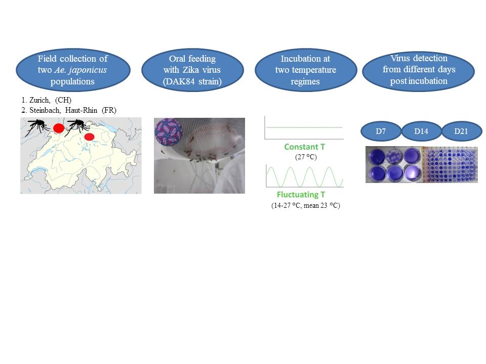
Field collected Ae. japonicus were used for this study. Eggs were collected with standard ovitraps baited with germination paper as oviposition substrate [31]. Two different locations were chosen for collection. The first was located in Zürich, Switzerland, with two collection sites, the Schwamendingen cemetery (47°24'5"N 8°34'28"E) and a private garden close to the University of Zürich - Irchel Campus (47°23'45.3"N 8°33'04.6"E). The second location was a private garden (47°49'16.02"N 7°8'59.20"E) in Steinbach (Haut-Rhin, France). Overall, 30 ovitraps were placed in Zürich (10 in the private garden and 20 in the cemetery) and 20 in Steinbach during June-August 2017. Germination papers were changed weekly, stored semi-dry in plastic zip bags for 7 days at room temperature and then placed in plastic trays with 2 liters of deionized water. One day post immersion, hatched larvae were counted, split (400 larvae/tray) and supplemented with yeast tablets (Gayelord Hauser Superlevure, Gayelord Hauser, France) as larval food (2 tablets/tray). If necessary, half a tablet was added few days later. Larvae were incubated at 27 °C with 85% relative humidity (RH) and the adults obtained were kept into polyester cubic netted cages (32.5 × 32.5 × 32.5 cm) (Bugdorm 43030F, MegaViewScienceCo. Ltd., Taichung, Taiwan) under long daylight conditions (16L:8D) including 1 h dusk and 1 h dawn. A 10% sucrose solution was provided daily to the adults as carbohydrate source.
Mosquito infection
Lyophilized ZIKA virus Dak84 strain, isolated in Dakar (GenBank KU955592 [32]) (7.57 log10 TCID50/ml) and provided by Dr. Failloux, Institute Pasteur, Paris (France). The virus was re-suspended into 400 µl of distilled water, then mixed (1:3) with washed heparinized rabbit blood (not older than 24 h) obtained from the slaughterhouse of a private company (H.R. Kyburz AG, Dorfstrasse 32, Lupfig, Switzerland) to obtain a final a titer of 7.1 log10 TCID50/ml. Finally, phagostimulant (ATP at 5 × 10⁻³ M) was added to each blood meal. Seven to 9 days old females were deprived of sugar 24 h before their exposure to virus-spiked blood as previously described [14]. Briefly, immediately after the preparation of the infectious blood mixture, 3 ml were transferred into a Hemotek feeder (Hemotek Ltd., Lancashire, UK) and covered with a pork intestine membrane fixed with a rubber ring. Mosquitoes were aspirated from the rearing cages and transferred into 500 ml plastic bottles (approx. 60 females/bottle) with the top side covered with a fine net through which the mosquitoes were exposed to the Hemotek feeder. After 20 min of exposure to the infectious blood, mosquitoes were anesthetized by placing the bottles at -20 °C for about 4-5 minutes and then transferred onto a petri dish previously layered with a filter paper. The petri dish was kept on an ice pack the whole time to keep the mosquitoes anesthetized. Fully engorged females were collected and placed into a cardboard box cylinder (12 cm diameter and 15 cm length) covered with nets at both sides which in turn was allocated into a bugdorm cage. Freshly engorged mosquitoes (two females/experiment) were collected immediately after blood feeding (Day 0) as well as a small aliquot of the infectious inoculum for further analysis. All the engorged females were incubated under two different climatic conditions: I) constant temperature (27 °C and relative humidity 85%); II) fluctuating temperature (21±7 °C [average 23 °C] with 45–90% relative humidity), reflecting a typical day in northern Switzerland in mid-summer (www.meteoswiss.admin.ch). The photoperiod for both temperatures was the same as above described. Accordingly, there were four different groups of engorged mosquitoes: Steinbach i) at constant and ii) fluctuating temperature; Zürich iii) at constant and iv) fluctuating temperature. At different time points (day 7, 14 and 21 post oral feeding), 30 females from each infection group were collected. Cardboard boxes were placed at -20 °C for 4-5 minutes to anesthetize the survived mosquitoes after which they were transferred onto a petri dish with ice pack as above described and processed for virus detection. From each time point collection we investigated the rate of infection (IR, proportion of females with infected abdomen among tested ones), dissemination (DR, proportion of females with infected heads among infected ones), transmission (TR, proportion of females with infected saliva among the ones with disseminated infection) and transmission efficiency (TE, proportion of females with infectious saliva among all tested ones). All the feeding, manipulation and incubation of ZIKV infected mosquitoes were done in biosafety containment level 3 (BSL3).
Virus detection in abdomen (infection) and head (dissemination)
After the incubation period, females were dissected for removal of wings and legs with sterile forceps and the rest of the body (head and abdomen&thorax) stored dry in 1.5 ml Eppendorf tubes at -80 °C until further investigation. Infection and virus dissemination, confirmed by the presence of virus particles in the tissues of abdomen&thorax (body) and heads respectively, was determined from body parts’ homogenates. A Tissue Lyser® II instrument (Qiagen, Hilden, Germany) was used for homogenization, at 25 Hz for 1 minute, followed by 5 min centrifugation at 13,000g at 4 °C as described [14]. Briefly, 300 µl of Eagle’s Minimum Essential Medium (EMEM) (LGC Standard, GmbH, Wesel, Germany) supplemented with 1% antibiotics and fungizone (1000 IU/ml penicillin/streptomycin; 4 µg/ml amphotericin) (Gibco, Thermo Fisher Scientific, Reinach, Switzerland) (EMEM complete), 2% fetal bovine serum [FBS], and one stainless steel bead (3 mm diameter) were added to each tube containing either the head or the body of the individual mosquitoes. 96-well plates layered with VERO cells (30 000 cells/100µl/well) and EMEM complete supplemented with 10% FCS were prepared 1 day prior to cells infection, and incubated at 37 °C with 5% CO2. When cells were 75-80% confluent, media was removed and 100 µl of serial dilutions of body part homogenates (neat, 1:10, 1:100) was inoculated into the monolayer of VERO cells. After the incubation period, cells were stained with a crystal violet solution (0.2% of crystal violet,10% formaldehyde and 10% ethanol) in order to identify positive wells. Briefly, two ml of the crystal violet solution were added to each well followed by 30 minutes incubation at room temperature after which the wells were washed two times with deionized water and the presence of viral particle assessed by detection of cytopathic effects (CPE) under a microscope. The whole body of day 0 females was also homogenized and titrated to confirm virus titre. Briefly, a serial dilution (1:10, 1:100, 1:1000, 1:10000, 1:100000) was loaded on 96 well plate layered with VERO cells as above described for the abdomen&thorax and heads. After seven day incubation, cells were checked and titre calculated based on the presence of plaques.
Virus detection and quantification in saliva (transmission)
For saliva collection, after the removal of wings and legs from each survived individual, the proboscis was inserted into 20 µl pipette tips filled with five µl of FBS. After 30 minutes salivation, the five µl of FBS with collected saliva were transferred into 1.5 ml Eppendorf tubes containing 45 µl of EMEM complete giving a final volume of 50 µl. Saliva was held on ice until all the samples were collected and then frozen at -80 °C.
The quantification of infectious virus particles was determined by plaque forming unit assay and expressed as PFU/saliva. Briefly, six-well plates layered with 75-80% confluent Vero cells (800‘000 cells/2ml/well) were inoculated with EMEM complete supplemented with 10% FBS 24 hours before incubation with saliva. For the infection, the media was removed from each well and 265 µl of EMEM complete supplemented with 2% FBS were added to each well, followed by 35 µl of the saliva sample giving a total volume of 300 µl/well. The remaining 15 µl of the saliva samples were kept at -80 °C as a back up. After one hour incubation at 37 °C, four ml of a 0.5% agarose solution (UltraPure™ Agarose, Invitrogen Life Technologies, UK) in EMEM complete were added to each well without the removal of the inoculum, and all plates incubated at 37 °C with 5% CO2. At day seven post incubation, the agarose gel was removed and cells were stained by adding two ml of the crystal violet solution per well. After 30 min incubation at room temperature, cells were rinsed with water and plaques enumerated according to the volume of the sample tested and expressed as PFU/saliva.
Statistical analysis
Differences in the rates of infection, dissemination, and transmission between the two temperature conditions (fluctuating 21±7 °C and constant 27 °C) and the two populations of Ae. japonicus were analysed by logistic regression.
There were therefore 3 logistic regression models. The first considered infection of mosquitoes as a binomial dependent variable (virus detect in the abdomen as infected or no virus detected as not infected). The independent variables were site (a categorical variable, France or Zürich), time (continuous variable n days post oral feedings) and temperature (categorical variable constant or fluctuating).
The second logistic regression model considered only those in which infection was proven. In this case dissemination was the dependent variable (virus detected in the head as disseminated or not disseminated).
The third logistic regression model only considered those mosquitoes in which there was dissemination of virus. The detection of virus in the saliva (transmission) was the dependent binomial variable (positive or negative). The same independent variables for the first logistic regression model were used in the second and third regression model. Interactions between independent variables were also analysed. All proportions (infected / not infected, disseminated / not disseminated, infectious / not infectious) are reported with exact 95% binomial confidence intervals. The association of viral growth (copy number) with time or temperature was analysed according to negative binomial Generalized Linear Model (GLM) with a log link function. This assumed that copy number was an integer dependent variable, with a minimum value of zero that was over dispersed. Hence the negative binomial model was considered the most appropriate model for analysis. For all regression models backward stepwise elimination was used to remove non-significant variables in the model. All possible interactions in the models were also examined. Regression models were also undertaken taking data from the two sites in separate models for an insight into variables that may only have an association at one of the two study sites. Significant variables remaining within models are reported as the p value and corresponding Z statistic. All analysis was undertaken in R [16].