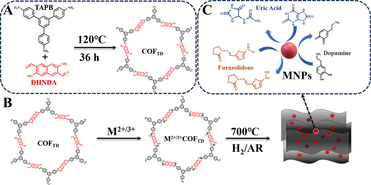
3.2 Characterization of CuNPs/CN
As shown in Fig. S1, the CN with an obvious two-dimensional nanosheet structure was formed after carbonization of COFTD, indicating that the process of carbonization did not destroy the two-dimensional structure of COFTD. TEM image showed that CuNPs/CN were two-dimensional nanosheets with a large number of nanoparticles uniformly (Fig. 2A), and the insert indicated these nanoparticles were 8 nm in size. As shown in Fig. 2B, there were obvious lattice fringes with a lattice spacing of 0.205 nm corresponded to (111) plane of CuNPs, demonstrating that the nanoparticles on CN were CuNPs [36].
Figure 2C showed the XRD of CN and CuNPs/CN. There was only a peak at around 25° on CN, which corresponds to the (001) crystal plane of carbon. The peaks at 43.19°, 50.30° and 73.89° on CuNPs/CN corresponding to the (111), (200) and (220) crystal planes of CuNPs (PDF#70-3038), which proved the successful formation of CuNPs on CN [35]. The BET area of CN and CuNPs/CN were 471 m2 g− 1 and 356 m2 g− 1, respectively (Fig. 2D). XPS spectrum showed there were C, N, O and Cu elements in CuNPs/CN (Fig. 2E). High-resolution XPS spectrum of CuNPs showed the peaks at 933.0 eV and 952.0 eV corresponded to Cu0 2p3/2 and Cu0 2p1/2 (Fig. 2F), which further proved that the successful formation of CuNPs on CN. In addition, the two satellite peaks could also confirm the existence of Cu in CuNPs/CN composites.
“Here in Fig. 2”
3.4 Electrochemical detection of DA and UA by using MNPs/CN nanozymes
Cyclic voltammograms (CV) curves in Fig. 4A were measured in 0.1 M KCl solution with 5 mM [Fe(CN)6]3−/4− at 50 mV s− 1. Compared with other materials, the peak current of CuNPs/CN/GCE was significantly increased, which showed that the electrochemical performance had been significantly improved after carbonization because of the synergistic effect of CN and CuNPs. Electrochemical impedance spectroscopy (EIS) of CuNPs/CN/GCE, CN/GCE, GCE and COFTD/GCE clearly showed that CuNPs/CN/GCE owned the lowest resistance (Fig. 4B), consistent that of CV.
The fast electron transfer manifested that CuNPs/CN/GCE could be used for further electrochemical sensors, such as the electrochemical detection of DA and UA. The DPV curve of CuNPs/CN/GCE, CN/GCE, GCE and COFTD/GCE toward 10 µM DA and 50 µM UA in N2- saturated PBS showed that CuNPs/CN/GCE obtained the largest response current toward DA and UA (Fig. 4C), which proved that the CuNPs/CN had the best detection performance. The good performance could be ascribed to the synergistic effect of CuNPs and CN. COFTD as a precursor had a strong chelating effect on Cu2+, and a large amount of Cu2+ are evenly dispersed in the pores of COFTD. After carbonization, the COFTD accumulation was eliminated, and CuNPs with a diameter of 8 nm were obtained, so that more active sites were exposed and the catalytic effect on DA and UA was improved. In order to study the kinetics of CuNPs/CN/GCE toward DA and UA, the electrochemical behavior of CuNPs/CN/GCE toward DA and UA in N2-saturated PBS with different pH (6.0–8.0) were investigated (Fig. 4D). The oxidation peak potential of DA and UA gradually shifted negatively with the increase of pH, and the peak oxidation current was the largest when the pH was 7.0. The illustration showed a linear fitting curve of pH and Epa, the slopes of the fitted curves were − 57.5 mV pH− 1 and − 61.4 mV pH− 1, approaching to -59 mV pH− 1 of the isoelectronic and proton process. It showed that oxidation of DA and UA catalyzed CuNPs/CN was an isoelectronic and proton process.
“Here in Fig. 4”
Then, under the condition of 0.2 M N2-saturated PBS (pH = 7.0) in presence of DA and UA, the CVs at different scan rates were explored. As shown in Fig. S2A and B, the redox process of DA occurred at about 0.15 V, and the effect of scan rates on peak position was not obvious, which demonstrated that the reversibility was good. Through the fitting graph, it could be found that the peak current density was directly proportional to the scan rate, which proved that what was happening on the electrode surface was a classic surface control process. The peak current density of UA was proportional to the scan rate, so it was also the dominant electron transfer process controlled by the surface (Fig. S2C and D).
Thanks to the good conductivity and excellent catalytic properties of the material, the CuNPs/CN/GCE electrochemical sensor was constructed to detect DA and UA. To ensure the performance of the sensor, a series of optimization of testing conditions were carried out. Under the condition that the mass of COFTD was 40 mg to coordinate with 0.1 mmol Cu2+, the resulted sensor achieved the best catalytic effect (Fig. S3A). The calcination temperature of Cu2+/COFTD was optimized from 500°C to 800°C (Fig. S3B), the results revealed that 700°C was the best temperature for obtaining the highest activity of CuNPs/CN. As discussed above, when the pH was 7.0, the peak current density of DA and UA detection reached the maximum, so pH = 7.0 were selected as the optimal detection pH value (Fig. S3C). Finally, because the concentration of the material on the electrode had a great influence on the catalytic effect, the concentration of CuNPs/CN was optimized. When concentration of CuNPs/CN was increased from 1 mg mL− 1 to 2 mg mL− 1, the catalytic current of DA and UA increased gradually. However, when the concentration was greater than 2 mg mL− 1, excess CuNPs/CN might accumulate on GCE, reducting the catalytic current, so 2 mg mL− 1 CuNPs/CN was used to construct electrochemical CuNPs/CN sensor (Fig. S3D).
Next, CuNPs/CN/GCE was used to separately measure DA and UA through the DPV technique. Figure 5A and B were the DPV diagrams of DA and UA respectively detected under N2 protection and the insets were the corresponding linear fitting diagram. With the gradual increase of DA and UA concentrations, the corresponding oxidation peak current density also gradually increased. The sensitivity of DA detection was 0.805 µA µM− 1 cm− 2, and the linear range was 55 nM-250 µM. The sensitivity of UA detection was 0.73 µA µM− 1 cm− 2, and the linear range was 62 nM-800 µM. This result indicated that CuNPs/CN could be used as a good catalytic material to construct electrochemical enzyme-free DA and AA sensors with good performances.
As we all know, the oxidation peaks of DA and UA are very close or even overlap, and they usually interfere with each other when using ordinary electrodes for detection. In order to prove the excellent catalytic effect of CuNPs/CN/GCE, DA and UA were measured simultaneously by DPV. In 0.2 M N2-saturated PBS (pH = 7.0), the DA was detected after adding quantitative 200 µM UA (Fig. 5C), the UA was detected after adding quantitative 50 µM DA (Fig. 5D) and the insets were corresponding curves of the peak current density versus concentration. Under the condition that the peak current of UA was basically constant, with the increase of DA concentration, the peak current linearly increased in the range of 0.042 µM-200 µM, and the sensitivity was 1.050 µA µM− 1 cm− 2. In the presence of DA, as the concentration of UA increased, DA increase was within the allowable range of error. The anode peak current of UA was proportional to the concentration from 0.12 µM to 1100 µM, and the sensitivity was 0.38 µA µM − 1 cm− 2. In summary, the oxidation of DA and UA on the CuNPs/CN/GCE occurred independently, with different oxidation peak positions, without any mutual interference. In the mixed solution, the concentrations of DA and UA were changed at the same time. As shown in Fig. 5E, the oxidation peak of DA was at 0.12 V, the oxidation peak of UA at 0.25 V, and the peak current densities of DA and UA all were directly proportional to the concentration. The linear range of DA detection was 0.015 µM-140 µM, the linear range of UA detection was 0.03 µM-175 µM, and the sensitivity of DA and UA were 1.03 µA µM− 1 cm− 2 and 0.52 µA µM− 1 cm− 2 (Fig. 5F). As shown in Table S1, the sensor exhibited a wider linear range and better sensitivity than other works, which proved the advantages of CuNPs/CN materials in the construction of sensors.
Subsequently, the repeatability, reproducibility and stability of sensors based on CuNPs/CN/GCE were explored. The CuNPs/CN/GCE was placed for 30 days, and DA and UA were continuously detected. As shown in Fig. S4A, the detection peak current densities of the two substances don’t change much within one month, the relative standard deviation (RSD) of DA is only 10.1%, and the RSD of UA is 11.41%, indicating that the sensor has good repeatability for DA and UA detection. Six CuNPs/CN/GCE were prepared to detect DA and UA at the same time, and the peak current densities were basically unchanged, indicating that the CuNPs/CN/GCE also has good repeatability (Fig. S4B). In the presence of DA and UA, the addition of interfering substances caused the changes in the oxidation peaks of DA and UA to be less than 10% of their original current density, which was sufficient to prove that the sensor had good selectivity (Fig. S4C and D). In conclusion, the CuNPs/CN/GCE biosensor has good stability, repeatability and selectivity for DA and UA detection.
“Here in Fig. 5”
In view of the excellent performance exhibited by CuNPs/CN/GCE sensor, the MNPs/CN nanozymes synthesized above were used to construct sensors, and the catalytic effects of the nanozymes were verified by detecting DA and UA. In 0.2 M N2-satruated PBS in the presence of UA, FeNPs/CN/GCE (Fig. 6A), CoNPs/CN/GCE (Fig. 6C) and NiNPs/CN/GCE (Fig. 6E) were used to detect DA. In 0.2 M N2-satruated PBS in the presence of DA, FeNPs/CN/GCE (Fig. 6B), CoNPs/CN/GCE (Fig. 6D) and NiNPs/CN/GCE (Fig. 6F) were used to detect UA. The illustrations corresponded to the relationship between peak current density and concentration. It can be observed that three nanozymes could clearly distinguish the oxidation peaks of DA and UA, and the oxidation process of DA and UA could occur independently and did not affect each other when DA and UA existed at the same time. The sensitivity of FeNPs/CN/GCE, CoNPs/CN/GCE and NiNPs/CN/GCE to detect DA were 1.30 µA cm− 2 µM− 1, 1.07 µA cm− 2 µM− 1 and 0.88 µA cm− 2 µM− 1, the linear ranges were 35 nM-200 µM, 42 nM-250 µM and 52 nM-250 µM. The sensitivity of FeNPs/CN/GCE, CoNPs/CN/GCE and NiNPs/CN/GCE for detecting UA were 0.310 µA cm− 2 µM− 1, 0.587 µA cm− 2 µM− 1 and 0.360 µA cm− 2 µM− 1, the linear ranges were 145 nM-900 µM, 77 nM-700 µM and 125 nM-800 µM. The performance of this series of sensors were compared with the related sensors in Table S1, and they all showed excellent performance.
“Here in Fig. 6”
3.5 Electrochemical detection of FZ based on CuNPs/CN/GCE
In view of CuNPs/CN/GCE high catalytic performance, an electrochemical sensor for FZ detection was constructed. Fig S5 compared DPV curves of different materials in 0.2 M PBS containing FZ. These materials showed a reduction peak of FZ around − 0.4 V. For the same concentration of FZ detection, the sensor constructed by CuNPs/CN/GCE not only had a higher current density, but also the reduction peak of FZ was positively shifted. This indicated the sensitivity of FZ on CuNPs/CN/GCE was higher and the reduction process was easier.
As mentioned above, CuNPs/CN was excellent material for detecting FZ. In order to study the kinetic process of FZ catalyzed by CuNPs/CN/GCE, the CVs at different scanning speeds in 0.2 M PBS containing 20 µM FZ were investigated (the scanning speed was 50 mV s− 1 to 500 mV s− 1) (Fig. S6). With the increase of the scan rate, the reduction peak current also gradually increased. It can be obtained from the illustration that the FZ reduction peak current had a linear proportional relationship with different scan rates, and the correlation coefficient was R2 = 0.99. This result confirmed that for CuNPs/CN/GCE, the electrochemical reduction of FZ was a surface-controlled electron transfer process.
Subsequently, the FZ was measured by DPV technique using CuNPs/CN/GCE, and the results were shown in Fig. 7A and B. Within a certain range, with the gradual increase of FZ, the concentration and reduction peak current showed a good linear relationship, and the correlation coefficient was R2 = 0.99. The sensitivity of CuNPs/CN/GCE biosensor to detect FZ was 0.732 µA µM− 1 cm− 2, and the lowest detection limit was calculated by (LOD) = 3 SD/SCP, where “SCP” is the slope of the calibration and the standard of “SD” regression difference. Based on this, the LOD was calculated to be 20.5 nM, and the linear range was 61.5 nM-200 µM, which exceeded the previously reported FZ sensors. Table S2 comprehensively compared the relevant electrochemical sensor parameters for detecting FZ. This result once again proved the potential of CuNPs/CN/GCE as a catalytic material to construct electrochemical enzyme-free sensors in clinical treatment and environmental protection.
“Here in Fig. 7”
When building sensors, selectivity played a very important role in practical applications. By adding different bioactive molecules and nitrogen-containing organic compounds, the anti-interference of CuNPs/CN/GCE was studied. Fig. S7 showed that CuNPs/CN/GCE had good selectivity for FZ detection. Next, the repeatability and stability of the FZ electrochemical sensor were studied. For 30 consecutive days, the same CuNPs/CN/GCE was used to measure the current response of a PBS containing 50 µM FZ, and its RSD was 7.58%, so the biosensor had good stability (Fig. S8A). Under the same conditions, five CuNPs/CN/GCE modified electrodes were used to detect 50 µM FZ, and the RSD was only 8.62%, so the CuNPs/CN/GCE sensor had good repeatability (Fig. S8B). Consequently, the FZ electrochemical sensor constructed with CuNPs/CN/GCE had an excellent performance.