USF1 deficiency ameliorates depression-like and anxiety-like behaviors.
Adult female and male USF1 KO and WT littermate controls were behaviourally characterized in a battery of standard paradigms, with special emphasis on tests assessing emotional behaviours relevant to depression and anxiety. In the Forced-Swim Test (FST) where immobility reflects behavioural despair in response to exposure to an acute inescapable stress situation, USF1 KO mice of both sexes were less immobile than their WT littermates (Fig. 1A; main effect of genotype F(1,59) = 35.46; p < 0.0001; n = 13–17/group). Further, in the Novelty Suppressed Feeding Test (NSF), in which animals are faced with the stressful conflict of pursuing food under anxiogenic conditions, both female and male USF1 KO mice presented with shorter latencies to begin eating in the centre of a brightly illuminated novel arena than their WT counterparts. This suggests that USF1 deficient animals are more prone to resolve the ambivalent situation in favour of the active behaviour to feed despite a fear inducing environment (Fig. 1B; main effect of genotype F(1,57) = 18.46; p < 0.0001 n = 12–17/group). However, food consumption in the home cage measured immediately after the NSF did not reveal any difference between genotypes of either sex, confirming that the phenotype of USF1 KO mice in the NSF was not due to a baseline alteration in the drive to eat (Fig. 1C). Notably, the displays in the FST and the NSF are considered as distinctive depression-related behavioural dimensions [52].
Exposure to the Elevated Plus Maze (EPM) allows for the determination of anxiety-like behaviour under fear-inducing conditions [53, 54]. Here a significant main effect of genotype was noted with female and male USF1 deficient mice displaying less anxiety face to the more aversive illuminated arms than WT littermates (Fig. 1D-E; F(1,58) = 8.734; p = 0.0045; n = 13–17/group).
Interestingly, in the Sucrose Preference Test (SPT), which probes hedonic behaviour when animals are left undisturbed in their home cage, female and male USF1 KO mice were undistinguishable from WT controls, suggesting no effect of USF1 deficiency under baseline conditions in the absence of an external stressor (Fig. 1F).
USF1 KO mice of both sexes travelled similar total distances in the Open Field Test (OFT) as their WT littermates evidencing no differences in exploratory and locomotor activity between genotypes (Fig. 1G). Similarly, motor coordination was unaltered in female and male KO mice as determined by the latency to fall in the Rota Rod (RR) (Fig. 1H), jointly dismissing unspecific biases through general behavioural alteration on the performance of USF1 KO mice in the depression-related and anxiety-related tests.
The surgical removal of interscapular BAT depots (iBATX) does not alter the behavioural phenotype of USF1 deficient mice.
In order to investigate whether the reduction in stress-related negative valence behaviors directly resulted from the highly active BAT of USF1 KO mice, interscapular BAT (iBAT), the largest BAT depot in rodents [55] corresponding to supraclavicular BAT in adult humans [56, 57] was surgically removed in female USF1 KO and WT mice (Fig. 2A-B). 6 weeks post iBATx or sham surgery all animals were tested in the FST and the EPM. No effect of iBATx was found in either genotype. Instead, the previously observed phenotype of USF1 KO mice was confirmed, with reduced immobility in the FST (Fig. 2C; F(1,24) = 9.53; p = 0.<01; n = 5–9/group) and a higher percentage of entries into the open arms in the EPM (Fig. 2D; F(1,23) = 32.64; p < 0.0001; n = 5–9/group) in USF1 KO mice. These results indicate that the behavioural phenotype resulting from USF1 deficiency does not require the presence of iBAT in adult animals.
Adult hippocampal neurogenesis is not affected by USF1 deficiency.
Progenitor cell proliferation in the subgranular zone of the adult hippocampal dentate gyrus and survival of newly generated neurons has been strongly related to depression-like behaviour in different animal models [58]. We therefore sought to examine whether the reduction of depression-like and anxiety-like behaviour in USF1 KO mice was also reflected in an alteration of adult hippocampal neurogenesis. We used Bromodeoxyuridine (BrDU)-dependent immunofluorescence histochemistry and evaluated proliferation of progenitor cells and differentiation and survival of newly born cells in the dentate gyrus (Fig. 3A). The number of BrDU + cells was comparable between USF1 KO and WT littermate controls both 24 h (Fig. 3B) and 14 days after BrDU administration (Fig. 3C). Similarly, the rate of differentiation of newly born cells into neurons (as indicated by the presence of NeuN; Fig. 3D and representative image 3D’) or astrocytes (as indicated by GFAP staining; Fig. 3E and representative image 3E’) was not significantly different between genotypes, suggesting that the lack of USF1 did not impact on the processes of adult hippocampal neurogenesis, and that neurogenic effects may likely not account for the behavioural phenotype of USF1 KO mice.
Several members of the xlr gene family are upregulated in the USF1 KO hippocampus.
Considering that USF1 is a ubiquitously expressed transcription factor and given that the behavioural phenotype of USF1 KO mice appeared independent of the signals acutely deriving from BAT activity, we next decided to employ a hypothesis-free approach to reveal the molecular signature in the brain, paralleling the behavioural repercussions of USF1 deficiency. To this end, hippocampal transcriptomic profiles were generated by mRNA sequencing and compared between USF1 KO and WT controls (Fig. 4A). 901 genes were found to be significantly differentially expressed (DEG) between genotypes in the hippocampus USF1 deficient mice (Fig. 4B, full data set in Supplementary Table 1).
A closer look at the list of DEGs revealed that the expression of several members of the xlr (X-linked lymphocyte-regulated) gene family was significantly upregulated in the USF1 KO hippocampus: xlr5c, xlr3a, xlr3b, xlr4b, xlr3c and xlr4a (Supplementary Table 1). The xlr gene family comprises closely related genes encoding similar proteins considered to be relevant to chromatin modification and recently discovered as important regulators of neuronal structure and function in the mouse brain [59, 60]. qRT-PCR confirmed the significant increase in the expression of xlr3b (Fig. 4C, p < 0.05, n = 5/group) and xlr4b (Fig. 4D, p < 0.0001, n = 4–5/group), which had been specifically identified as regulators of dendritic complexity and spine number and morphology [59].
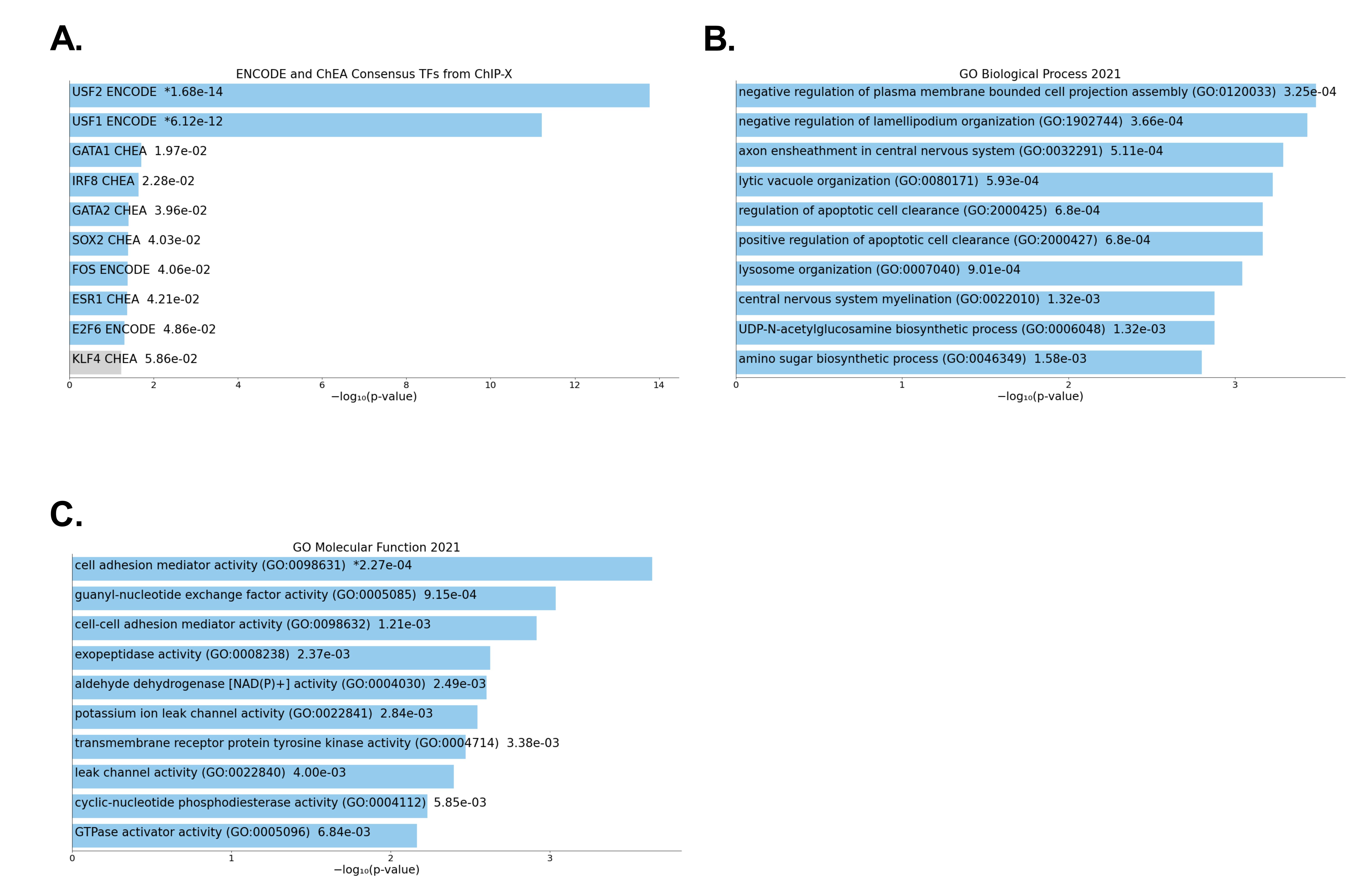
Additional bioinformatic analysis of the top 100 DEGs confirms most of the affected genes to be direct targets of USF-mediated transcription (Suppl. Figure 1A) and suggests alterations in biological functions and molecular processes with relevance to dendritic outgrowth and complexity (Suppl. Figure 1B and 1C).
USF1 deficiency leads to stubbed dendritic length and complexity.
We then went on to explore whether enhanced levels of xrl transcripts also related to alterations in neuronal structure in the USF1 KO brain. To this end, we investigated neuronal morphology in in USF1 KO and WT mice using single-cell reconstructions of Golgi-Cox stained hippocampal sections (Fig. 5A-B).
We focussed on pyramidal cells in the CA1, as previously the effects of xlr genes on neuronal structure in the cortex had also been examined in pyramidal cells [59]. We found a significant decrease in the cumulative dendritic length (Fig. 5C, p < 0.001, n = 14–27/group), specifically resulting from a highly significant reduction in the length of non-apical dendrites (Fig. 5D, p < 0.0001, n = 14–27/group), in line with previous observations related to increased expression of xlr3b and xlr4b [59]. Next, we asked whether USF1 deficiency also impacted on dendritic arborization and spine density. In comparison with WT controls, USF1 KO neurons had a significantly lesser number of nodes (Fig. 5F, p < 0.01, n = 14–27/group), whereas no difference in spine density was observed (Fig. 5G). These observations demonstrate that lack of USF1, concomitantly to the dysregulation of xlr gene expression, alters neuronal morphology in the brain.