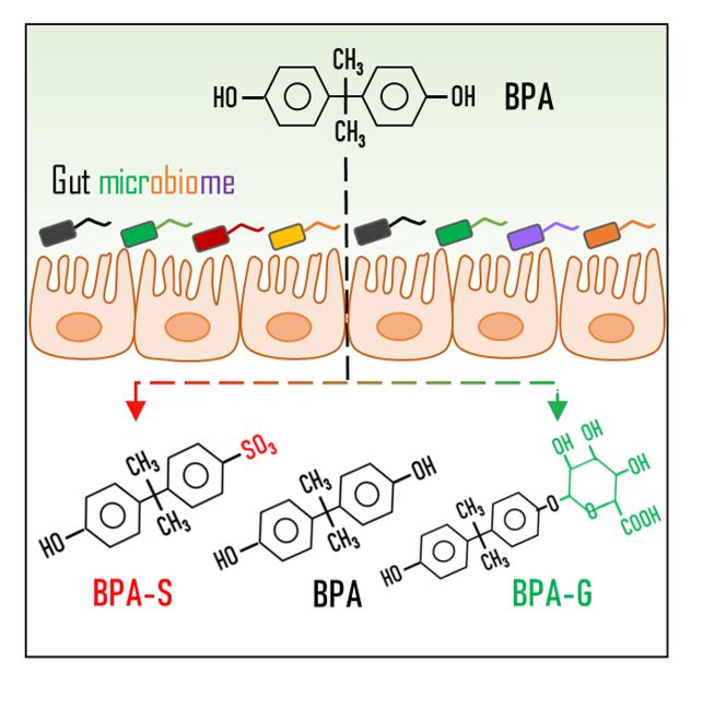
Standards and Reagents. BPA (2,2-(4,4’-dihydroxydiphenyl)propane; purity > 98%), bisphenol A mono-β-d-glucuronide (BPA-G; > 98%), bisphenol A mono-sulfate (BPA-S; > 98%), bisphenol A bis-β-d-glucuronide (BPA-BG; > 97%), bisphenol A di-sulfate (BPA-DS; > 97%), 13C12-BPA, 13C12-BPA β-d-glucuronide (13C12-BPA-G; > 98%), and D6-BPA mono-sulfate (D6-BPA-S; > 98%) were purchased from Toronto Research Chemicals (North York, ON, Canada). Dimethylsulfoxide (DMSO), methanol, physiological saline (0.9% NaCl solution), pure water, and ammonium acetate were obtained from Sigma-Aldrich (Oakville, ON, Canada).
Animal and Experimental Design. Sprague-Dawley rats (ten weeks old, body weight 230 − 280 g) were obtained from SLRC Animal Laboratory (Shanghai, China). All animals were housed (5 rats/cage) in a temperature (22–25°C), humidity (40–50%), and light (12/12 hr light/dark cycle) controlled house under specific pathogen-free conditions, and given ad libitum access to standard food (Medicience Ltd., Jiangsu, China) and water. Before conducting the experiment, all animals were allowed to acclimate for seven days. All SD rats were humanely treated throughout the experiment, following protocols approved by the Zhejiang University of Technology Animal Ethics Committee.
A first experiment was conducted in 10 SD rats to investigate whether the metabolism of BPA was changed under stable gut microbiota community. On day 1, 8, 15, 22, and 29 (7-day interval), all SD rats were administered with BPA (reconstituted in 50% DMSO/water) once only by gavage at a dose of 500 µg/kg bw. After oral BPA intake, SD rats (n = 10) were individually placed into metabolic cages, and total rat feces and urine samples were collected after the natural micturition until 24 h after BPA administration. After that, rat whole blood samples (1 mL) were collected from vena caudalis (24 h after BPA administration).
A second experiment was performed in another 10 SD rats to assess whether the altered gut microbiota could change the metabolism of BPA. All SD rats were continuously administered with BPA (reconstituted in 50% DMSO/water) at 500 µg/kg bw by oral gavage once a day for 29 days. On day 0, 1, 3, 5, 7, 9, 15, and 29, BPA-exposed rats (n = 10) were individually placed in metabolic cages after dosing for 24 hours to collect urine and feces samples. After that, rat whole blood samples (1 mL) were collected from vena caudalis (24 h after BPA administration).
In both experiments, SD rats (n = 10), dosed with the same amount of 50% DMSO/water and given ad libitum access to clean water and food, were set as control groups. The total volume and weight of urine and feces collected from individual rats were accurately measured. Rat blood was collected in BD Vacutainer® tubes (embedded with sodium heparin; NJ, USA). Dry ice was placed surrounding the urine and feces collection glass vessels during the sample collection period. After collection, rat feces samples were transferred to RNase-free microfuge tubes (15 mL; Conical, Thermo Fisher; ON, Canada), and stored at -80 oC until extraction and analysis.
Sample Extraction. Prior to extraction, all samples were spiked with 13C12-BPA, 13C12-BPA-G, and D6-BPA-S (5 ng each), working as internal standards. Rat urine samples were diluted with pure water, and then extracted by solid-phase extraction (SPE), following the method of Liao and Kannan (2012). Rat blood samples (500 µL) were directly extracted with methanol. Feces samples were freeze-dried, homogenized, and then extracted using 80% methanol/water, with additional purification using Supelco Envi-Carb cartridges (Sigma-Aldrich; ON, Canada). In additional, spike and recovery experiments were conducted to evaluate the extraction efficiency of analytes. Detailed extraction procedures and recovery experiments are provided in the Supporting Information (SI).
Instrumental Analysis. All sample extracts were analyzed with a ACQUITY liquid chromatography coupled with a triple quadrupole mass spectrometer (XEVO_TQS; Waters; Milford, MA, USA) (Jin et al. 2020, Zhao et al. 2021). The liquid chromatography was performed using an HSS T3 column (1.8 µm, 2.1 × 50 mm; Waters, MA, USA), with the mobile phase composed of methanol and water (2.0 mM ammonium acetate, pH = 7). The column temperature and flow rate of mobile phase were maintained at 40 oC and 0.2 mL/min, respectively. The gradient elution was initially held at 20% methanol for 0.5 min, increased to 40% methanol by 1.0 min, and ramped to 95% methanol by 6.0 min, which was held for 2 min, and then returned to the starting condition. The mass spectrometer was operated in negative ionization mode, and spectral data was record by multiple reaction monitoring (MRM; 2 transitions per analyte). Detailed MRM transitions of target analytes are provided in the SI, Table S1.
QA/QC. Pure methanol (10 µL) was analyzed between every ten samples to monitor carryover contamination, and no obvious cross contamination between injections was found. Several steps were taken to achieve a very low or undetectable background BPA pollution. Despite glassware was used in the whole extraction procedures, procedural blanks analyzed along with every ten real samples still contained stable and low concentrations of BPA, which was possibly originated from SPE cartridges. BPA and BPA conjugates in collected samples were quantified by the internal calibration method. Background BPA concentrations were subtracted from quantified BPA concentrations in real rat samples. Limits of detection (LODs) were defined as the concentrations of analytes correspond to a signal-to-noise ratio of 3 in sample extracts from control rats, and were in the range of 0.047 − 0.088 ng/mL, 0.039 − 0.077 ng/g, and 0.039 − 0.15 ng/mL in rat blood, feces, and urine, respectively. Extraction recoveries of target analytes in rat blood, feces, and urine ranged from 72 to 114%. Detailed LODs and extraction recovery of analytes are shown in the SI, Table S2 and S3.
To avoid the in-source fragmentation of BPA conjugates, the capillary voltage of ion source was set at a low level (-1.0 kV) at a little sacrifice of detection sensitivity. Fragmentation of BPA conjugates in the second quadrupole could generate BPA, which greatly increased the quantified BPA concentrations. This interference was minimized by the baseline separation of BPA and its conjugated metabolites. A T3 column having strong retention capacity with hydrophilic compounds was used to reduce the serous tailing and shifting retention time of the peak of BPA-DG. Typical chromatograms and molecular structure of target analytes in the standard solution and rat blood are shown in Fig. 1 and SI, Figure S1. Given the instability of BPA conjugates (Waechter et al. 2007), special care was taken to avoid deconjugation during rat sample collection and extraction. Rat blood, feces, and urine samples spiked with 13C12-BPA-G and D6-BPA-S (at 10 or 100 ng/mL) were analyzed along with real samples. No measurable 13C12-BPA and D6-BPA were detected in these fortified samples, demonstrating the negligible deconjugation of BPA metabolites in the sample analysis process. BPA metabolites were not detected in any control rat blood, urine, and feces samples. In control rats only BPA was detected in feces at levels around LOD (mean 0.07 ng/g), which was subtracted from BPA concentrations in feces of exposed rats.
Fecal Bacterial DNA Extraction and 16S rRNA Sequencing. Bacterial DNA was extracted from rat fecal samples using an E.Z.N.A.® Stool DNA Kit (50T, Omega Bio-Tek; Norcross, GA, USA), according to the manufacturer’s protocol. The purity of extracted DNA was determined with NanoDrop 2000 (Thermo Scientific; USA). After that, the DNA was PCR-amplified with barcoded primers (27F: 5’-AGRGTTYGATYMTGGCTCAG-3’ and 1492R: 5’-RGYTACCTTGTTACGACTT-3’), targeting the V1 − V9 regions of the bacterial 16S rRNA gene. The PCR reaction mixture (20 µL; performed in triplicate) included 4 µL of 5 × FastPfu buffer, 2 µL of 2.5 mM dNTPs, 0.8 µL of each primer (5 µM), 0.4 µL of FastPfu polymerase, and 20 ng of extracted DNA. The PCR (TransGen AP221-02: TransStart Fastpfu DNA Polymerase) program began with a denaturation step (94°C; 3 min), followed by denaturation (20 cycles of 1 min; 94°C), annealing (1 min; 65°C to 57°C with a 1°C reduction every two cycles, one cycle at 56°C, and one cycle at 55°C), elongation steps (72°C; 1 min), and a final 6 min extension at 72°C. The PCR products were pooled, purified with MiniBest DNA Fragment Purification Kit Ver 4.0 (TaKaRa; Tokyo, Japan), and then sequenced on an Illumina Miseq (Illumina; San Diego, CA, USA). Prior to sequencing, the DNA library was quantified using Library Quantification Kits (KAPA Biosystems; Merck, USA).
Sequencing Data Analysis. The obtained raw fastq files were demultiplexed (using 8-bp barcodes) and quality-filtered using the QIIME software (Caporaso et al. 2010). Reads with any unknown bases, > 2 mismatches to the primers, > 1 one mismatch to the barcode, or < 50 bp length were discarded. The DNA reads having 3 consecutive low-quality bases calls were truncated. After that, overlapped paired-end reads were merged to tags (USEARCH; http://www.drive5.com/usearch/), which were further stepwise clustered to Operational Taxonomic Unit (OTU) with ≥ 97% sequence similarity (UPARSE; http://drive5.com/uparse/). A representative sequence from each OTU was taxonomically classified, using the Ribosomal Database Project (RDP) classifier (http://rdp.cme.msu.edu/), against the SILVA database (http://www.arb-silva.de) with a confidence threshold of 80%. In this study, taxonomical classification was primarily focused on gut microbiota at phylum and genus levels. Unknown, unclassified, and unassigned classifications were omitted from the final dataset.