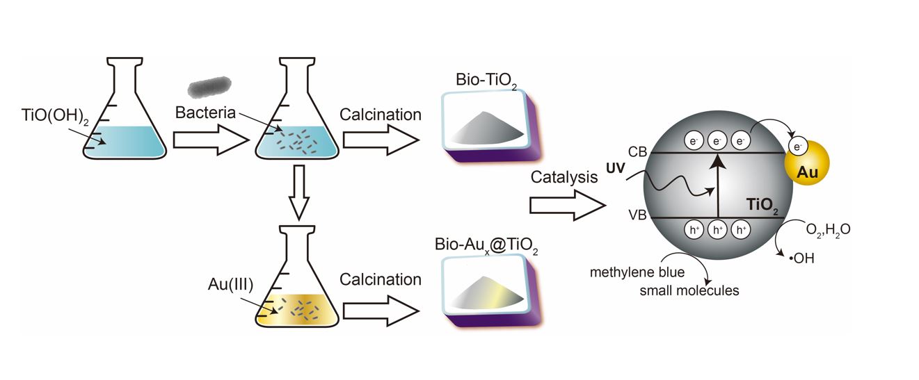
3.1 Materials Characterization
The morphology and structure of bio-TiO2 and bio-Aux@TiO2 materials synthesized by the IMH strain were characterized using multiple complementary techniques. XRD spectra (Fig. 1) resolved the characteristic diffraction peaks for bio-TiO2 at 25.33°, 37.53°, 47.87°, 53.53°, 54.86°, 62.36° and 74.63°, corresponding to (101), (004), (200), (105), (211), (204) and (215) crystal planes of the standard anatase TiO2 (JCPDS-01-071-1168), respectively.(Nassar et al. 2017) In the XRD spectra of bio-Aux@TiO2, the diffraction peaks at 38.19°, 44.39°, 64.58°, 77.57° and 81.73° correspond to the (111), (200), (220), (311) and (222) crystallographic planes of standard Au metal (JCPDS-01-089-3697), respectively.(Tran et al. 2022) And other major peaks from bio-Aux@TiO2 spectra corresponded to the planes of standard anatase (JCPDS-01-071-1168). (Nassar et al. 2017) In addition, the peak positions of TiO2 did not shift from bio-TiO2 to bio-Aux@TiO2, indicating that bio-TiO2 still kept the anatase crystalline phase during the deposition of Au NPs. XRD spectra results demonstrated that the IMH strain could successfully synthesize bio-TiO2 and bio-Aux@TiO2 materials.
The FE-SEM (Fig. 2a) micrograph and EDX spectrum showed that bio-TiO2 consists of aggregated irregular spheres with diameters of about 50–100 nm. The SEM images (Fig. 2b-2e) of bio-Aux@TiO2 exhibited the Au NPs as bright dots were dispersed on the surface of bio-TiO2 support. Meanwhile, the Au NPs of bio-Au0.5@TiO2 and bio-Au0.75@TiO2 were spheres with particle size of 5 ~ 10 nm; the diameter of Au NPs in bio-Au1@TiO2 was mainly 10 nm; and the Au NPs of bio-Au2@TiO2 were spheres with a diameter of 5 ~ 15 nm. With the increase of concentration of Au ions in cell solution, the quantity and dimension of biosynthesized Au NPs notably increased, and the nanoparticles distribution tends to be even.
HR-TEM in Figure S1 showed that Au NPs (5–20 nm) were evenly dispersed in the bio-TiO2 layers. The EDS elemental mapping in Figure S1c shown that the bio-Au2@TiO2 nanocomposite synthesized by the IMH strain consisted of elemental Au, Ti, O, and C.
The valence state of Au and Ti in bio-Aux@TiO2 was analyzed using XPS as shown in Fig. 3. Specifically, the peaks at 83.7 ~ 84.4 eV and 87.4 ~ 87.9 eV were ascribed to the 4f7/2 and 4f5/2 of Au(0), respectively(Liang et al. 2012). XPS results further confirmed that the IMH strain could completely reduce Au(III) to synthesize Au NPs. The high resolution XPS scan resolved two peaks of Ti 2 p electrons at 459.3 eV and 465.05 eV, which were respectively assigned to Ti 2p3/2 and Ti 2p1/2 of oxidized state Ti4+, respectively.(Khalid et al. 2016) The energy difference (5.7 eV) between the peaks of Ti 2p3/2 and Ti 2p1/2 is corresponding to the Ti4+ state of anatase phase TiO2, which is consistent with the previous XRD results.(Trino et al. 2018)
The FTIR spectra of the IMH strain, bio-TiO2 and bio-Aux@TiO2 were shown in Figure S2. The IMH strain spectra displayed typical bacterial characteristic peaks, which were mainly located at 3310 cm− 1 (-OH stretching), 1610 cm− 1 (C = C stretching), 1540 cm− 1 (N-H bending) and 1040 cm− 1 (C-N stretching).(Bosch et al. 2008, Fischer et al. 2006, Schmitt &Flemming 1998) While in the FTIR spectra of bio-TiO2 and bio-Aux@TiO2, the broad band around 1000 cm− 1 represented the stretching vibration of Ti-O-Ti bond.(Li et al. 2022) Meanwhile, the bands at 3310 cm− 1 and 1610 cm− 1, which were ascribed to -OH group and C = C bond stretching, were also appeared in the spectra of bio-TiO2 and bio-Aux@TiO2. Combined with previous literature,(Ni et al. 2021) the hydroxyl functional groups in IMH strain were speculated to react with metatitanic acid to dehydrate, and finally to produce bio-TiO2.
Our previous research reported that the strain IMH used diverse strategies to reduce Au(III) to Au NPs(Lengke et al. 2006, Liu et al. 2018a), including reduction of Au(III) by exopolysaccharides in extracellular polymeric substances (EPS) and protein/enzymatic reduction in the cytoplasm involving fucO and glutathione relevant proteins.(Liu et al. 2018b) In this work, cells reduce Au(III) to form AuNPs and then AuNPs were deposited on the surface of bio-TiO2, thus successfully synthesizing bio-Aux@TiO2.
3.2 Catalytic performance
The bio-TiO2 and bio-Aux@TiO2 materials were applied to the degradation of methylene blue. After adding the bio-TiO2 and bio-Aux@TiO2 materials, the absorbance at 664.5 nm (Abs664.5 nm) indicative of methylene blue decreased.(Zou et al. 2016) As shown in Fig. 4a, the bio-TiO2 degraded 79% of methylene blue molecules within 60 mins, but the degradation does not continue with the further increase of reaction time to 90 mins. Increasing concentrations of Au(III) in bio-Aux@TiO2 from 0.5, 0.75, 1, to 2 mM bleached the blue color of the methylene blue in 50, 50, 40, and 30 min, respectively, indicating the completion of the reaction. For a quantitative comparison of kinetics, the first-order kinetics rate constant kapp was calculated to evaluate the catalytic activity of bio-TiO2-relevant materials. Among others, bio-Au2@TiO2 exhibited highest degradation efficiency on methylene blue with the highest kapp (0.195 min-1) (Fig. 4f). Meanwhile, bio-TiO2 and other bio-Aux@TiO2 materials also have good photocatalytic degradation effect on methylene blue. And the degradation effect is directly proportional to the loading contents of Au in bio-Aux@TiO2 system.
Moreover, compared with Au@TiO2-containing catalysts synthesized by chemical or biological methods reported in the previous literature (Table S2), the bio-Aux@TiO2 material, particularly bio-Au2@TiO2 had comparable or even higher catalytic performance in the degradation of methylene blue.
3.3 Hydroxyl Radical analysis
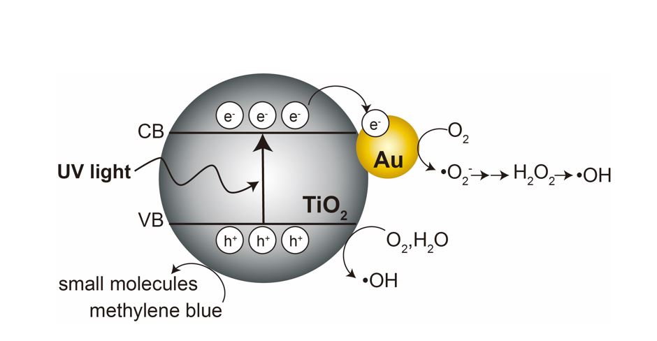
Highly reactive oxygen species, especially hydroxyl radical (•OH), plays a vital role in the organic pollutants degradation procedure in TiO2/UV system. To evaluate the content of ·OH in our study, the terephthalic acid (TA) fluorescent probe method was employed.(Nakabayashi &Nosaka 2015) The TAOH concentration in the experiments could be calculated on the basis of the linear relationship between the concentration of TAOH and its fluorescence intensity at 426 nm. Since OH radical is the only reactant for the formation of TAOH, the concentration of ·OH was calculated through the formula [·OH] = [TAOH]/0.45. Fluorescence experiments result (Fig. 5) shows that signal intensity for ·OH was the highest for bio-Au2@TiO2. Meanwhile, the amount of ·OH produced in solution followed the order of bio-Au2@TiO2 > bio-Au1@TiO2 > bio-Au0.75@TiO2 > bio-Au0.5@TiO2 > bio-TiO2, which is in accordance with the catalytic degradation speed of methylene blue.
In the photocatalytic process, electron-hole pairs are created by excitation of electron from valence band to the conduction band. The photogenerated electrons (e−) and hole (h+) pairs could react with adsorbed O2 and H2O to generate •OH. In the irradiation of UV light, biosynthesized TiO2 can be photoexcited and produce electron-hole pairs to form hydroxyl radicals, thereby realizing the degradation of methylene blue (Scheme 1).
More importantly, Au NPs in bio-Aux@TiO2 play a vital role in the photocatalytic effect. The UV irradiation by the Au NPs resulted in the positive charges in the lower 5d band of Au, which can capture electrons from the organic pollutants adsorbed on AuNPs, ensuing in the oxidation of methylene blue. Furthermore, as Au is added to TiO2, its band gap energy decreases below the values in anatase and rutile TiO2. The decrease in the band gap energy of TiO2 resulted in the stronger interaction between Au and TiO2, and increased charge separation between the excited electron (e−) and hole (h+), causing the enhancement of photocatalytic activity of TiO2. Besides, the biosynthesized materials, particularly Au NPs, are nanoscale with large specific surface area, which are capable to adsorb more methylene blue molecules, contributing to the high catalytic activity.
Thus, bio-Aux@TiO2 materials, especially bio-Au2@TiO2, displayed the superior catalytic activity in the degradation of methylene blue.