We found that LRRC1 was upregulated in HCC tissues at the transcriptional and protein levels. Abundant LRRC1 expression in HCC indicates a poor prognosis. The DEGs between the LRRC1-high and –low groups were significantly enriched in pathways associated with cancer, amino acid metabolism, carbohydrate metabolism, and the immune system. We identified 15 differentially infiltrated immune cells between the LRRC1-high and -low groups and found that LRRC1 expression correlated with 14 of them, including macrophages, Tregs, and NK cells. Based on the differentially infiltrated immune cells, we used WGCNA to identified 83 immune cell-related genes, of which 27 were prognosis-related. A PPI network revealed that ANXA5, MMP9, and NCF2 were hub genes. Transcription factor CREB1 regulated ANXA5, MMP9, and LRRC1 in the TF regulatory network.
Although LRRC1 shares 60% identity with human SCRIB, its role in the development of human cancers remains largely unknown [6]. The cell polarity regulator proteins DLG1 and PSD-95 and LRRC1 interact on the basolateral side of human epithelial cells [29]. Loss of epithelial cell polarity has been implicated in liver cancer progression [30]. The idea that LRRC1 is aberrantly overexpressed in HCC tissues and cell lines compared to normal controls is reasonable [6]. The expression of LRRC1 is associated with stem cell markers in normal and tumor breast epithelial cells [31]. The present study also found that LRRC1 was upregulated in HCC tissues at the transcriptional and protein levels, which was consistent with previous findings. Moreover, high LRRC1 expression indicated low rates of disease-free survival, overall survival, and a poor clinical prognosis. Thus, LRRC1 might be a risk factor for HCC.
To further explore the potential regulatory mechanisms of LRRC1 in HCC, we analyzed DEGs under high and low LRRC1 expression. These DEGs were significantly enriched in pathways associated with cancer, amino acid metabolism, carbohydrate metabolism, cell growth and death, and the immune system. Accumulated evidence indicates that interference with cellular metabolism predisposes humans to cancer. Amino acids are important in driving nucleoside synthesis, maintaining cellular redox homeostasis, and generating energy. Therefore, an abundant supply of amino acids is necessary for cancer cells to maintain their proliferative drive [32]. In addition to amino acid metabolism, carbohydrate metabolism also plays a crucial role in tumor progression [33]. Rates of aerobic glycolysis are high in many human cancers [34]. Increased glucose metabolism enhances lipogenesis and nucleotide biosynthesis and promotes tumor cell proliferation by providing essential bioenergy molecules [35, 36]. Taken together, increased LRRC1 expression may promote HCC progression by regulating amino acid and carbohydrate metabolism [33].
Mutations in TP53 and CTNNB1 are considered drivers of HCC development [37]. Wild-type p53 protein plays a key role in regulation of the cell cycle and apoptosis after DNA damage [38]. Cells with p53 mutations can escape apoptosis and transform into cancer cells after DNA damage [39]. A TP53 mutation is the most common genetic change in HCC, with an average mutation frequency of 30% [38], that can increase to 60% when HCC is associated with the hepatitis B virus [40]. Mutations in TP53 correlate with tumor differentiation, vascular invasion, and tumor stage in HCC [38], and the most frequently mutated oncogene in HCC, CTNNB1, correlates with elevated glutamine synthetase levels and vascular invasion [41]. Mutations in TP53 and CTNNB1 appear to be mutually exclusive [37]. The present findings found that the mutation frequencies of TP53 were 42% and 18%, and those of CTNNB1 were 13% and 37% in the LRRC1-high and -low groups, respectively. These findings suggested that mutation landscapes differ between HCC with high and low LRRC1 expression.
Immune cell infiltration plays a critical role in tumorigenesis [42]. The present study revealed 15 differentially infiltrated immune cells between the LRRC1-high and –low groups, 14 of which were significantly associated with LRRC1 expression. For instance, macrophages and Treg cells positively correlated with LRRC1 expression. Due to their roles in immune invasion, increased numbers of tumor-associated Tregs and macrophages are associated with a poor prognosis for patients with HCC [43, 44]. The DEGs between the LRRC1-high and –low groups were significantly enriched in pathways associated with the immune system.
Therefore, attenuated antitumor immunity could explain the poor prognosis of patients in the LRRC1-high group, and LRRC1 could be a potential immune-associated prognostic biomarker for HCC.
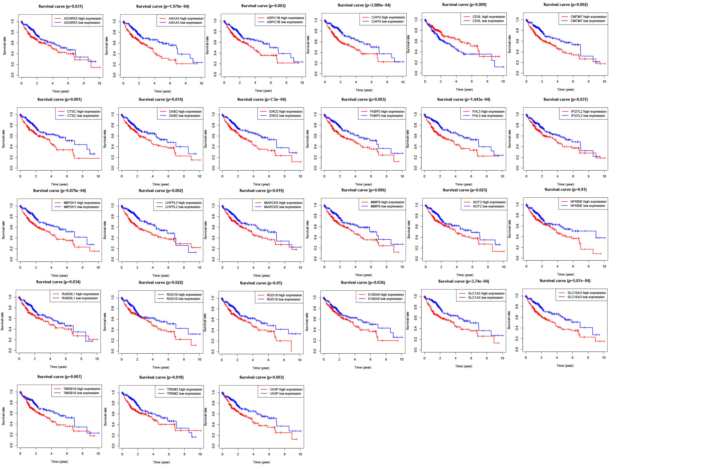
Based on the differentially infiltrated immune cells between LRRC1-high and -low groups, we identified 83 genes associated with immune cells, of which 27 were associated with prognosis. A PPI network revealed that ANXA5, MMP9, and NCF2 were hub genes. The TF CREB1 regulated ANXA5, MMP9, and LRRC1 in the TF regulatory network. The overexpression of ANXA5, which is a calcium-dependent phospholipid binding protein, potentially facilitates lymphatic metastasis of HCC [45]. This protein has been applied as an immune checkpoint inhibitor in cancer treatment [46]. MMP9 in the MMP family participates in inflammation, immunity, wound repair, and embryonic development under normal conditions. Under external stimulation, MMP9 can degrade the extracellular matrix of local tissues to promote tumor invasion and metastasis [47]. The CREB1 of the leucine zipper family is essential for DNA repair, cell cycle, and apoptosis [48]. Once activated, CREB1 can regulate its downstream target genes, including MMP9 [49]; this result is consistent with ours. The regulatory relationship between CREB1 and LRRC1 has not been reported to the best of our knowledge. Given that ANXA5 and MMP9 were identified as DEGs in the LRRC1-high and -low groups, we speculated that a regulatory mechanism functions between LRRC1 and the CREB1-MMP9/ANXA5 axis during the immune response to HCC. Further studies are needed to understand the underlying mechanisms.
In conclusion, our findings suggested that LRRC1 expression is upregulated in HCC tissues, and that upregulated LRRC1 expression indicates a poor prognosis for patients with HCC. The DEGs between LRRC1-high and -low groups were associated with the immune system, suggesting that LRRC1 could be a potential immune-associated prognostic biomarker of HCC.