The present study describes the histopathological patterns of response of CRLMs to preoperative NACT followed by liver resection. To our knowledge, this is the first report to focus on change of histopathological heterogeneity comparing between primary tumor sites and CRLMs.
Over the past decade, NACT has been widely recommended for the management of initially resectable CRLMs with the aim of inducing tumor shrinkage to identify optimal candidates for subsequent surgical removal. The assessment of tumor regression has been gradually used to quantify the histopathological response to NACT and has served as an early parameter predicting prognosis (7, 8).
Discrepancies in the tumor regression patterns in response to different chemotherapy regimens have been revealed in previous literature. Rubbia-Brandt et al. reported that an oxaliplatin-based regimen improved histopathological response compared with 5-fluorouracil-, and irinotecan-based regimens (9, 10). In terms of monoclonal antibodies, a bevacizumab-containing regimen provided a better histopathological response than chemotherapy alone or in combination with cetuximab (11, 12).
Poultsides et al. published a large retrospective analysis of 366 patients (68% treated preoperatively and 32% not) who underwent CRLM resection. In that study there was no increase in the degree of necrosis after chemotherapy (13). Nevertheless, it should be noted that only 69 out of 249 (28%) patients received bevacizumab as part of the preoperative treatment, and the results in terms of necrosis for that subgroup were not reported. In other experience, increase in necrosis seemed to be due to a bevacizumab-related effect (14, 15). Additionally, the tumor histopathological response rate was defined as the proportion of patients with grade ≥Ib.
Taken together, all of the above-reported observations of reduced viable cells, fibrosis, and necrosis from a pathological perspective explained the typical pattern of CLMs detected using computed tomography scanning in patients receiving chemotherapy and bevacizumab. Before treatment, the lesions showed different types of enhancement, a heterogeneous degree of attenuation, and ill-defined borders that were transformed into hypo-attenuated and homogeneous metastases with well-defined borders after treatment (16). Such histological and morphological characteristics strengthen the hypothesis that the RECIST criteria are not completely adequate for evaluating response in patients receiving bevacizumab (17).
In contrast, the anti-EGFR agent cetuximab in combination with chemotherapy has been reported to increase the response rate and yield a good curative hepatectomy. In the PEAK (18), FIRE–3 (19) and CALGB/SWOG 80405 (20) randomized controlled trials performed to compare bevacizumab and anti-EGFR therapy for the progression of recurrent CRC, anti-EGFR was also confirmed to have a positive effect on survival extension in the presence of wild-type RAS.
Recently, the multicenter, randomized, phase II ATOM trial from Japan was designed to evaluate the efficacy and safety of mFOLFOX6 plus bevacizumab and mFOLFOX6 plus cetuximab in patients with liver-limited metastasis from wild-type all-RAS CRC. After study treatment followed by surgical resection of tumors with R0/R1 status, the median progression-free survival of the bevacizumab-treated arm was 6.5 months [95% confidence interval (CI) = 4.0–13.6 months], whereas that of the cetuximab-treated arm was 13.8 months (95% CI = 8.4 months-not reached; hazard ratio = 0.610, 95% CI = 0.298–1.245). Of the 57 tumors for which the histopathological analysis was assessable, the histopathological response rate (grade 1b/2/3) was 66.6% (20/30) in the bevacizumab-treated arm and 92.6% (25/27) in the cetuximab-treated arm (p = 0.0229) (16), indicating that the rate tended to be better in the cetuximab-treated arm (21).
Falcão et al. reported three categories of tumor growth: (a) Replacement growth pattern, in which the tumor permeates between the liver hepatocytes without disrupting the normal architecture; (b) desmoplastic growth pattern, in which the tumor is separated from the liver parenchyma by a band of fibrous tissue that contains tumor-infiltrating lymphocytes; and (c) pushing growth pattern, in which the tumor expands and compresses the surrounding hepatocytes. They reported the pushing growth pattern to be an independent risk factor for reduced survival (22).
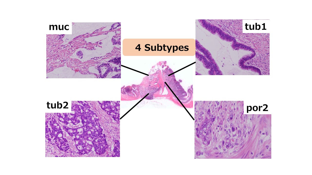
Recently, a tumor-heterogeneity concept that considers a single tumor to consist of many tumor cell sub-clones has become an important topic in cancer genomics (23). It is hypothesized to play a critical role in the progression of many cancer types and is a major obstacle to precision cancer therapy. During this process, sub-clones continuously arise via genomic mutation. The presence of sub-clones has been shown to adversely affect outcome in chronic lymphocytic leukaemia, head and neck cancer, and lung adenocarcinoma. However, the full complement of factors that lead to tumor heterogeneity during CRC progression is unknown.
Several promising CRLM treatments have been reported, including chemotherapy and molecular agents that target EGFR and VEGF. Anti-EGFR drugs resulted in high response and resection rates in the CELIM phase II trial and other studies for initially unresectable CRLM with wild-type KRAS (24,25).. Anti-VEGF regimens, such as mFOLFOX6 or CAPEOX plus bevacizumab, have also shown high response and resection rates in phase II studies (26).. These reports suggest that the combination of targeted agents and chemotherapy can increase the response rate.
A previous study reported a higher pathological response rate to bevacizumab than for cetuximab. This study suggested that the loss of intratumoral heterogeneity markedly affects the response to chemotherapy.