Mouse model of OIR
Experiments using mice were performed following the recommendations in the Guide for the Care and Use of Laboratory Animals of the National Institutes of Health and the United States Department of Agriculture Animal Welfare Act (9 CFR, Parts 1, 2, and 3). All experimental procedures were approved by the Institutional Animal Care and Use Committee at Augusta University.
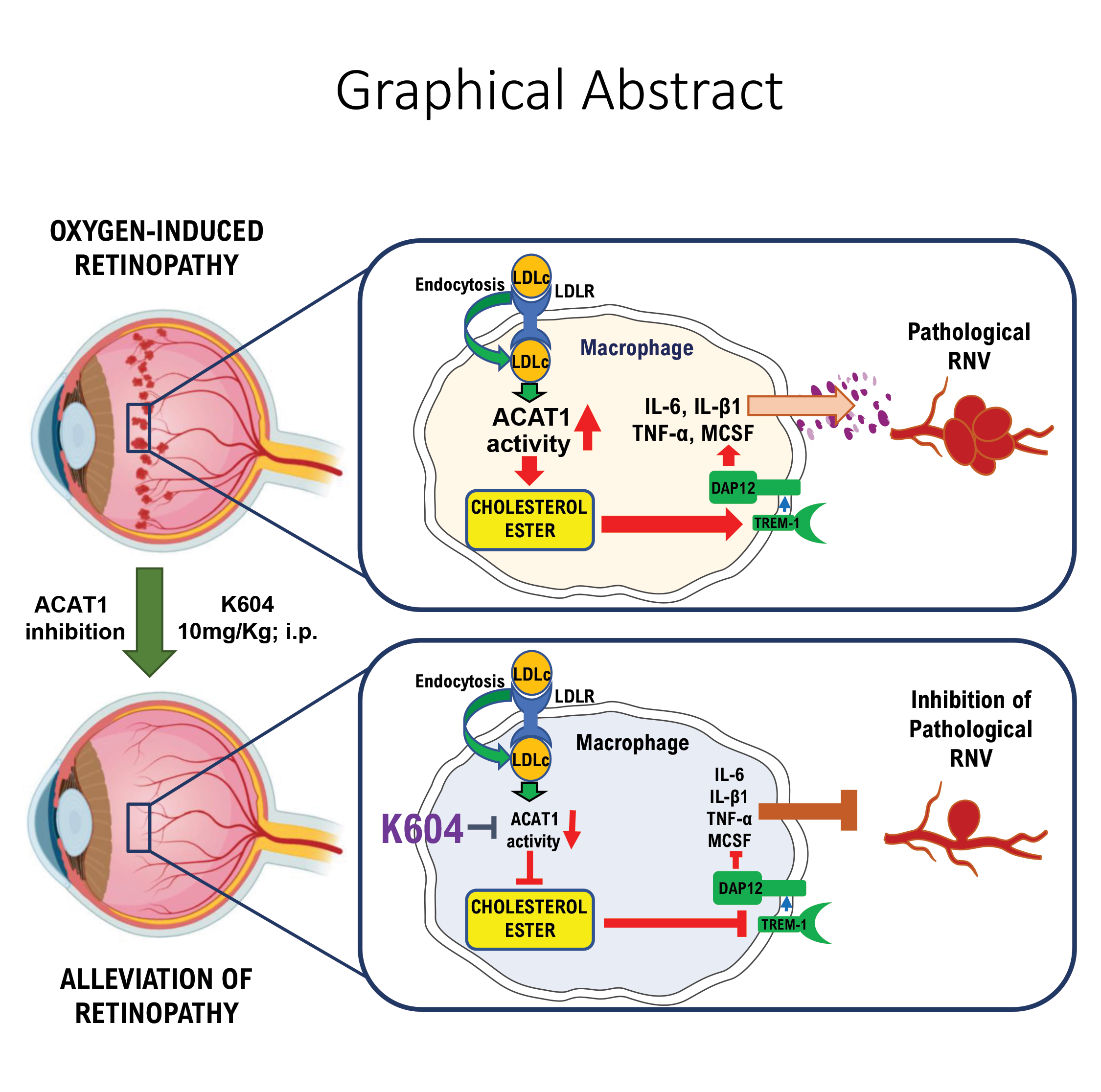
Litters of neonatal C57BL/6J and LDLR-/- mice and nursing dams were maintained in hyperoxia (75% oxygen) from postnatal day 7 (P7) until P12 and then returned to normoxia until P17 and retinas were collected for analysis (Supplementary Fig.1a). One group of wildtype pups was treated with a cinnamic acid derivative (N-[3-(4-hydroxyphenyl)-1-oxo-2-propenyl]-L-phenylalanine, methyl ester, Santa Cruz Biotechnology, Dallas, Tx) that inhibits both ACAT1 and ACAT2 isoforms equally [26]. This drug was administered via i.p. injections (10mg/kg in 50 µl PBS) every two days from P7 to P16 and mice were sacrificed on P17 (Supplementary Fig.1b). Additional groups of wildtype pups were treated with daily injections of a specific ACAT1 inhibitor (K604, BioVision, Milpitas CA, 10mg/kg in 50 µl PBS) from P7 to P16 and sacrificed on P17, from P7 to P11 and sacrificed at P12, or from P12 to P16 and sacrificed on P17 (Supplementary Fig.1b, c and d). The drug dosage was based in previous reports [27]. Vehicle control groups received PBS alone. Retinas from all groups were collected and prepared for analysis of RNV and vaso-obliteration as well as, expression of TREM1, MCSF, VEGF, ACAT1, and LDLR and levels of cholesterol esters as outlined below.
Oil Red O staining for detection of neutral lipids
Cholesterol and other neutral lipids were detected as described previously [28]. Retina frozen sections were reacted with 0.5% Oil Red O (Sigma-Aldrich, St. Louis, MO, dissolved in 1,2-isopropanol, 15 min, room temperature), rinsed in 60% 2-propanol for 5 min and rinsed in distilled water (twice 5 min each time). Sections were mounted in aqueous mounting media to capture the images using a Zeiss Axioplan 2 fluorescence microscope (Carl Zeiss Meditec, Inc., Dublin, CA) equipped with a 420 nm excitation filter, 520 nm barrier filter, and a 20x lens.
Filipin staining for detection of cholesterol ester (CE)
Retinal frozen sections were reacted with the fluorescent polyene antibiotic filipin (Sigma-Aldrich, St. Louis, MO) to detect CE. This compound binds specifically to sterols and interacts with the 3-β hydroxyl group of cholesterol. For CE detection, we followed the protocol described by Rudolf and Curcio [29]. Native unesterified cholesterol (UC) was extracted from cryosections by rinsing in 60% ethanol for 10 minutes. Native CE was hydrolyzed with cholesterol esterase (1.65 units/ml) in PBS for 3 hours at 37°C (C9281, Sigma St. Louis, MO). The newly released UC by the hydrolysis of CE was stained with 50ng/ml filipin in PBS.
Determination of RNV, AVA and tip cells in retina flat mount
Retinal flat mounts were labeled with Isolectin B4 (IB4) as described before [30]. Images of the retinal flat mounts were constructed by capturing a series of 12 pictures from all samples using a 5x lens. The images were then regrouped to make a retina map. Next, areas of vaso-obliteration (AVA) and RNV were quantified using NIH ImageJ software as previously described [30]. Retinas flat mount stained with IB4 were also used to quantify tip cells by direct observation under fluorescence microscope with a 20x lens. Representative images from each group were taken with a 40X lens. Images were captured with a 20x lens using a Zeiss Axioplan2 fluorescence microscope (Carl Zeiss Meditec, Inc.).
Immunolocalization
Retinal frozen sections and flat mounts were processed for immunolabeling according to our standard protocol [31]. The samples were blocked with normal goat serum or donkey serum and incubated overnight with IB4, rat anti-mouse TREM1, rabbit polyclonal anti-mouse MCSF1, rat anti-mouse F4/80, rabbit anti-mouse Iba1, goat anti-mouse ACAT1, or rabbit anti-mouse LDLR antibodies (Abcam, Cambridge, MA). The samples were washed 3 times with PBS and incubated with secondary antibodies (Invitrogen, Waltham, MA). Images were captured with a 20x lens using a Zeiss Axioplan2 fluorescence microscope or Zeiss 780 inverted Confocal microscope (Carl Zeiss Meditec, Inc. Dublin, CA).
Macrophage
THP1 human macrophages were cultured as described previously [32]. For experiments, cells were incubated in DMEM for 2 hr and then switched to RPMI medium containing 5 mM glucose, 2% FBS, and 1% penicillin. Macrophages were treated with K604 (10 ug/ml) or vehicle and subjected to hypoxia (1% O2) or normoxia (21% O2). After 16 hr, the cells were collected and processed for protein quantification and western blot following our established protocol [25].
Western blot assays
Lysates from retina samples and THP1 cells were prepared for protein quantification and western blot analysis following our established protocol [33]. The samples were homogenized in modified RIPA buffer (20 mM Tris-HCl, 2.5 mM EDTA, 50 mM NaF, 10 mM Na4P2O7, 1% Triton X-100, 0.1% sodium dodecyl sulfate, 1% sodium deoxycholate, 1 mM phenylmethylsulfonylfluoride, pH 7.4). Samples containing equal amounts of protein were separated by 10% or 12% sodium dodecyl sulfate polyacrylamide gel electrophoresis, transferred to polyvinylidene difluoride (PVDF) or nitrocellulose membrane, and reacted for 24 h with monoclonal rat anti-mouse TREM1, polyclonal rabbit anti-mouse MCSF1, VEGF, LDLR, and goat anti-mouse ACAT1 antibodies (Abcam, Cambridge, MA) in 2% BSA, followed by incubation with corresponding horseradish peroxidase-linked secondary antibodies (GE Healthcare Bio Science Corp., Piscataway, NJ). Bands were quantified by densitometry and the data were analyzed using ImageJ software and normalized to loading control. Equal loading was verified by stripping the membranes and reproving them with a mouse monoclonal antibody against β actin (Sigma-Aldrich, St Louis, MO).
Quantitative real time RT PCR (qRT PCR) analysis
The total RNA from mouse retina samples was extracted with an RNAqueous 4PCR total RNA isolation kit (Invitrogen, Carlsbad, CA, US), and qRT PCR was performed as described previously [34]. Briefly, a 0.25 μg sample of RNA was utilized as a template for reverse transcription using M-MLV reverse transcriptase (Invitrogen). qRT PCR was performed on an ABI 7500 Real Time PCR System (Applied Biosystems, Foster City, CA) with the respective gene-specific primers listed in Table 1. The relative gene expression is calculated using the comparative threshold cycle (ΔΔCt) method against the internal control, hypoxanthine phosphoribosyl-transferase (HPRT). Expression levels for all genes are reported as fold change to room air controls.
Measurement of cholesterol ester
Plasma and retinas are collected from RA, OIR and K604-treated OIR pups at P17. The levels of cholesterol esters were measured using luminescence-based Cholesterol/Cholesterol Ester GloTM Assay (J3190, Promega, Madison, WI) as per manufacturers’ protocol. Briefly, retinas were homogenized, and blood plasma were diluted (1:10) in Cholesterol lysis solution and incubated at 37°C for 30 mins. Equal amounts of extracts were incubated in cholesterol detection reagent with and without cholesterol esterase enzyme in a 96 well white bottom plates at room temperature for one hour. Luminescence was recorded using Polar Star Omega microplate reader (BMG Labtech Inc, NC). Total and free cholesterol concentrations were measured by comparing the luminescence of samples with and without cholesterol esterase, respectively. Cholesterol ester concentrations were calculated as the difference between total and free cholesterol concentrations.
Detection of K604 in retina samples by LC-MRM MS analysis (Liquid chromatography- multiple reaction monitoring -mass spectrometry)
K604 is an small molecule capable of penetrating the blood-brain-barrier in experimental models [35]. To verify its ability to penetrate the blood-retinal-barrier, mice were treated with K604 (10mg/kg) from P7 to P16 and retinas were collected for analysis on P17 (18 hours after the last K604 injection). LC-MRM MS analysis was performed to detect the K604 spectrum. Retina samples were processed as follows: 200µl extraction buffer (acetonitrile: isopropanol: water = 4:4:2) was added to the retina sample tube together with 0.1ml zirconium oxide bead (0.5mm diameter, Next Advance). The sample tube was placed in a Bullet Blender (BBX24, Next Advance) and blended using Speed 8 for 3 minutes. Sample tubes were then centrifuged at 16,000g for 10 minutes (room temperature) and the supernatant was transferred into a glass vial for LC MS analysis.
Separation of extracted retina samples (5µl) was performed using a Thermo Hypersil C8 column (50x2.1mm, 1.9um) on a Shimadzu Nexera UHPLC system at a flowrate of 0.2ml/min using a gradient elution from 5% to 95% acetonitrile (with 0.1% formic acid) in 6 minutes. The effluent was ionized using positive electrospray and analyzed on a TSQ Quantiva triple-quadrupole mass spectrometry with the following instrument settings: ion spray voltage 3500V, sheath gas 35, ion transfer tube temperature 325, aux gas 10, and unit resolution for Q1/Q3. The optimal collision energy and RF lens setting were determined using purchased standards. The transitions monitored for K604 were as 503/353, 503/241, and 503/226.
The integrated peak areas for these transitions were calculated for each sample using Skyline software (version 20.0, University of Washington) and the most intense one (503/353) was used for quantification. The internal standard for K604 was performed at 1 pmol and the spectrum was detected at an intensity of 5,530.124 equal to 1 pmol at 4.9 minutes. The K604 concentration for retinal samples detected at 4.9 minutes ranged from retina 0.3 pmol to 1.07 pmol (mean+SEM=0.66+0.24, n=3), indicating that the drug reaches the retina and persists over time.
Statistical analysis
Differences among the groups were compared by an independent two sample t-test for two groups or one-way ANOVA followed by a post hoc test for multiple comparisons. Values are represented as means ± standard error of the means (SEM). P values of less than 0.05 were considered significant.