Animal experiments
This research followed National Institutes of Health (NIH) criteria and Xi'an Medical University's animal care committee's approval. In C57BL/6 mice, streptozotocin (50mg/kg/d) was administered intraperitoneally for five successive days 15. A week following the last administration of STZ, experimental animals with fasting glucose levels > 11.1 mmol/L were determined to develop diabetes. Mice serving as controls had a similar injection of vehicle every day. Vehicle or BNP (GenScript, Piscataway, NJ, USA) was randomly assigned and was administered to control or diabetic mice for 4 weeks using subcutaneous osmotic pumps at a rate of 0.25 µL/hour (Alzet, model 2004; Cupertino, CA, USA). All experimental models were placed in conventional settings (12h/12h light/dark cycle) and fed a recommended mouse chow with ad libitum access to water. Animals were anesthetized by 3% isoflurane and maintained with 1% isoflurane via inhalation. Euthanasia was carried out using carbon dioxide, in accordance with the AVMA Guidelines for the Euthanasia of Animals 80 (2020) and with the approval of local animal welfare committees.
Human study
The clinical research was reviewed by the Ethics Committee of Xi'an Medical University, which ultimately gave its approval for the project. Diabetes mellitus (DM) was diagnosed according to World Health Organization (WHO) criteria before being included in this experiment, all subjects provided written informed consent. Participants with coronary artery disease, hypertrophic cardiomyopathies, restrictive cardiomyopathies, dilated cardiomyopathies, arrhythmogenic right ventricular dysplasia, or other micro- or macrovascular consequences of diabetes were excluded from the current study. Additionally, patients with renal failure, serious psychological problems and cancer were excluded. Final enrollment included 30 control volunteers, 30 diabetics, and 30 diabetics with DCM. The plasma samples were obtained and kept at -80℃. The patient data were included in Supplemental Table S1.
Echocardiography
Echocardiography was performed in M-mode using VEVO 2100 system (Visual Sonics, Toronto, Canada) 14. Mice were anaesthetized with isoflurane. The images of left ventricular (LV) dimensions were recorded to measure left ventricular ejection fraction (LVEF) and left ventricular fractional shortening (LVFS).
Biochemical and Histological Analysis
An automated biochemical analyzer was used to determine the levels of blood glucose, total cholesterol (TC), and triacylglycerol (TG) in fasting plasma (Chemray 800, Rayto, China). We measured plasma BNP concentrations using an ELISA kit (Raybiotech, Norcross, GA, USA). PKG activity assays were performed using the PKG Kinase Enzyme assay kit from Promega.
Heart morphology were stained with hematoxylin and eosin (Beyotime, Jiangsu, China). Staining cardiomyocytes with FITC-labeled WGA (Servicebio, Wuhan, China) allowed us to determine their cross-sectional area. Masson's trichrome stain (Servicebio, Wuhan, China) was used to examine the myocardium's interstitial fibrosis.
Measurement of blood pressure
Systolic blood pressure and diastolic blood pressure was measured using the non-invasive tail-cuff system (Kent scientific corporation, Torrington, CT, USA). Briefly, mice were placed on the pre-warmed platform (30°C) and tails were inserted into the tail cuffs for 5 consecutive days. At each session, at least 10 reads were obtained and averaged as the blood pressure at that time point.
Transmission electron microscopy (TEM)
The cardiac tissues were fixed in a 2.5% glutaraldehyde and 1% cacodylate buffer at pH 7.4 for 2 days at 4°C. The tissues were first rinsed several times with 0.1 M cacodylate buffer, followed by the addition of 0.1% tannic acid to deionized water, and finally osmium tetroxide to deionized water. The detailed steps were carried out as described before 14. An image of each slice was taken with a charge-coupled device (CCD) camera equipped with a JEM-1230 TEM (JEOL Ltd., Tokyo, Japan). The mitochondrial images were analyzed using Image J software.
Cell culture
Primary cardiomyocytes were separated from the hearts of neonatal Sprague-Dawley mice via collagenase I (gibco) digestion, as previously described 16. Cells were cultured in Dulbecco's modified Eagle's medium (DMEM) containing 10% fetal bovine serum (FBS) and normal glucose (5.5 mmol/L, NG). Cardiomyocytes were exposed to normal glucose (5.5 mmol/L, NG) or HG (33 mmol/L, HG) for 48 hours 17 with the vehicle or BNP supplement.
Mitochondrial morphology in the cardiomyocytes
To assess mitochondrial morphology in the cardiomyocytes, the mitochondria were stained using the fluorescent probe MitoTracker Green FM (Ex/Em: 490/516 nm, M7514, Invitrogen, USA) in accordance with the methods of the manufacture. The cardiomyocytes were plated in a confocal dish and treated with 50 nM MitoTracker Green FM for 30 minutes. We examined the images under a confocal laser scanning microscope (Nikon A1R MP + Confocal Microscope, Nikon, Japan) and analyzed mitochondrial morphology as previously described 18.
Measurement of mitochondrial membrane potential
As described previously 19, we measured the mitochondrial membrane potential using JC-1 dye from Beyotime (Jiangsu, China). FACScan flow cytometry (BD Facscalibur, Franklin Lakes, NJ, USA) was used to measure the fluorescence of JC-1 after incubation of primary cardiomyocytes at 37°C for 15 min with 1.0 µM JC-1.
Quantitative real-time PCR (RT-qPCR)
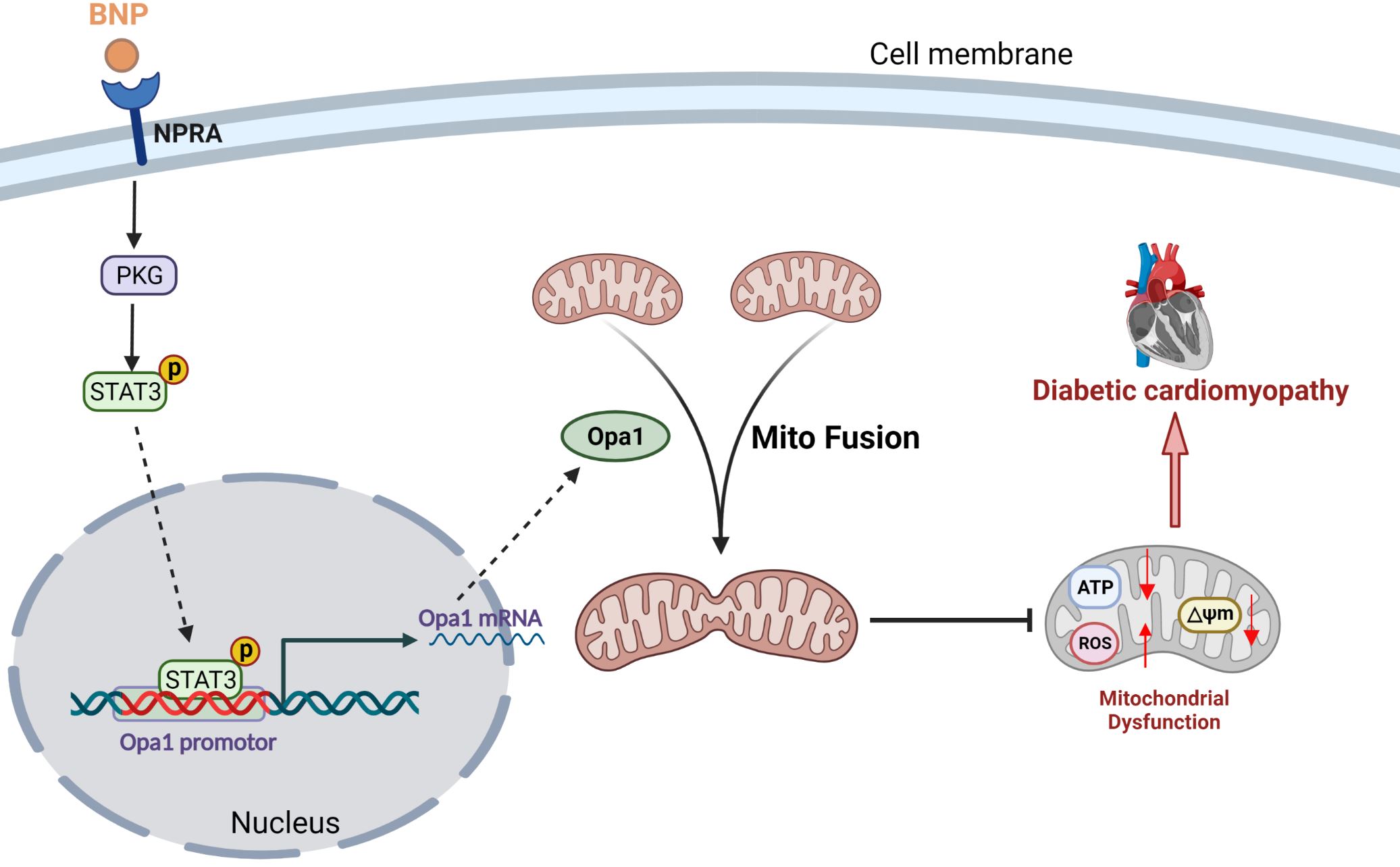
RT-qPCR was used to detect the gene expression levels for Opa1 and the relative mtDNA content to nuclear DNA content as described previously 20,21. The primer sequences were: Opa1 forward CAACCCCGCAGGAACTTTTG; reverse GGTGTACCCGCAGTGAAGAA; mtDNA forward AACATACGAAAAACACACCCATT; reverse AGTGTATGGCTAAGAAAAGACCTG; β-actin forward CCCTGGCTCCTAGCACCAT; reverse AGAGCCACCAATCCACACAGA.
Reactive Oxygen Species (ROS) generation in the cardiomyocytes and hearts
Cardiomyocytes were seeded in a confocal dish and were loaded with 5 µM MitoSOX Red (Thermo Fisher Scientific, Waltham, MA, USA) for 10 minutes in the dark at 37°C. After several washes, images were visualized under a confocal laser-scanning microscope (Nikon A1R MP + Confocal Microscope, Nikon, Japan).
Dihydroethidium (DHE) staining was used previously to assess the production of superoxide anion in mouse heart tissue 16. Briefly, 2 µmol/L fluorescent dye DHE (Thermo Fisher Scientific, Waltham, MA, USA) was added to the 6 µm thick snap-frozen heart sections and incubated at 37℃ for 30 min in a humidified dark chamber. ImageJ analysis software was used to analyze the DHE-positive cells. MnSOD activity was measured using MnSOD assay kits (S1013, Beyotime Biotechnology, Jiangsu, China) according to the manufacturer's protocol.
Mitochondrial oxygen consumption rate (OCR) analysis
Mitochondrial respiratory activity was measured employing an XF24 Extracellular Flux Analyzer (Agilent Seahorse Bioscience, Santa Clara, CA, USA). Primary cardiomyocytes were seeded at 2× 10⁴ per well in the XF24 Cell Culture Microplate. We used Seahorse XF Cell Mito Stress Test Kit (Agilent Technologies, Santa Clara, CA, USA) to record the OCR with sequential injection of 1 µM oligomycin A, 1 µM FCCP, and 0.5 µM antimycin A 22. Pierce BCA Protein Assay Kit (Thermo Fisher Scientific, Waltham, MA, USA) was used to adjust all raw results to the protein content.
Transfection of siRNAs against BNP, Opa1, STAT3, PKG and NPRA
SiRNAs against BNP (#sc-62023, Santa Cruz Biotechnology), Opa1 (sense: CCAGCAAGGUUAGCUGCAATT; antisense: UUGCAGCUAACCUUGCUGGTT), STAT3 (#sc-270027, Santa Cruz Biotechnology), PKG (sense: CGAAGAUUCUCAUGCUCAA; antisense: UUGAGCAUGAGAAUCUUCG), NPRA (#sc-40126, Santa Cruz Biotechnology) and negative control siRNA were transfected into primary cardiomyocytes by using Lipofectamine RNAiMAX reagent (Invitrogen). Following 48 h of transfection, cells were used for subsequent tests in NG or HG media.
Assay for apoptotic cell death
Using Annexin V-FITC and PI apoptosis detection kits (BD Biosciences, CA, USA), the apoptotic frequency of primary cardiomyocytes was determined by flow cytometry. A terminal deoxynucleotidyl transferase-UTP nick end labeling (TUNEL) assay kit (Roche Applied Science, Penzberg, Germany) and the caspase-3 activity test kit (KeyGEN Biotech, Jiangsu, China) were used to evaluate apoptotic rate in cardiac tissues according to the manufacturer's recommendations. Confocal fluorescence microscopy (Leica, Heerbrugg, Switzerland) was used to evaluate the TUNEL and DAPI-stained slices.
Adeno-associated virus transfection in vivo
For the Opa1 or BNP knockdown studies in vivo, adeno-associated viral (AAV) vectors carrying a scrambled sequence (AAV-Con) or a short hairpin RNA (shRNA) directed against the mouse Opa1 RNA (AAV-Opa1-shRNA) or BNP RNA (AAV-BNP-shRNA) were constructed by Hanbio Biotechnology (Shanghai, China). Isoflurane (2.5%) was used to anesthetize the mouse models, and the hearts were exposed. A total amount of 40 µL (approximately 1 × 10¹¹ PFU/mL) AAV-Con shRNA, AAV-Opa1-shRNA, or AAV-BNP-shRNA was injected at four different sites of each left ventricle free wall, as we have described previously 16. The mice were administered with STZ (50 mg/kg/d) intraperitoneally for 5 consecutive days one week after transfection to establish a diabetes model 15, and then the vehicle or BNP was administered.
Western blotting
Total proteins in mouse heart tissue or primary cardiomyocytes were determined with a Bradford protein assay (Beyotime, Jiangsu, China) on lysates obtained with RIPA (Beyotime, Jiangsu, China). The extracted proteins were electrophoresed on SDS-PAGE and transferred to PVDF membranes. After blocked with 5% milk, the membranes were incubated with Drp1 antibody (#14647, Cell Signaling Technology), Fis1 antibody (#GTX111010, Genetex), Opa1 antibody (#80471, Cell Signaling Technology), Mfn1 antibody (#13798-1-AP, Proteintech), Mfn2 antibody (#9482, Cell Signaling Technology), STAT3 antibody (#9139, Cell Signaling Technology), phosphorylated STAT3 Tyr705 antibody (#9145, Cell Signaling Technology), PGC1α antibody (#66369-1-Ig, Proteintech), BNP antibody (#ab239510, Abcam), NPRA antibody (#ab14356, Abcam), PKG antibody (#3248, Cell Signaling Technology) or β-actin antibody (#3700, Cell Signaling Technology) under 4°C overnight. Afterwards, the membrane was treated with an anti-rabbit or anti-mouse secondary antibody that had been coupled with horseradish peroxidase (#A0208, #A0216, Beyotime Biotechnology, Jiangsu, China) at room temperature for 1 hours and the blot was exposed by Supersignal chemiluminescence detection kit (Thermo Fisher Scientific, Waltham, MA, USA). The blot densities were analysed by Quantity One software (Bio-Rad, San Diego, CA, USA).
Chromatin immunoprecipitation (ChIP) analysis
A Simple ChIP Plus Enzymatic Chromatin IP kit (#9005; Cell Signaling Technology, Danvers, USA) was used to perform ChIP in accordance with the manufacturer 's recommendations as described in our previously report 23. Briefly, after fixation with 1% formaldehyde, the cardiomyocytes were homogenized in lysis buffer. Following that, antibodies against STAT3 (#12640, Cell Signaling Technology) and protein G magnetic beads were added. Normal IgG was used as the negative control. DNA was extracted from the precipitation and then analyzed using primers for the Opa1 promoter (forward: 5´-TCCAGTTAGGTTTTGGGCCTT-3´ and reverse: 5´- TCCTTTTATGAGCCCCAATTTCCTT-3´) in RT-qPCR.
Co-immunoprecipitation assays
Co-immunoprecipitation was performed using the Pierce Co-Immunoprecipitation Kit (Thermo Fisher Scientific, Rockford, IL, USA) as described previously 24. Briefly, the cells were prepared in Lysis/Wash Buffer and incubated overnight with specific antibodies at 4°C. The immunocomplexes were precipitated and performed with agarose resin slurry for 1 h. Those beads were washed and then subjected to western blot procedures.
Statistical analysis
All the data are expressed as the mean ± SEM. For the statistical analysis of two groups, the unpaired Student's t-test was used. More than two groups were compared using one-way analysis of variance (ANOVA) or two-way ANOVA followed by Bonferroni's multiple comparison tests using GraphPad Prism 8.0. P values less than 0.05 were considered statistically significant.