Isolation and growth of human primary lung fibroblasts
IPF primary human lung fibroblasts (IPF-HLF) were acquired from diagnostic biopsies of IPF patients (n=5) and normal primary human lung fibroblasts (N-HLF) were acquired from healthy donor lung tissues failed for transplantation (n=5). The diagnosis of IPF was supported by history, physical examination, pulmonary function tests, and typical high-resolution chest computed tomography findings of IPF. The IPF patients (n=5) and healthy donors (n=5) were all male and aged from 40 to 65 years. Exclusion criteria included current or recent use of immunosuppression; chronic infection such as HIV or hepatitis; known pulmonary hypertension; cardiovascular, renal, or neoplastic disease; and inability to provide informed consent. This study was approved by the Ethics Committees of The First Affiliated Hospital of Zhengzhou University and written informed consent was obtained on all patients prior to the procedure being performed.
The human primary lung fibroblasts were isolated as previously described [9]. Briefly, lung tissues were cut into small pieces and subjected to enzymatic dissociation in Hank's balanced salt solution (containing 600 U/ml collagenase I, 2 U/ml papain, 2 U/ml protease, and 3.8 mM calcium chloride) at 37℃ for 1 hour. Next, tissues were ground by glass pipette trituration, and the supernatant was centrifuged at 800g for 5min to collect the cells. Then, cells were redispersed in high-glucose Dulbecco's Modified Eagle Medium (DMEM) containing 10% fetal bovine serum (FBS), 50 U/ml strepto-ECM, and 50 μg/ml penicillin. The phenotype of human primary lung fibroblasts was confirmed by positive immunocytochemistry for vimentin (data not shown). In all experiments, the lung fibroblasts were harvest in passages four to six for the test.
Cell culture
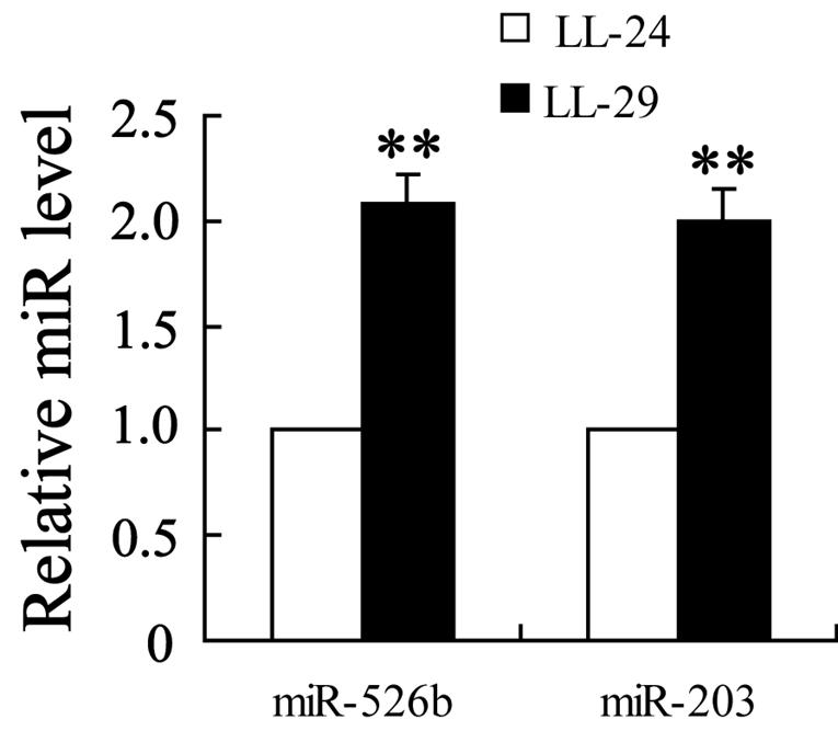
The human IPF fibroblastic cell lines (LL-97A and LL-29) and normal human fibroblastic cell line (LL-24) were obtained from the American Type Culture Collection (USA), maintained in Kaighn’s Modification of Ham’s F-12 Medium (F-12K Medium) containing 15% FBS and cultured with 5% CO2 at 37℃. The hyperproliferation and activation of normal human lung fibroblasts were performed as previously described [4]. Briefly, LL-24 cells were starved for 48 hours and incubated with fetal calf serum (FCS; 2% or 5%), PDGF-BB (30 or 60 ng/ml), and IGF-1 (100 or 200 ng/ml) for 6 h to promote fibroblasts proliferation and incubated with TGF-β1 (5 or 10 ng/ml) for 6 h to activate fibroblasts. All growth factors and cytokines were purchased from Solarbio (China).
Cell transfection and infection
Cells were cultured in 6-well plates with a concentration of 4×10⁵ cells/well. When the cells were cultured to 70% confluence, cells were transfected with RNAi-vector [si-circTADA2A, si- Caveolin 1 (Cav-1), si-Cav2] or micro RNA inhibitor (miR-526b inhibitor, miR-203 inhibitor) or their relative negative controls (si-control, NC) using Lipofectamine 2000 (Invitrogen, USA). Cells were incubated with 2 ml Opti-MEM medium (GIBCO, USA) containing plasmids (1 μg) and Lipofectamine 3000 (2.5 μl). The medium was changed after 6 h, and the RNA extraction was performed at 48 h to verify the transfection efficiency.
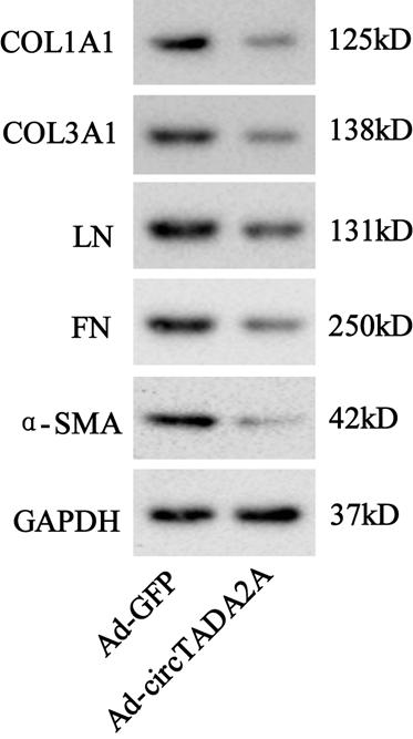
To overexpress circTADA2A or Cav1 or Cav2, the adenovirus expressed circTADA2A or Cav1 or Cav2 (Ad-circTADA2A or Ad-Cav1 or Ad-Cav2) were produced by Ribobio (China). The appropriate volume of virus particles calculated by the multiplicity of infection (MOI) was added in the cell culture medium. Forty-eight hours later, virus infection efficiency was monitored by GFP expression using the fluorescence microscope. Ad-GFP was used as a negative control. The sequences of the transfected components were shown in Table 1.
Cell Proliferation Assay
Lung fibroblasts proliferation was detected using the BrdU incorporation assay kit (Sigma Aldrich, USA). Cells were cultured in 6-well plates with a concentration of 4×10⁵ cells/well. When the cells reached confluence, cells were incubated with BrdU (10 μm) for 40 min. After that, cells were fixed in 4% paraformaldehyde (PFA) for 10min and permeabilized with 0.5% Triton X-100 for 15 min. Then, cells were orderly treated with a blocking buffer, a primary antibody against BrdU, secondary antibody, and DAPI. The BrdU-positive cells were counted using the fluorescence microscope (Nikon, Japan).
Quantitative RT-PCR
Total RNAs were isolated from cells or lung tissues of lung fibrosis mice using TRIzol Reagent (Invitrogen, USA). The quality of total RNA samples was evaluated by spectrophotometer and the high-quality RNAs (1.8 < OD260/280 < 2.0) were inversely transcribed into cDNA using a cDNA synthesis kit (Thermo Fisher). Quantitative RT-PCR was performed to measure circRNAs and miRNAs expressions using the Thunderbird SYBR qPCR mix (Toyobo, Japan). Gene expressions were calculated by the 2-∆∆CT method, the relative expressions of circRNA and miRNA were normalized to GAPDH and U6 respectively. The sequences of qRT-PCR primers were shown in Table 1.
Western blot
The determination of protein levels of collagen 1a1 (COL1A1), collagen 3a1 (COL3A1), α-smooth muscle actin (α-SMA), laminin (LN), fibronectin (FN), Caveolin-1 (Cav1) and Caveolin-2 (Cav2) were done by western blot with total protein purified from cell lysate or lung tissues of lung fibrosis mice by RIPA lysis buffer. Proteins were subjected to 10% sodium dodecyl sulfate-polyacrylamide gel electrophoresis and then transferred to PDVF membrane (ThermoFisher Scientific, USA). After being blocked with 5% skim milk for 30 min, membranes were incubated with primary antibodies (against COL1A1, COL3A1, α-SMA, LN, FN, Cav1 and Cav2; all purchased from Abcam). After the night, the membranes were incubated with the secondary antibodies specifically. Immunoblots were visualized in IBright FL1500 Intelligent Imaging System (ThermoFisher, USA) and GAPDH was used as an internal control.
The primary antibodies used in the experiment were as follows: anti-COL1A1 (1:1000; sc-59772, Santa Cruz Biotechnology, USA), anti-COL3A1 (1:1000; sc-271249, Santa Cruz Biotechnology, USA), anti-α-SMA (1:1000; ab32575, Abcam, UK), anti-LN (1:2000; ab11575, Abcam, UK), anti-FN (1:1000; ab268021, Abcam, UK), anti-Cav1 (1 µg/ml; ab2910, Abcam, UK), anti-Cav2 (1:5000; ab133484, Abcam, UK), and GAPDH (1:2500; ab9485, Abcam, UK). The secondary antibody used in the experiment were m-IgGκ BP-HRP (1:10000; sc-516102, Santa Cruz Biotechnology, USA) and Goat Anti-Rabbit IgG H&L (1:5000; ab205718, Abcam, UK).
Dual-luciferase reporter gene assay
To verify the combination of circTADA2A and miR-526b/miR-203, the sequence of circTADA2A was amplified and inserted into pGL3-basic plasmids. 0.5 μg plasmid and 20nM miR-526b mimic or miR-203 mimic or mimic-negative control (Pre-NC) were co-transfected in well-grown 293T cells by using lipofectamine 2000 (ThermoFisher, USA). Forty-eight hours after transfection, the cells were lysed and the activities of Renilla luciferase and firefly luciferase were measured with Dual-luciferase Reporter Assay Kit (Promega, China) following the manufacturer’s protocol. To verify the combination of miR-526b and Cav1/miR-203 and Cav2. The luciferase activities of Cav1-3’-UTR-mutant (Cav1 UTR WT), Cav2-3’-UTR-mutant (Cav2 UTR WT), and Cav2-3’-UTR-mutant (Cav2 UTR WT) were measured in the same way. The primer sequences were shown in Table 2.
RNA pull-down assay
The combination of circTADA2A and miR-526b/miR-203 was determined by RNA pull-down assay. The circTADA2A probe and its negative control [oligonucleotide probe (Oligo probe)] were constructed by Ribobio (China). LL-29 cells (1.5×10⁷) were collected and lysed using 100 μl lysis buffer. The lysate was then incubated with 50 pmol biotin-labeled circTADA2A probe and 50 μl streptavidin agarose magnetic beads for 1 h at 4℃. miR-526b and miR-203 in circTADA2A probe pull-down complex were detected by qRT-PCR using the Oligo probe pull-down complex as a negative control. The probe sequences were shown in Table 2.
Fluorescence in situ hybridization (FISH)
LL-29 cells were cultured on the coverslips, fixed with 4% paraformaldehyde for 15 min., incubated with proteinase K, and washed with alcohol solutions. Then, the slides were incubated with hybridization solution for 30 min at 37℃. Cy3-labeled circTADA2A probe and FAM-labeled miR-526b/miR-203 probes were denatured for 8 min at 73℃ and hybridized to the slides for 24 h at 42 °C. Then, blocking was performed and 4,6-diamidino-2-phenyl-indole (DAPI) was used to counterstain the cell nuclei. Finally, the images were obtained with a confocal microscope (Carl Zeiss, Germany).
Mouse model of IPF
Male C57BL/6 mice (4-6 weeks old) were purchased from Laboratory Animal Resources, Chinese Academy of Sciences (Beijing, China). The Ad-circTADA2A and its negative control (Ad-vector) were produced by Ribobio (China). Before the establishment of a lung fibrosis mice model, 50 μl saline containing 11011 virus particles of Ad-circTADA2A or Ad-vector were injected in mice intratracheally. After 2 days of injection, mice were divided into four groups. In bleomycin (BLM; n=6), BLM+vector (n=6), and BLM+ circTADA2A (n=6) groups, BLM (3 U/kg) in 50 μl saline was administered intratracheally in mice using trachea cannula. In the saline group (n=6), 50 μl saline was administered intratracheally in mice using the same way. Two weeks later, lung function measurements (total lung capacity, lung compliance, and tissue resistance) were detected by Resistance and Compliance Plethysmographs (Yuyan Instruments, China), and then mice were sacrificed for the following experiment. All protocols in this study have been approved by the Ethics Committee of The First Affiliated Hospital of Zhengzhou University.
Lung Histological Examination
For H&E staining, fresh lung tissues were fixed in 4% PFA, embedded in paraffin, and sectioned using an automatic slicing machine (Leica, Germany). The slices were undergone deparaffinating and rehydration and then stained with hematoxylin and eosin (Nanjing Jiancheng Bioengineering Institute, China).
To evaluate the deposition of collagen in lung tissues, Masson trichrome staining was performed as previously described [10]. Briefly, the slices were orderly stained with hematoxylin, ponceau acid fuchsin, and aniline blue. Finally, images were captured using a microscope (Nikon, Japan).
Hydroxyproline Assay
The assessment of hydroxyproline content in lung tissues was performed using a Hydroxyproline Assay Kit (Abcam, USA) under the manufacturer’s protocol. The absorbances of samples at 550 nm were obtained utilizing a microplate reader. The results were expressed as µg/mg lung tissues.
Statistical analysis
Experimental results were expressed as mean ± standard deviation (SD) and analyzed using GraphPad 7.0 Prism. The differences were analyzed by Student t-test or one-way analysis of variance (ANOVA) with the Newman-Keuls post hoc test. Results were considered statistically significant when P<0.05.