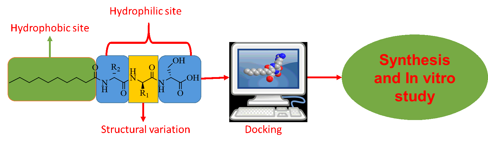
Peptide design rationale and screening simulations
Thirty-eight amphiphilic peptides were designed by selecting amino acid from each group, i.e., group 1= (RHK), group 2 = (DE) and group 3= (STNQ) and hydrocarbons chain (Table 1). The binding affinity of all amphiphilic peptides (1–38) with active site of urease were investigated through docking and it was observed that 1, 3, 18, 30 has good binding affinity with active site of enzyme, while 12 and 33 has excellent binding affinity as shown in Table 2. The high potential of 12, and 33 might be due to carboxylic and guanidino group, respectively, at the side chain of second amino acid, which donates some of its electron density via inductive effects to nickel ions and London dispersion forces between hydrophobic tail of AP and nonpolar amino acids in the vicinity of active site. The molecular interaction of the peptides 1, 3, 12, 18, 30, and 33 are shown in Fig. 2.
Table 1
Designed amphiphilic peptides (1–38)
| No. | Sequence | No. | Sequence |
| 1 | N-Dodecanoyl-Arg-Ser-Ser | 20 | N-Dodecanoyl-Arg-Val-Ser |
| 2 | N-Dodecanoyl-Arg-Asp-Ser | 21 | N-Dodecanoyl-Arg-Ala-Ser |
| 3 | N-Dodecanoyl-Arg-Thr-Ser | 22 | N-Dodecanoyl-His-Met-Ser |
| 4 | N-Dodecanoyl-Arg-Gln-Ser | 23 | N-Dodecanoyl-His-Ile-Ser |
| 5 | N-Dodecanoyl-His-Ser-Ser | 24 | N-Dodecanoyl-His-Val-Ser |
| 6 | N-Dodecanoyl-His-Asn-Ser | 25 | N-Dodecanoyl-His-Ala-Ser |
| 7 | N-Dodecanoyl-His-Thr-Ser | 26 | N-Dodecanoyl-Lys-Met-Ser |
| 8 | N-Dodecanoyl-His-Gln-Ser | 27 | N-Dodecanoyl-Lys-IIe-Ser |
| 9 | N-Dodecanoyl-Lys-Asn-Ser | 28 | N-Dodecanoyl-Lys-Val-Ser |
| 10 | N-Dodecanoyl-Lys-Thr-Ser | 29 | N-Dodecanoyl-Lys-Ala-Ser |
| 11 | N-Dodecanoyl-Lys-Gln-Ser | 30 | N-Dodecanoyl-Arg-Lys-Ser |
| 12 | N-Dodecanoyl-Arg-Asp-Ser | 31 | N-Dodecanoyl-His-Lys-Ser |
| 13 | N-Dodecanoyl-Arg-Glu-Ser | 32 | N-Dodecanoyl-Lys-Lys-Ser |
| 14 | N-Dodecanoyl-His-Asp-Ser | 33 | N-Dodecanoyl-Arg-Arg-Ser |
| 15 | N-Dodecanoyl-Glu-His-Ser | 34 | N-Dodecanoyl-His-His-Ser |
| 16 | N-Dodecanoyl-Lys-Asp-Ser | 35 | N-Dodecanoyl-Lys-Arg-Ser |
| 17 | N-Dodecanoyl-Lys-Glu-Ser | 36 | N-Dodecanoyl-His-His-Ser |
| 18 | N-Dodecanoyl-Arg-Met-Ser | 37 | N-Dodecanoyl-Lys-His-Ser |
| 19 | N-Dodecanoyl-Arg-Ile-Ser | 38 | N-Dodecanoyl-Arg-His-Ser |
Table 2
Inhibition constant (ki) and Gibb’s free binding energy (ΔG) of amphiphilic peptides calculated through MOE
| No. | ΔG (kcal mol− 1) | ki (µM) |
| 1 | −6.31 | 36.12 |
| 3 | −6.65 | 20.80 |
| 12 | −7.34 | 5.00 |
| 18 | −5.58 | 64.74 |
| 30 | −5.12 | 256.60 |
| 33 | −7.63 | 4.25 |
| Std = Thiourea | −0.89 | 236287.03 |
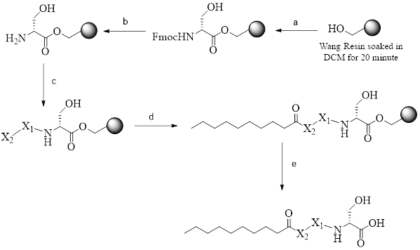
Synthesis
The targeted amphiphilic peptides (Fig. 3) were synthesized by Fmoc protocol (Scheme 1). In brief, Fmoc-Serine (3 equiv.) was treated with Wang resin (2 gm) in presence of tripyrrolidino phosphonium hexafluoro phosph- 77 as a coupling reagent (3 equiv.) and N, N-diisopropylethylamine base (3 equiv.). Similarly, second and third amino acids were coupled. After the formation of polar head by Fmoc protection and deprotection strategy [27], peptide was further treated with decanoic acid to introduce nonpolar nature. 95% trifluoro acetic acid was used for the cleavage of AP from Wang resin.
Structural studies of 1, 3, 12, 18, 30, and 33
The FAB MS spectra for 1, 3, 12, 18, 30, and 33 shows molecular ion [M + H] + peak at 503.2, 517.3, 547.3, 544.1, 572.1 and 544.2, respectively. The FAB MS-MS spectra further confirmed fragmentation pattern of the peptides. The fragment ion peak at 416, 430, 457, 460, 457, and 485 in FAB MS spectra (see supporting information) of amphiphilic peptides 1, 3, 12, 30 and 33 respectively showed the c1 cleavage i.e., between alpha carbon and amine groups of serine, while the peaks at 328 showed c2 cleavage (between alpha carbon and amine groups of second amino acid). Similarly, the peaks at 311 in FAB MS spectras of all the amphiphilic peptides are due to the formation of b2 ion, i.e., loss of arginine + decanoic acid (Figure-4).
The amphiphilic peptides were further characterized by 1H-NMR (400 MHz) and 13C- NMR (125 MHz, cryoprobe), by using CD3OH as solvent to record the data. The 1H-NMR spectra of amphiphilic peptides 1 showed three alpha protons in the region δ 3.90–4.50, Sixteen methylene protons of decanoic acid appeared as a multiplate at δ 1.39–2.06 and methyl proton between δ 0.90–1.15 (t). 13C-NMR data were recorded on 125 MHz (Cryoprobe) NMR machine, to obtained better 13C-NMR signals. 13C- NMR spectras of 1, 3, 12 18, 30 and 33 showed carbonyl carbons between δ 170.0-174.0. (for detail 1H-NMR and 13C-NMR data see supporting information Table 1).
In vitro urease inhibitory activity
Inhibition of urease by amphiphilic peptides (1, 3, 12, 18, 30, 33) at different concentrations were evaluated. It was observed that among the six amphiphilic peptides only 12 and 33 showed excellent activity having IC50 values 20.5 ± 0.01 µM, and 28.1 ± 0.03 µM respectively, as compared to standard thiourea (IC50 21.0 ± 0.2 µM) at (p < 0.001), which support MOE data (Table 3).
Table 3
In vitro urease inhibitory activity of Amphiphilic Peptides (1, 3, 12, 18, 30, 33)
| AP. No. | IC50 µM ± SEM |
| 1 | 51.7 ± 0.01 |
| 3 | 48.4 ± 0.17 |
| 12 | 20.5 ± 0.02 |
| 18 | 58.0 ± 0.14 |
| 30 | 76.0 ± 0.03 |
| 33 | 28.1 ± 0.03 |
| Std = Thiourea | 21.0 ± 0.21 |
| SEM standard error of the mean, Std standard inhibitor or urease enzyme |