3.1 Fabrication and structural characterization of SFPUHE-HTF and SFPUHE-HTF-CNTs
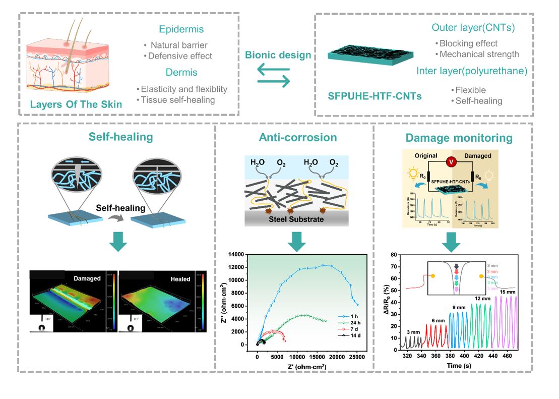
Inspired by the multi-layered structure of human skin, the bilayer skin-like coating SFPUHE-HTF-CNTs was constructed by scraping and spraying techniques in this paper. As shown in Fig. 1a, the design of the skin-like coating is based on two principles: First, the similarity of material properties and structure. The skin consists of a thinner, harder epidermal layer and a thicker, softer dermal layer[27]. Correspondingly, the skin-like bilayer coating designed here consists of a thinner rigid CNTs layer and a thicker flexible polyurethane layer. Secondly, the similarity of functions, the epidermis layer of skin provides a barrier for the substrate, and the dermis layer confers good flexibility and elasticity to the skin, providing insulation, buffering external shock and self-healing function of skin tissue damage[28]. Similarly, the polyurethane layer composed of flexible molecular chains containing dynamic hydrogen and disulfide bonds provides flexibility, elasticity and self-healing functions to the coating, while the CNTs layer is relatively rigid and dense, which endows the polyurethane elastomer layer with a significant barrier effect and mechanical strength[29]. Figure 1b shows that the SFPUHE-HTF-CNTs have a distinct layered structure, with the inner layer composed of self-healing polyurethane and the outer layer composed of CNTs. Besides, the outer layer is enlarged as shown in Fig. 1c, from which it can be seen that the CNTs are densely tangled and thus lays excellent foundations for the subsequent shielding protection. The preparation process of skin-like SFPUHE-HTF-CNTs coating is shown in Fig. 1d, and no organic solvent is used in the preparation process, which is a green and clean production approach.
The polyurethane inner layer imparts flexibility and self-healing properties to SFPUHE-HTF-CNTs, and whether it is successfully synthesized will affect the overall performance of the material. Therefore, the structure of self-healing polyurethanes were firstly characterized, and their infrared spectras are shown in Fig. 2a. The absorption peaks at 3332 cm-1, 3331 cm-1 and 3328 cm-1 corresponded to the stretching vibrations of the N-H bond in SFPU-HE0, SFPU-HE and SFPUHE-HTF, respectively. It is evident that the N-H absorption peaks shifted to lower wavenumbers, indicating an increase in the degree of hydrogen bonding with the addition of HEDS and HTF. Moreover, the absorption peaks at 2945 cm-1and 2862 cm-1 were attributed to the stretching vibration of -CH3 and -CH2- bonds in self-healing polyurethanes. The absorption peaks appeared at 1705 cm-1 and 1232 cm-1 belonging to C = O and C-O-C bonds. As shown in the curve of SFPUHE-HTF, the appearance of a new minor peak at 795 cm-1 was assigned to Si-CH3. All these observations demonstrated that HTF was successfully grafted onto the SFPU-HE molecular chain and the SFPUHE-HTF was successfully synthesized. Figure 2b illustrates the Raman spectroscopy of the self-healing disulfide-bonded polyurethanes. No sulfur-containing bonds appeared in SFPU-HE0, while SFPU-HE and SFPUHE-HTF both showed the corresponding absorption peaks of S-S and C-S bonds at 512 cm-1 and 640 cm-1, respectively. Besides, it was found that after some of the disulfide bonds were replaced by HTF, the intensity of the peaks corresponding to disulfide bonds in the SFPUHE-HTF spectrum decreased relative to that of SFPU-HE, indicating that the disulfide bond had been successfully cross-linked to the PU molecular chain. In polyurethane macromolecules, hard segments dominated by isocyanates and chain expanders and soft segments dominated by polyols are thermodynamically incompatible, and a microphase separation state will occur, resulting in poor crystallinity of polyurethane[30]. The crystallization properties of the self-healing polyurethanes were analyzed by XRD, and the results are presented in Fig. 2c. The peak positions and shapes of the SFPU-HE0, SFPU-HE and SFPUHE-HTF samples were basically the same, with no obvious sharp peaks, suggesting that the samples were in an amorphous state. A broad "diffuse scattering peak" appeared at 2θ = 20°, which can be ascribed to the short-range ordered crystallization peak in the hard segment region. The formation of this peak is due to the presence of a large number of microcrystals or subcrystals in the SFPU-HE and SFPUHE-HTF systems, which intermix to inhibit the crystallization of the soft segments[31].
Thermogravimetric analysis (TGA) was performed to evaluate the thermal stability of the self-healing polyurethanes, as depicted in Fig. 2d, the initial decomposition temperature of SFPU-HE0 increased from 261 ℃ to 308 ℃ of SFPU-HE. This phenomenon was ascribed to the increase of the content of the hard segments in SFPU-HE by the introduction of disulfide bonds, leading to higher cohesion energy and eventually to higher thermal stability. However, the initial decomposition temperature of SFPUHE-HTF was reduced to 295°C, which was due to the lower thermal stability of HTF with a decomposition temperature of around 230°C, and its introduction lowered the overall decomposition temperature of SFPUHE-HTF. The micro quotient thermogravimetric curve (DTG) of self-healing polyurethanes is displayed in Fig. 2e. The SFPUHE-HTF exhibited two inflection points, indicating two decomposition process stages[32]. The first inflection point occurred at 298 ℃ because of the decomposition of carbon chains in the polyol soft segment. A second inflection point could be observed when the temperature reached 413 ℃, which was attributed to the decomposition of some functional groups on the hard segment, such as the -NHCOO- bonds. It is worth noting that the weight loss temperature was higher than 286 ℃ of SFPU-HE0, indicating that the introduction of disulfide bonds and HTF has improved the thermal stability of SFPUHE-HTF. To further investigate the thermal reversibility of polyurethanes with disulfide bonds and HTF in the main chain, the variations of SFPU-HE0, SFPU-HE and SFPUHE-HTF under the effect of temperature were tested by DSC. As illustrated in Fig. 2f, all three curves display two steps, representing the glass transition temperatures of the soft and hard polyurethane segments, respectively. The glass transition temperature of the soft segment determines the thermal properties of polyurethane. It can be observed that the glass transition temperatures of the soft segments of SFPU-HE and SFPUHE-HTF were lower than room temperature, indicating that the polyurethanes are highly elastic at room temperature. Their chain segments are highly mobile, which is conducive to the dynamic reversible disulfide bond translocation exchange reaction in the molecular chain, thus facilitating the self-healing process. Additionally, SFPUHE-HTF presented a prominent heat absorption peak near 60 ℃, which was attributed to the exchange reaction of disulfide bonds affected by temperature, thus reassembling the original broken bonds into new disulfide bonds, causing the system to absorb heat[33]. More importantly, the appearance of this heat absorption peak robust a powerful basis for the temperature selection of further self-healing.
The CNTs outer layer provides protection and functionality for SFPUHE-HTF-CNTs, and the preparation of dense and homogeneous CNTs layers helps to maximize their functionality. During the spraying process, the dispersion of CNTs in the polymer matrix is the key issue[34]. As shown by the SEM images of SFPUHE-HTF-CNTs composites (Fig. 2g-h), CNTs are uniformly dispersed in the polymer matrix without apparent aggregates, which is conducive to the formation of perfect and effective barrier channels and conductive pathways, and thus improving the anti-corrosion and sensing performances of SFPUHE-HTF-CNTs coatings. Figure. 2i-k show the physical pictures of SFPUHE-HTF-CNTs before and after self-healing and the schematic diagram of the healing mechanism. The surface of the original SFPUHE-HTF-CNTs showed a uniform black color, and a bright crack appeared in Fig. 2i after breaking. When heated at 60 ℃ for 16 min, the scratches on the surface of the coating almost disappeared and only slight traces of healing residue could be observed (Fig. 2k). Notably, when human skin is injured, blood clots on the wound and gradually self-heals. This process is dominated by the upward diffusion of active keratinocytes[35], and similar to it, dynamic chemical bonding promotes self-healing of the coating. The self-healing of SFPUHE-HTF-CNTs is due to the translocation exchange reaction of disulfide bonds on the one hand, and HTF can form dynamic hydrogen bonds with the polar groups in the polyurethane chain segments on the other hand. The two synergistically drive the repair of the upper CNTs, thus facilitating the self-healing process, which provides favorable conditions for the subsequent realization of long-lasting corrosion and damage sensing properties that can be self-healed.
3.2 Self-repair performance and repair mechanism of SFPUHE-HTF and SFPUHE-HTF-CNTs
The self-healing properties of SFPUHE-HTF and SFPUHE-HTF-CNTs were systematically investigated by UV irradiation, polarized light microscopy and ultra-deep field microscopy. The self-healing process of SFPUHE-HTF-CNTs under UV irradiation is illustrated in Fig. 3a. Firstly, half of the sample was stained and then cut along partition line and placed in the UV chamber at an energy value of 200 for 48 h. From Fig. 3a, it can be seen that the two parts of the section were fused into one. The weight-bearing capacity of the repaired section was tested using two 100 g weights and it was not pulled off, indicating that SFPUHE-HTF-CNTs can self-repair under UV light. Significantly, light-responsive self-healing coating has the advantages of eco-friendly, rapid, remote and precise repair of damaged areas, which is more convenient for the coating repair outdoors[36].
According to the DSC curve in Fig. 2f, SFPUHE-HTF showed a distinct heat absorption peak at 60°C. Therefore, the self-healing behaviors of SFPU-HE, SFPUHE-HTF and SFPUHE-HTF-CNTs were observed at 60°C under a polarized light microscope equipped with a heating stage. All three samples were cut with a sharp knife in the same size (1 cm long, 20 µm wide). Figure 3(b1-b4) show that the surface cut of SFPU-HE gradually disappeared and was utterly repaired after 16 min at 60 ℃, but some bubble structures appeared. As displayed in Fig. 3(c1-c4), the cut marks on the surface of SFPUHE-HTF disappeared in the same time and no bubbles appeared on the surface, probably due to the leveling effect of the low surface energy HTF. Notably, the hydrophobicity also showed favorable recoverability, with a static water contact angle of 116° before cutting to 114° after repair at the damaged location. Similarly, the cut marks disappeared after SFPUHE-HTF-CNTs were repaired at 60 ℃ for 16 min, and the sample surface remained flat and smooth. The water contact angle changed from 117° before the cut to 115° after the repair (Fig. 3(d1-d4)). It was seen that all samples could achieve self-healing at 60°C for 16 min, which will be favored for its further application in self-healing anti-corrosion coatings.
The morphologies of SFPU-HE, SFPUHE-HTF and SFPUHE-HTF-CNTs before and after self-repair were analyzed by super-field microscopy. The e1、e2, f1、f2 and g1、g2 in Fig. 3 correspond to the morphologies of SFPU-HE, SFPUHE-HTF and SFPUHE-HTF-CNTs before and after self-healing, respectively, where f1、f2 and g1、g2 are embedded with contact angle test plots. As shown in Fig. 3 (e1, f1, g1), the cracks were obvious before repair, and the results were shown in Fig. 3(e2, f2, g2) after 16 min of repair at 60 ℃. The contact angles of f1 and f2 at the same position changed from 119° before cutting to 117° after repair, the contact angles of g1 and g2 at the same place changed from 118°to 115°, and all specimens showed a relatively flat state at the healed area. These results indicated that all samples had favorable self-healing properties, which are consistent with the findings revealed by UV irradiation and polarized light microscopy. Moreover, the coating has a mild thermally stimulated responsive self-healing condition, thus avoiding the risk of material degradation due to localized concentrated heating. The environmental friendliness and precision of light-stimulated responsive self-healing allows for contactless repair over long distance, thus promising for a broader range of applications.
To quantify the self-repair performance of SFPUHE-HTF-CNTs, tensile tests were carried out to campare the tensile strength of the healed samples and the intial, and the self-healing effiency were calculated by Eq. 1[37]. As presented in Fig. 4a, the maximum stress of SFPUHE-HTF-CNTs gradually decreased with the increase of the number of cycles, and the repair efficiency of the 1st cycle and the 4th cycle is 88.4% and 65.7%, respectively (Fig. 4b). The static contact angle tester was used to measure the water contact angle to evaluated the hydrophobicity of SFPUHE-HTF-CNTs, and the results are shown in Fig. 4c-d. The static water contact angle ranged from the original 118° to the fourth repaired 95°, all cycles showed good hydrophobicity, with the repair efficiency of 97% in the first cycle and 80% in the fourth cycle.
The self-healing mechanism of SFPUHE-HTF-CNTs is shown in Fig. 5a. On the one hand, the broken disulfide bond can be reconnected with the other one to generate a new disulfide bond, i.e., the translocation exchange reaction[38]. On the other hand, the introduction of HTF replaces part of the disulfide bond. Although the content of the disulfide bond is reduced, the introduction of HTF increases the degree of hydrogen bonding of polyurethane, and Studies have shown that the existence of dynamic hydrogen bonding will promote the healing process of polyurethane[39–41]. Figure 5b shows the properties of the FT-IR spectral bands of the vibrations of the C = O group in SFPUHE-HTF. The vibrational region of C = O was obtained by Gaussian splitting into three peaks: the stretching vibrational region of the C = O group with free hydrogen bonding appears at 1738 cm− 1, the C = O group with ordered hydrogen bonding at 1693 cm− 1, and the C = O group with disordered hydrogen bonding at 1719 cm− 1, in general agreement with the literature[42, 43]. The FTIR spectra of the samples were calculated using Gauss Lorenz curves, and SFPUD-HTF had a high degree of hydrogen bonding. In addition, the hydrophobic self-healing mechanism of SFPUHE-HTF-CNTs is shown in Fig. 5c. The good hydrophobic self-healing of SFPUHE-HTF-CNTs originates from the migration of fluorinated polysiloxane chain segments to the surface. The glass transition temperature of HTF is much lower than 60°C, so the macromolecular chain segments can undergo thermal movement at this temperature. When the fluorine-containing groups on the surface of the coating are damaged and the hydrophobic property of the coating decreases, the fluorinated polysiloxane chain segments can migrate from the inside to the outside, so that the fluorine-containing groups with low surface energy can be enriched on the surface of the coating again[44, 45]. The hydrophobic property of the material can be regained, thus completing the hydrophobic self-healing process.
3.3 Anti-corrosive properties of SFPUHE-HTF-CNTs
The skin-like bilayer structure design grants the coating the same self-healing function as human epidermis and has shielding and anticorrosive effects. The anti-corrosion protective layer of SFPUHE-HTF-CNTs coating consists of carbon nanotubes, which can form zigzag barrier channels to shield corrosive media due to their large aspect ratio[46]. SFPUHE-HTF-CNTs were coated on steel sheets, and the effect of CNTs contents on the corrosion resistance of the composite coating was explored by EIS in 3.5 wt% NaCl solution. Figure 6a shows the tafel polarization curves of the bare and the coated steel sheet with different CNTs contents. The corrosion current density (Icorr) and corrosion potential (Ecorr) of SFPUHE-HTF-CNTs with different CNTs contents can be obtained from the polarization curves, as shown in Table S1. As shown in Fig. 6a and Table S1, the bare steel sheet had the highest corrosion current density and the lowest corrosion potential with Icorr and Ecorr of 1.47×10− 6 A·cm-2 and − 0.68 V, respectively. After coating SFPUHE-HTF-CNT0, Icorr decreased to 5.24×10− 7 A·cm-2 and Ecorr increased to -0.54 V. Past research have shown that smaller Icorr and more positive Ecorr represented lower corrosion trends and dynamic corrosion rates[47, 48], indicating that the SFPUHE-HTF-CNTs coating could effectively improve the corrosion resistance of the steel sheets. Additionally, as the content of CNTs sprayed increases, the Icorr of the coated steel sheet shows a trend of decreasing and then increasing, while the Ecorr is the opposite. The SFPUHE-HTF-CNTs-3 coated steel sheet has a relatively excellent anticorrosive effect, and its Icorr is reduced by two orders of magnitude relative to SFPUHE-HTF-CNTs-0; Ecorr is shifted by 0.16 V in the positive direction relative to SFPUHE-HTF-CNTs-0. It is thus clear that, due to the barrier and shielding effect of the CNTs layer, the bilayer SFPUHE-HTF-CNTs have better anti-corrosion performance compared with the single-layer coating. However, the corrosion protection ability of SFPUHE-HTF-CNTs decreased when the CNTs content was further increased, which might be due to the tendency of agglomeration when the CNTs content was too high, thus reduced the interaction with the polymer chain segments and made it easier for the corrosive media to penetrate into the steel substrate. Subsequently, the corrosion behavior of the bare steel sheet and the coated SFPU-HTF-CNTs-3 immersed in 3.5 wt% NaCl solution for 1 h, 24 h, 7 d and 14 d was further monitored by EIS. From Fig. 6b and Fig. 6c, it can be seen that the coated steel sheet had a higher capacitive circuit diameter (Rct=2.51×10⁴ Ω·cm2, 1 h) and a higher impedance modulus (|Z|=4.79×10⁵ Ω·cm2, 1 h) after immersion in NaCl solution. It is shown that the skin-like bilayer structure of SFPUHE-HTF-CNTs has effective anticorrosive properties, which can be attributed to the good nano-size effect of CNTs to prolong the path of corrosive media (water, oxygen, etc.) into the steel substrate surface[49]. Figure 6d shows that the coated steel sheet still has a high impedance modulus (|Z| = 4.54 × 10⁴ Ω·cm2, 1 h) after self-healing, which proves the excellent anticorrosive self-healing properties of the composite coating. Similarly, the Tafel curve (Fig. S1) confirms this conclusion.
Further, the surface corrosion of bare steel sheets and steel sheets coated with SFPUHE-HTF-CNTs-3 before and after self-repair were observed by optical microscopy after immersion in 3.5 wt% NaCl solution for 1 h, 24 h, 7 d, and 14 d. From Fig. 6(e1-e4), it can be seen that with the extension of soaking time, corrosive ions enter, so the oxidation and corrosion on the steel sheet surface become more and more serious. Figure 6(f1-f4) show that when coated with SFPUHE-HTF-CNTs-3, thanks to the excellent barrier ability of the organic coating itself and the zigzag barrier channels formed after spraying CNTs on the surface, compared with the bare steel sheet, the corrosion on the surface of the metal steel becomes less after coating for the same soaking time. From Fig. 6(g1-g4), it can be seen that the layer after damage and self-repair still has anti-corrosion performance after soaking. In summary, the SFPUHE-HTF-CNTs-3 coating significantly improved the corrosion resistance of the steel sheet and provided some anticorrosive self-healing capability. This process is due to the synergistic action of the inner and outer layers of the skin-like bilayer structure coating. The outer CNTs constitute a barrier channel to provide anticorrosion protection, when the outer layer is damaged, the inner polyurethane layer can achieve self-healing of the overall coating driven by dynamic bonding. Meanwhile, the shielding effect of the outer layer provides a suitable environment for self-healing the inner layer[50]. Under the combined effect of protection and self-healing function, the anticorrosive self-healing process is completed (Fig. 6h-j). The synergy between the inner and outer layers of the skin-like bilayer structure coating is of great significance to achieving intelligent self-healing corrosion protection.
3.4 Sensing performance of SFPUHE-HTF-CNTs and its application in real-time damage monitoring
Although self-repairing anticorrosion coatings can extend service life by repairing damage, many damages are difficult to detect initially, so timely detection of damage and repair is essential to reduce maintenance costs and achieve long-lasting corrosion protection. Combining the outer layer of CNTs with the inner layer of the elastic matrix has become a commonly used strategy. When CNTs are sprayed onto the matrix, conductive pathways can be formed[51]. The damage can be responded to with the change in resistance of the conductive network, which is conducive to the monitoring of the defects (Fig. 7a). Based on this design concept, SFPUHE-HTF-CNTs skin-like coatings were prepared, and thanks to the excellent conductivity of the outer CNTs, the SFPUHE-HTF-CNTs exhibited good electrical conductivity. Among them, the conductivity of SFPUHE-HTF-CNTs-3 could still reach 112.92 S·m-1 after damage and self-repair (Table S2), and the good conductivity provided an excellent basis for the sensing performance of the composite coating.
Usually, the damage is caused by external forces. Considering that SFPUHE-HTF-CNTs-3 has relatively excellent conductive self-healing properties, the pressure sensing performance of SFPUHE-HTF-CNTs-3 was examined by simulating pressure damage to the coating during actual application by the pressure generated by weights. The normalized relative resistance change (ΔR/R0) was used to evaluate the responsiveness of the composite coating, where ΔR is the change in resistance in the pressure state and R0 is the original resistance in the unstressed state. Figure 7b shows that SFPUHE-HTF-CNTs-3 can respond to pressures from 10 kPa to 90 kPa, with ΔR/R0 of 25.03%, 34.23%, 44.82%, 54.32%, and 65.90% when subjected to different levels of pressure, respectively, with a linear increasing trend and good repeatability. Figure 7c presents that SFPUHE-HTF-CNTs-3 has fast response time and recovery time of 400 ms and 200 ms, respectively, indicating that SFPUHE-HTF-CNTs-3 has the potential to be applied for rapid detection of microcrack damage. Further, to simulate different sizes of coating damage in practical applications, different sizes of cracks (3 mm, 6 mm, 9 mm, 12 mm, 15 mm) were cut with a blade, and their electrical signal changes during the scratching process were examined. Figure 7d shows that SFPUHE-HTF-CNTs-3 can respond to different degrees of crack scratches with a linear increasing trend of ΔR/R0 as the scratch size increases. Figure 7e shows the sensitivity curve of SFPUHE-HTF-CNTs-3, which can be divided into two linear regions with different slopes, corresponding to two GFs, 3.55 for the fracture range from 0 to 9 mm and 2.29 for the fracture range from 9 to 15 mm. The excellent sensing sensitivity indicates that SFPUHE-HTF-CNTs-3 can respond quickly to different levels of damage, which will be beneficial for its application in more complex environments.
Good sensing self-repair performance is a necessary prerequisite to determine whether the material can be continuously monitored for damages. To investigate the sensing self-repair ability of SFPUHE-HTF-CNTs-3, the changes in resistance signals before, during and after repair were examined under constant pressure (Fig. 8a). From Fig. 8b, it can be seen that the original SFPUHE-HTF-CNTs-3, due to the intact conductive path of CNTs, when connected to the circuit, can make the small bulb glow with an initial resistance(R0) of 6 400 Ω, and the electrical signal remains relatively stable under constant pressure. In contrast, the R0 of the broken SFPUHE-HTF-CNTs-3 increased to 7595 Ω and the electrical signal was unstable due to the destruction of the conductive network (Fig. 8c). When it was heated at 60 ℃ for 16 min, due to the conductive network was restored, the bulb was lit again. R0 was restored to 6600 Ω, and the electrical signal was relatively stable under constant pressure (Fig. 8d). Usually, the conductive network will be rearranged after the self-healing material is damaged and self-repaired, which will affect the overall electrical properties of the material[52], but the skin-like SFPUHE-HTF-CNTs bilayer coating prepared here has excellent recovery of sensing signals. The good sensing self-healing ability facilitates the material to continuously detect internal cracks and external scratches, making its application and damage detection in complex environments possible.