2.1 Characterization of Se NPs and Se@PDA
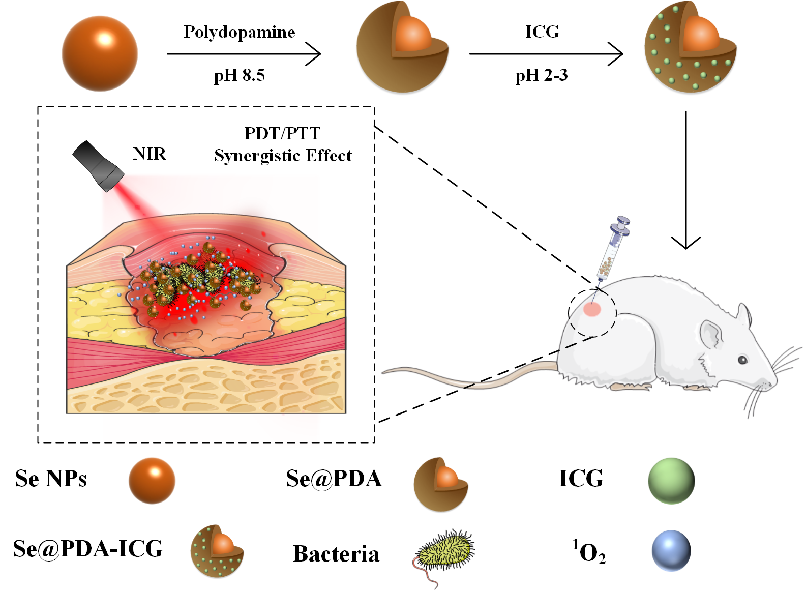
We first synthesized selenium nanoparticles (Se NPs) by referring to the preparation of homologous tellurium nanowires [26], and then functionalized the surface to form a stable system using the PDA (Se@PDA). The PDA wrap improves the dispersion, biocompatibility and photothermal properties of the Se NPs. Se NPs and Se@PDA NPs were characterised in series, as shown in Fig. 1A-I. Figure 1A shows transmission electron microscopy (TEM) images of Se NPs, from which it can be seen that the Se NPs are spherical and well dispersed with a uniform size of about 90 nm (Fig. S1A). Figure 1B shows X-ray diffractograms (XRD) of Se NPs, matching the standard card PDF#86-2246, indicating good crystallinity and a relatively complete morphological structure of the material. Figure 1C shows the X-ray photon spectra (XPS) of Se NPs, where the presence of the element Se provides evidence that the synthesised nanomaterials are Se NPs and that the elements C, N and O are probably derived from the surfactant. The Se@PDA nanomaterials are shown in Fig. 1D, a uniform transparent polymer shell layer of approximately 30 nm thickness can be observed on the surface of the Se NPs (Fig. S1B). In addition, the elemental distribution of Se@PDA was characterized by elemental mapping images, which showed that Se NPs were mainly concentrated in the nucleus of the nanoparticles, while N were mainly distributed on the outer edge of the nanoparticles to envelop Se NPs (Fig. 1G-I). The fourier transform infrared spectroscopy (FTIR) and ultraviolet-visible spectroscopy (UV-Vis) spectra of Se@PDA are shown in Fig. 1E and F. The colour of the aqueous solution of Se NPs is orange as shown in the physical illustration, while the colour of the aqueous solution of Se@PDA to brown-black after the coating of the PDA shell layer. The successful preparation of the nanocomposite was demonstrated.
2.2 Photothermal conversion efficiency of Se@PDA
The synthesized Se@PDA has good NIR absorption and the photothermal effect was then evaluated. Aqueous solutions of Se@PDA with different concentrations of Se@PDA were placed under a NIR light source (808 nm 2 W/cm2) to observe the temperature rise within 10 min. As shown in Fig. 2A, the temperature of the sample solution gradually increased with increasing concentration, and Se@PDA showed an excellent warming effect and strong concentration-dependent photothermal effect compared with the water. In addition, the power density of the irradiated laser had a significant effect on the photothermal effect. As shown in Fig. 2B, the amount of temperature change of the sample solution increased from 6.3°C to 18.3°C as the laser strength increasing from 1 W/cm2 to 3 W/cm2, which indicated that the higher the power density the higher heating up velocity. The photothermal stability of Se@PDA was studied by photothermal cycling. There was no significant decrease in the warming effect after four cycles, indicating the good photothermal stability of Se@PDA (Fig. 2C). Figure 2D shows the cooling curve of Se@PDA, and the calculated photothermal conversion efficiency (η) of Se@PDA was as high as 81.98%. Se@PDA has a high photothermal conversion efficiency compared with other similar PDA-modified materials, such as PDA/Au colloidal hollow nanoparticle (19.88%), Pt-PDA hybrid nanocomposite (44.5%) and PDA-Ce6 (60.4%) [27–29]. These results suggested that the synthesized Se@PDA have good photothermal properties.
2.3 Photothermal and photodynamic effects of Se@PDA-ICG
PDA shells can be loaded with near-infrared photosensitizer ICG through hydrophobic interaction and π-π stacking [30]. Herein, Se@PDA and ICG were assembled with different feeding ratios, and the UV absorption spectra were then measured. As shown in Fig. 3A, the characteristic absorption peak of ICG could be definitely observed at 780 nm after loading, and the loading rate of ICG was 8.11% when the feeding ratio of Se@PDA and ICG was 1:0.8. The color of the solution changed from black brown to dark green after loading ICG, and the average Zeta potential decreased from − 25 mV to -37.6 mV (Fig. 3B), further indicating the successful loading of ICG. ICG could get turned on to produce both heat and ROS under 808 nm NIR irradiation. Figure 4A shows the graphs of the photothermal warming data of Se NPs, Se@PDA and Se@PDA-ICG at a concentration of 200 µg/mL after irradiation with the 808 nm NIR at a power density of 2 W/cm2 for 10 min. More broadly, the temperature of Se NPs alone was not evident, the temperature of Se@PDA NPs changed slightly, while Se@PDA-ICG showed abrupt ascending temperature speed. The temperature of Se@PDA-ICG increased by 38.9°C after 10 min of irradiation, exhibiting excellent warming effects at lower concentrations and power densities. The infrared thermal images of each component sample under the same conditions were presented in Fig. 4B. Obviously, the temperature of Se@PDA-ICG changed significantly with increasing irradiation time, which was in line with the temperature change curve. To assess the ability of Se@PDA-ICG to produce singly linear oxygen (1O2), DPBF was used as a fluorescent probe. DPBF can react irreversibly with 1O2 and undergo a fluorescence quenching occurs, resulting in a reduction of the UV absorption peak at 419 nm. The absorbance spectrum of Se@PDA-ICG incubated with DPBF under 808 nm laser irradiation was demonstrated in Fig. 4C. The characteristic absorption peak (419 nm) of Se@PDA-ICG gradually decreased with increasing irradiation time, indicating that Se@PDA-ICG could be triggered to effectively produce 1O2. Figure 4D shows the variation of absorption at 419 nm with prolonging the irradiation time of solutions containing different samples and Se@PDA-ICG exhibited the most effective ROS production, probably due to the more active molecular motion of ICG accompanied by the heating of Se@PDA. The above results indicated that the photothermal effect and photodynamic effect of Se@PDA-ICG were significantly enhanced compared to free ICG and Se@PDA, indicating that Se@PDA-ICG had the potential to take on the role of a photoresponsive antimicrobial therapeutic agent.
2.4 Se@PDA-ICG for in vitro antibacterial
The antibacterial activity of Se@PDA-ICG was next assessed in vitro by plate counting with E. coli and S. aureus. PBS, Se@PDA, ICG, and Se@PDA-ICG were co-incubated with bacterial suspension for 2 h and then irradiated with the laser for 10 min (808 nm 1 W/cm2). We can see in the Fig. 5A-B, that after the treatment of E. coli in each group, the survival rate of Se@PDA was 94.8%, and that of Se@PDA + NIR was 67.17%, indicating that Se@PDA featured an inhibitory effect on bacterial growth through the photothermal effect. The survival rate of the ICG group was 91.1%, while that of the ICG + NIR group was 3.4%, due to the exceptional photodynamic effect of ICG. The survival rate of Se@PDA-ICG in dark was 85.7%, however, the Se@PDA-ICG + NIR group annihilated the bacterial (100%). All in all, we could draw a conclusion that Se@PDA-ICG possessed excellent photothermal and photodynamic synergistic antibacterial effects compared with Se@PDA and ICG alone.
Next Fig. 5C-D shows the bacteriostatic efficacy of each treatment group for S. aureus. The antibacterial rate of the Se@PDA-ICG group was 52.7%, and that of Se@PDA-ICG + NIR was 100%, indicating an excellent sterilization effect on S. aureus of Se@PDA-ICG. More importantly, Se@PDA-ICG showed better in vitro antibacterial effects compared to similar type of nanomaterials, such as PDA-ICG-NPs encapsulated in mixed shell polymeric micelles [31]. The bacterial survival rate of S. aureus was approximately 40% after dealing with the PDA-ICG-NPs under the laser for 10 min (808 nm 1.3 W/cm2). And antimicrobial porous collagen scaffolds containing chitosan and Se NPs were only effective in inhibiting S. aureus but not E. coli at all [16]. Herein, Se@PDA-ICG was also more selective for S. aureus, which may stem from the structural differences in the bacterial outer membrane of Gram-negative and Gram-positive bacteria [23]. In other to further investigate the antibacterial mechanism of Se@PDA-ICG, the cell morphology of bacteria after Se@PDA-ICG treatment was observed by SEM. As can be seen in Fig. 6A-B, the growth status of bacteria in the PBS control group was well maintained and the bacterial morphology was uniform and plump, while the bacterial surfaces of both strains in the Se@PDA-ICG + NIR experimental groups showed obvious wrinkling or rupture, and the cell integrity was significantly impaired. The release of bacterial cytoplasm might be caused by a combination of hyperthermia and ROS generated by Se@PDA-ICG and the excellent sterilization effect of the synergistic treatment was obtained as expected.
2.5 In vitro antibacterial activity assay
Biological security is the primary requirement for further application in advance of antibacterials. Therefore, the safety of Se@PDA-ICG was verified by a series of experiments before in vivo antimicrobial assays. The effect of Se@PDA-ICG on haemoglobin was first assessed by haemolysis assays. As shown in Fig. 7A-B, the haemolysis rate was only 0.9% even at Se@PDA-ICG concentrations up to 600 µg/mL. The cytocompatibility of Se@PDA-ICG was then determined by MTT assay using mouse fibroblasts (L929 and NIH 3T3 cells) and the viability of both cells was above 90% when Se@PDA-ICG concentrations reached 300 µg/mL (Fig. 7C). These results indicated that the synthesized Se@PDA-ICG exhibited lower cytotoxicity and better biocompatibility than other polydopamine-modified nano-carrier of ICG. For instance, the cell survival rate was below 80% at a low concentration of 100 µg/mL for MONs@PDA-ICG (manganese oxide nanoflowers as the core) [32], while it was also below 80% at an even lower concentration of 40 µg/mL for NDs@PDA@ICG (nanodiamonds as the subject) [33]. The biocompatibility of Se@PDA-ICG was further evaluated in vivo. The liver and kidney function and blood parameters of the mice were assessed after 7 days by tail vein injection of Se@PDA-ICG (300 µg/mL 200 µL) and the results were shown in Fig. 7D-F. As expected, compared with the PBS control group the experimental group showed no significant abnormalities. The above experiments showed that Se@PDA-ICG was of excellent biocompatibility and safety.
2.6 In vivo antibacterial performance evaluation
To verify the antibacterial activity of the synthesized Se@PDA-ICG in vivo, a wound S. aureus infection model was established in which PBS buffered solution was used as the control group. The wound model was created by infecting mice with S. aureus at the back wound and then irradiated with NIR (808 1W/cm2). As shown in Fig. 8A, B and C, the results showed varying degrees of reduction in trabecular area from 0 day to 8 day in all groups. At 2 days of treatment, there was some yellow pus on the trabecular surface, indicating that the S. aureus infection model was successfully established. After 8 days of treatment, the wound closure rates in the PBS, PBS + NIR, Se@PDA-ICG and Se@PDA-ICG + NIR groups were 45.8%, 55.52%, 70.36% and 88.74%, and the wound healing effect in the Se@PDA-ICG + NIR treatment group was significantly better than other control groups. More importantly, the Se@PDA-ICG showed better wound closure rates than other similar light-responsive antimicrobial therapeutics on the 8th day of treatment [34, 35]. Se@PDA-ICG + NIR provides antibacterial action in the early stages of infection and promotes vascular remodelling during the wound healing phase. Furthermore, the bactericidal effect of the different groups of wounds was assessed by plate counting on the 8th day of treatment, and the Se@PDA-ICG + NIR group had the best bactericidal effect with almost no bacteria surviving in the tissue fluid (Fig. 8D). During the treatment, the body weight of the mice remained normal with almost no significant difference between different groups (Fig. 8E), suggesting no damage to the health status of the mice.
On day 8 of wound healing, skin tissue from the trauma site was removed for pathological analysis, and the wound healing effect of each group was evaluated. Figure 9A shows hematoxylin and eosin (H&E) and Masson staining in each group. It can be seen from the picture that there are still obvious inflammatory infiltration and fewer collagen fibres in the PBS group. Se@PDA-ICG group had neovascularisation and higher collagen deposition than the control group. In particular, the Se@PDA-ICG + NIR group showed intact capillaries and the most collagen deposition, most similar to the state of healthy tissue. In addition, the changes in cytokines during wound healing are closely related to cell metabolism and proliferation [36]. Vascular endothelial growth factors (VEGF) play an important role in vascular regeneration. Leukocyte common antigen CD45 is strongly expressed in inflammation and decreases with the degree of inflammation. Therefore, the changes in angiogenesis and inflammation in the stage of wound healing can be evaluated by double staining of VEGF and CD45. As shown in Fig. 9B, the expression level of VEGF was lower and the expression level of CD45 was higher in the PBS group. The VEGF and CD45 in the PBS + NIR group and Se@PDA-ICG group were alike. The expression level of the VEGF factor in the Se@PDA-ICG + NIR group was the highest, the CD45 antigen was the least, indicated the repair effect was the best. These results demonstrate that Se@PDA-ICG + NIR can promote new angiogenesis, reduce cellular inflammation and accelerate wound healing. At the end of the treatment, the stained section images of the heart, liver, spleen, lung and kidney of experimental groups showed no significant abnormalities compared with control groups, indicating that the synergistic photoresponse treatment was non-invasive to the internal organs with insignificant side effects (Fig. 9C).