Al is the most abundant metal in the Earth crust. Several studies have shown that the damaging effect of Al on the nervous system is often reflected through its close correlation with various neurodegenerative diseases, such as AD, amyotrophic lateral sclerosis, and dialysis encephalopathy53. Studies have shown that long-term exposure to Al can cause accumulation in the body. Al can accumulate in the brain through the blood-brain barrier and placental barriers, leading to a variety of neurodegenerative diseases54. Studies have shown that Al can cause neurotoxicity and plays an important role in the occurrence and development of AD55, 56.
Al is too chemically reactive to exist as a free metal in nature. In this study, maltol was used as a coupling agent to combine with Al to generate maltol Al that infects cells. Maltol is a by-product of sucrose hydrolysis or starch thermal degradation, which is less harmful to the human body and is often used as a food additive in the food processing industry. Maltol and Al can be combined to produce electrically neutral maltol Al, which is not easily degraded, has a stable structure, and is not easily hydrolysed at room temperature. Under normal physiological pH, maltol Al can release a large number of Al ions, which makes it highly bioavailable; it can thus be absorbed and accumulated in the brain tissue, which is suitable for relevant studies on neurotoxicity57. The PC12 cell line is a common nerve cell line derived from adrenal pheochromocytoma of Rattus norvegicus, which is a sympathetic nervous system tumour cell with a stable growth state that is often used in gene transfection studies58. Therefore, maltol Al was selected as the dye venom to infect the PC12 cells with for subsequent experimental detection.
AD has two iconic pathological features, namely senile plaques and neurofibrillary tangles, which are mainly formed by hyperphosphorylation of tau protein10. Studies have shown that hyperphosphorylation of tau protein plays a major role in the neuropathology of AD59. In patients with AD, the earliest damage occurs in the tangles of cerebral endodermis cells, and the formation of the tangles involves the interaction between Al and the highly phosphorylated tau protein60. Other studies have shown that Al is closely related to the abnormal phosphorylation of tau protein, and Al can cause the pathological reaction thereof16, 17. In this study, to verify whether Al can induce tau hyperphosphorylation, maltol Al was exposed to PC12 cells, and phosphorylated tau protein was detected. The results showed that the expression of phosphorylated tau protein changed with an increase in maltol Al exposure concentration, and the expression of phosphorylated tau protein increased significantly in the 200 and 400 µmol/L Al(mal)3 groups (P˂0.05). In addition, Buee et al.61 found that abnormal hyperphosphorylation of tau protein is one of the characteristics of adult neurodegenerative diseases (such as AD and PD). The results of our study are consistent with these findings; therefore, we propose that Al can lead to hyperphosphorylation of the tau protein.
CREB is the core component of the molecular switch that converts short-term memory into long-term memory and plays a key role in learning and memory34. CREB is a ubiquitous nuclear transcription factor that can regulate the transcription of target genes, and it must be modified and activated by autophosphorylation to play its role in regulating the transcription of target genes41. Studies have shown that the PKA/CREB pathway plays an important role in memory62. The tau gene is one of the target genes regulated by CREB, and downregulation of the PKA/CREB signaling pathway can lead to learning and memory defects in patients with AD and in mice63–66. In the brains of patients with AD, downregulation of CREB expression or activity can lead to overexpression of phosphorylated tau protein38, 42. Phosphorylation at Ser-133 is one of the main pathways involved in CREB activation. Phosphorylation at this site is controlled by PKA; PKA can promote the phosphorylation of CREB, and the transcriptional activity of CREB increases. Destruction of CREB function in the brain can lead to neurodegeneration, and CREB phosphorylation plays an important role in neuroprotection.
In this study, a maltol Al exposure model was constructed to detect the protein expression levels of PKA, p-CREB (Ser133), and p-Tau (Ser396). The results showed that with an increase in maltol Al exposure concentration, the protein expression levels of PKA decreased gradually (P˂0.05), the protein expression of P-CREB (Ser133) decreased (P˂0.05), and the expression of p-tau (Ser396) protein increased (P˂0.05). The results indicated that maltol Al exposure can inhibit the activity of the PKA/CREB signalling pathway, suggesting that the inhibition of PKA/CREB signalling pathway activity may be an upstream regulatory pathway leading to tau hyperphosphorylation. The detection results of the miR-200a-3p low expression model showed that compared with the control group, the protein expression levels of PKA and P-CREB (Ser133) increased (P˂0.05), the expression of p-tau (Ser396) protein decreased (P˂0.05), compared with the 200 µmol/L Al(mal)3 group. The protein expression levels of PKA and P-CREB (Ser133) in the positive transfection + 200µmol/L Al(mal)3 group increased (P˂0.05) and the expression of p-tau (Ser396) protein decreased (P˂0.05), which is consistent with previous studies that the transcriptional activity of CREB is reduced, and the destruction of CREB function in the brain can lead to the expression of the protein of neurodegeneration44. These results indicate that Al may lead to a decrease in the activity of the PKA/CREB pathway and an increase in the phosphorylation level of the tau protein.
miRNAs have gene regulatory functions and can negatively regulate target genes by pairing with the 3’-UTR of target messenger genes67, 68. miRNAs extensively regulate multiple aspects of the nervous system by participating in post-transcriptional regulation of biological gene expression. Studies have shown that miRNAs are involved in various neurodegenerative diseases. For example, in the pathological processes of AD and PD69, miRNA may be potential regulatory factors for AD-related target genes29. miR-200a-3p belongs to the miR-200 family, which regulates cell differentiation, apoptosis and proliferation and plays an important role in human diseases such as cancer and AD31, 70. Many studies have shown that miR-200a-3p plays an important role in AD, but there are variations in miR-200a-3p levels detected in various AD models. Some studies have shown that miR-200a-3p expression is down-regulated during AD29. However, other studies have shown that the expression of miR-200a-3p was increased in the hippocampus of patients with AD and APP/PS1 mice32, 33. The model was constructed to elucidate whether the expression level of miR-200a-3p in Al-exposed PC12 cells changed and whether the change in miR-200a-3p expression level affected the change in tau phosphorylation level. PC12 cells were exposed to various concentrations of maltol Al to construct the tau hyperphosphorylation model, and then miR-200a-3p was detected. The results showed that the expression level of miR-200a-3p increased with increasing maltol Al exposure.
To verify the relationship between miR-200a-3p levels and Al exposure, the miR-200a-3p low expression model was constructed by inhibiting the expression of miR-200a-3p in PC12 cells. RT-PCR results showed that the expression of miR-200a-3p in the positive transfection group was lower than that in the control group (P˂0.05). Compared with the 200 µmol/L Al(mal)3 group, miR-200a-3p expression in the positive transfection + 200 µmol/L Al(mal)3 group was decreased (P˂0.05), indicating that the low expression model of miR-200a-3p in PC12 cells was successfully constructed. Western blot analysis showed that the expression of p-tau (Ser396) protein was decreased compared with that of the control group (P˂0.05). Compared with the 200 µmol/L Al(mal)3 group, the expression of p-tau (Ser396) protein in the positive transfection + 200 µmol/L Al(mal)3 group was decreased (P˂0.05). Therefore, there was an interaction between the inhibition of miR-200a-3p and the expression of tau phosphorylated by 200 µmol/L Al(mal)3. These results indicate that miR-200a-3p is involved in regulating the mechanism of Al induced tau phosphorylation; inhibition of miR-200a-3p expression results in decreased expression of phosphorylated tau protein.
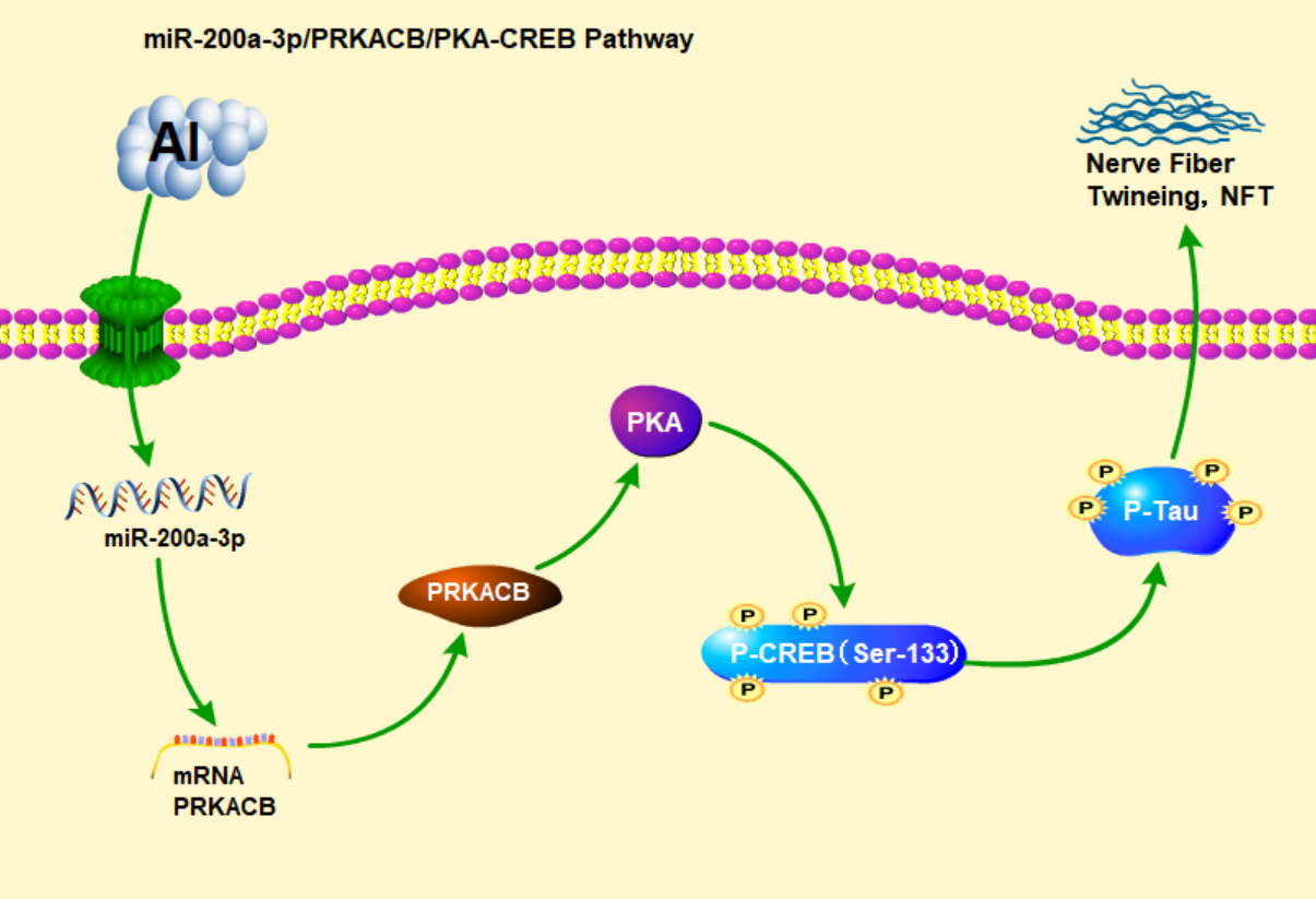
Phosphorylation of CREB at Ser-133 is controlled by PKA. When cAMP binds to the inactive PKA tetramer regulatory subunit to activate PKA, the activated PKA enters the nucleus, where it activates CREB by phosphorylating the CREB Ser-133 site43. CAMP binds to the two regulatory subunits of the inactive PKA tetramer, which separates the regulatory subunit from the catalytic subunit, and releases and activates the catalytic subunit51. The catalytic subunit of PKA can further act on CREB, resulting in its phosphorylation to produce a series of physiological activities.
To verify whether miR-200a-3p regulates the PKA/CREB pathway by targeting PRKACB, we analysed the constructed tau protein hyperphosphorylation model and miR-200a-3p low expression model. Analysis of the detection results of the tau hyperphosphorylation model showed that with the increase in maltol Al exposure concentration, the expression of miR-200a-3p increased (P˂0.05), mRNA-PRKACB and PRKACB protein expression decreased (P˂0.05), and the protein expression of P-CREB (Ser133) in the 200 and 400 µmol/L Al(mal)3 groups was significantly decreased (P˂0.05), compared with the control group. This suggests that Al targeted PRKACB to reduce its expression by increasing miR-200a-3p expression, and then participated in reducing PKA/CREB signalling pathway activity. To verify the hypothesis, the miR-200a-3p inhibition model was tested, and it was found that, compared with the control group, the expression of miR-200a-3p in the positive transfection group was decreased (P˂0.05); Compared with the 200 µmol/L Al(mal)3 group, miR-200a-3p expression in the positive transfection + 200 µmol/L Al(mal)3 group was decreased (P˂0.05); mRNA-PRKACB, PRKACB, PKA, and p-CREB (Ser133) protein expression increased (P˂0.05), compared with the 200 µmol/L Al(mal)3 group; and mRNA expression levels of PRKACB, PRKACB, PKA, and P-CREB (Ser133) in the positive transfection + 200 µmol/L Al(mal)3 group were increased (P˂0.05); These results showed that there was an interaction between the inhibition of miR-200a-3p and the expression of PRKACB, PKA, and CREB of the 200 µmol/L Al(mal)3 group. Therefore, Al may be involved in reducing the activity of the PKA/CREB signaling pathway by targeting PRKACB expression through increasing miR-200a-3p.