Data reporting
The sample size was predetermined according to the references without statistical methods5, 34, 35. The experiments were randomized and the investigators were blinded during data analysis for all experiments.
Rats
Rats (200-300 g weight) including Sprague Dawley (SD), Lewis and Brown Norway (BN) rats served as donors and recipients, they were purchased from Beijing Vital River Laboratory Animal Technology Corporation. All rats were housed and cared for in a temperature and light-controlled environment with free access to food and bottled water and the rats were fasted for 12 hours before LT. All experiments were conducted in compliance with the standards for animal use and care set by the Institutional Animal Care Committee of Henan Provincial People’s Hospital and were approved by the Ethics Committee of Henan Provincial People’s Hospital (No:HNTCMDW-20190304).
Orthotopic rat LT and experimental groupings
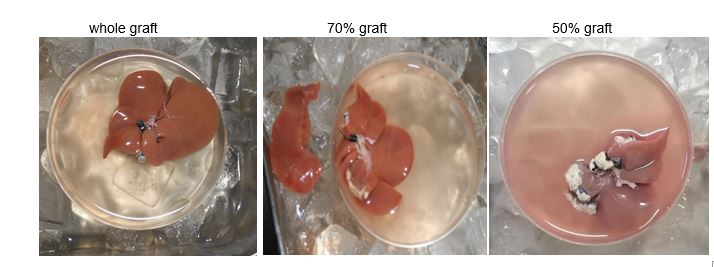
Rat LT was performed by one surgeon under a microscope. After opening the abdomen with a modified mask for isoflurane inhalation anesthesia we perfused the liver grafts first via the aorta with 5 ml of heparinized (50 U/mL) normal saline and then through the portal vein with 10 ml of cold lactated Ringer solution containing dexamethasone (24 mg/l). The cold storage time was less than 3 hours. The recipient's native liver was explanted out and the allograft was orthotopically implanted, followed by suprahepatic vena cava anastomosis with an 8-0 running suture. The two-cuff technique was used to reconnect the portal vein and the infrahepatic vena cava36,37. Arterial reconnection was made with a stent38. Biliary continuity was achieved by a polyethylene stent. The recipients were monitored daily for mobility, weight, posture, and urine color. A 50% liver graft consists of removal of the left lateral lobe, left portion of the middle lobe and caudate lobes; for a 70% graft, the left lateral lobe was removed39 (Table 1, Extended Data Table.1, Extended Data Fig.1).
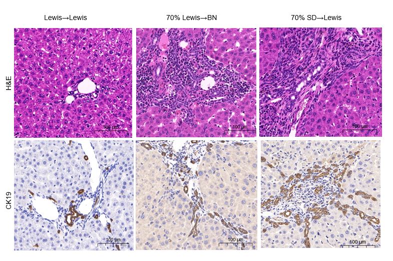
To formulate tolerance induction processes and investigate the potential mechanism of liver regeneration, we performed LT from Lewis to BN rats in the different groups, which is a well-established model of strong acute rejection. Whole LT was performed to compare half-size allograft LT with daily subcutaneous injection of CSA at 2 mg/kg for 9 d and r-GSF at 200 U/kg for 5 d and study the significance of liver regeneration. Half LT was performed with injection of CSA to emphasize the contribution of host bone marrow stem cells to the regenerating liver compared with the tolerant group (Table 1). 70% graft LT was performed for comparison with 50% graft LT to determine graft size (Extended Data Table 1). Other rat LT was performed to provide the controls for the histology images (Extended Data Fig.2).
Skin transplantation
We selected inbred Lewis rats as skin donors and post-LT BN rats as recipients for skin transplantation. A 1.5 cm × 1.5 cm full thickness allograft was prepared from the donor abdomen, and a graft bed (1.5 cm × 1.5 cm) was prepared on the back of the recipient rat40. When the skin allograft was attached to the back of the recipient with interrupted sutures of 5-0 silk, we covered the allograft with a protective tape and made the first inspection 3 days later and daily thereafter. Rejection was defined as a red–brown color, hard consistency, and necrosis and sloughing in the skin allograft.
Blood collection and liver sample preparation
After the subject rat was euthanized, the abdominal cavity was opened through a midline incision, the intestine was pulled out to expose the abdominal aorta, a blood collection needle pierced the aorta and was connected to a negative air pressure tube containing ethylenediaminetetraacetic acid (EDTA) for anticoagulation, and then the tube was inverted several times to prevent blood clotting. The plasma was collected at 2000 g centrifuge for 10 minutes (min) and the aliquots were labeled and cryopreserved until use. For spectrometry, the corresponding parameters were set on the automatic biochemical analyzer, the results were obtained after plasma was loaded, and ALT and AST levels were measured through the Substrate Method.
The liver allograft was perfused with normal saline through the mesenteric vein and extracted. The graft was weighed and cut into several small parts, and then immerged in 10% neutral paraformaldehyde solution for later use.
Liver histology
H&E and Masson trichrome staining were performed as previously described. Tissues were fixed in 10% neutral buffered formalin, and tissue processing, sectioning and staining was performed by Wuhan Servicebio Technology Ltd. The 3-µm paraffin sections were deparaffinized in xylene three times and rehydrated in a graded alcohol series. The slides were placed in citric acid (pH6.0) and heated several times for antigen epitope retrieval. Endogenous peroxidase was inhibited with 3% hydrogen peroxide and incubated for 25 min at room temperature in the dark. For CD3 staining, the slides were incubated first with normal goat serum, primary antibody was applied at a dilution of 1:1000 and incubated at 4 °C overnight, and the corresponding secondary antibody to primary antibody (hydrogen-peroxide oxidoreductase label) was applied to the slides and incubated at room temperature for 50 min. After being slightly dried, the slide was colored with freshly-prepared diaminobenzidine chromogen solution. Counterstaining was performed with hematoxylin. This immunohistochemistry staining procedure was also applied to CD44 (1:400), CD34 (1:500), CD133 (1:500), CK19 (1:2000) and IgG (1:500) (Servicebio Inc, Wuhan, China). Tissue sections were scanned and analyzed by Pannoramic confocal 3DHISTECH and with analyzed with Caseviewer software (The Digital Pathology Company, Budapest, Hungary).
Evaluation of liver regeneration
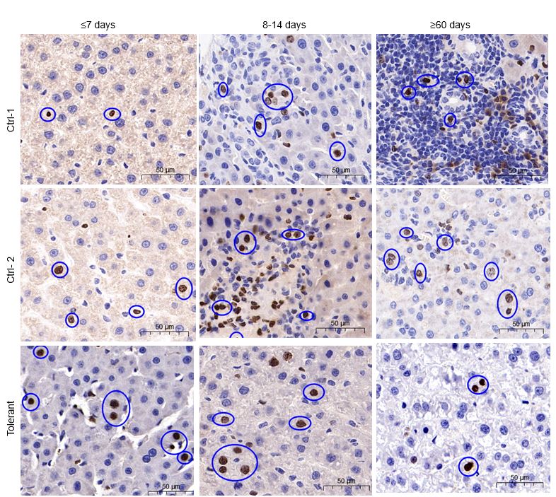
We studied hepatocyte replication by measuring the incorporation of BrdU with immunohistochemistry. BrdU (50 mg/kg) was injected intraperitoneally 24 hours before the liver tissue was harvested. The liver tissues were then cut into small pieces that were fixed in 10% neutral formalin, embedded in paraffin and sectioned. BrdU-incorporated hepatocytes were detected with an immunochemical system to monitor cell proliferation with a monoclonal anti-BrdU cell proliferation kit41-45 (Extended Data Fig.3), (GB12051, Servicebio Inc. Wuhan, China).
q-PCR analysis of rat Y chromosomes
Total DNA was extracted from isolated cells by using a TIANamp genomic DNA Kit (DP304-03, Hefei, China) according to the manufacturer’s protocol. The primer sets to amply rat Y chromosome were 5-ATTTATGGTGTGGTCCCGTGGAGA-3 and 5-TTCTGGTTCTTGGAGGACTGGTGT-320. The primer sets for control amplification of glyceraldehyde 3-phosphate dehydrogenase (GAPDH) were 5-AGGGAAATCGTGCGTGAC-3 and 5-CATACCCAAGAAGGAAGGCT-3. All primers used in our study were synthetized and purchased from General Biol Inc (Hefei,China). Twenty microliters (µl) of PCR reaction solution contained 10 µl of 2×ChamQ Universal SYBR qPCR Master Mix (Vazyme, NJ, USA),1 µl of cDNA, 0.4 µl of forward primer, 0.4 µl of reverse primer, 8.2 µl of RNase Free double distilled water. The thermal cycling conditions started with one cycle at 95 °C for 10 min. This was followed by 40 cycles at 95 °C for 10 seconds(s), 58 °C for 30 s, 72°C for 30 s, and 72 °C for final extension for 30 s. PCR products were electrophoresed on 2.5% agarose gels and visualized with ethidium bromide immunofluorescence staining (Zvast-bio Inc, Nanchang, China).
FISH
To further assess host bone marrow stem cell repopulation, we performed FISH of X and Y-chromosomes using rat a chromosome X Point Probe (Cat. No. FRWC-20P, Creative Bioarray Inc, USA) and a Y Point Probe (Cat.FRWC-21P, Creative Bioarray Inc, USA) according to the manufacturer’s protocol. The tissue slides were placed in 100% xylene for 5 min and this process was repeated 3 times. The slides were rehydrated in a graded alcohol series. After that, the slides were rinsed in distilled water for 1 min and in phosphate buffered saline (PBS) for 5 min, and then heated in distilled water at 100 °C for 15 min. The tissue was treated with pepsin solution for 10 min and rinsed in PBS. The slides were dehydrated by incubating in a gradient ethanol series each for 1 min and then air dried. Denaturing solution was equilibrated in an 88±2 °C water bath for approximately 30 min and the slides were immersed in the denaturing solution for 5 min. We dehydrated slides immediately in a gradient ethanol series each for 2 min, and air dried slides again. We denatured the FISH probes (10 μl of probe for each slide) in 88 °C for 5 min and kept the probes at 37 °C for 2 min. Finally we applied the denatured probes to the slides, applied a cover-slip, sealed them with rubber cement, and then hybridized them overnight at 40 °C in a humidified chamber. The rubber cement and cover slips were removed, and the slides were immersed in saline sodium citrate buffer (SSC)/0.3% NP-40 (Nonidet P-40) lysis buffer at 74±1 °C for 4 min. Then the slides were immersed in 2×SSC/0.3% NP-40 at room temperature for 5 min. Dehydrate slides by incubating slides in a gradient ethanol series each for 1 min and air dried the slides in the dark. Cell nuclei were stained blue with 4,6-diamidino-2-phenylindole (DAPI). Tissue sections were analyzed by confocal fluorescence microscopy (Creative Bioarray Inc, New York, USA).
Statistics and reproducibility
Statistical analysis was performed for survival curves generated by Kaplan-Meier analysis using GraphPad Prism 8. p≤0.05 was set as significant difference.
Data availability
All data generated and supporting the findings of this study are available within the paper, Additional information and materials will be made available upon request.
34.Shimizu, Y. et al. Restoration of tolerance to rat hepatic allografts by spleen-derived passenger leukocytes. Transpl. Int. 9,593-595 (1996).
35. Dresske, B., Lin, X., Huang, D.S., Zhou, X.& Fändrich, F. Spontaneous tolerance: experience with the rat liver transplant model. Hum. Immunol. 63, 853-861 (2002).
36. Lee, S., Charters, A.C., Chandler, J.G.& Orloff, M.J. A technique for orthotopic liver transplantation in the rat. Transplantation. 16,664-669 (1973).
37. Zhao, H.B. et al. Revisiting orthotopic rat liver transplant. Exp Clin Transplant. 9, 956-962 (2021).
38. Zhou, S.T. et al. New method of stent-facilitated arterial reconstruction for orthotopic mouse liver transplantation. J. Surg. Res. 187, 297-301 (2014).
39. Madrahimov, N., Dirsch, O., Broelsch, C.& Dahmen, U. Marginal hepatectomy in the rat: from anatomy to surgery. Ann. Surg. 244,89-98 (2006).
40. Barker, C. F. & Billingham, R. E. Skeletal muscle as a privileged site for orthotopic skin allografts. J. Exp. Med. 138, 289-299 (1973).
41. Forbes, S. J. et al. A significant proportion of myofibroblasts are of bone marrow origin in human liver fibrosis. Gastroenterology 126,955-963(2004).
42.Dalakas, E. et al. Bone marrow stem cells contribute to alcohol liver fibrosis in humans. Stem. Cells. Dev. 19,1417-1425 (2010).
43. Chen, Y.H. et al. Contribution of mature hepatocytes to small hepatocyte-like progenitor cells in retrorsine-exposed rats with chimeric livers. Hepatology. 57,1215-1224 (2013).
44. Higashiyama, R. et al. Negligible contribution of bone marrow-derived cells to collagen production during hepatic fibrogenesis in mice. Gastroenterology 137,1459-1466 (2009).
45. Assy, N. & Minuk, G.Y. Liver regeneration: Methods for monitoring and their applications. J. Hepatol. 26, 945-952 (1997).