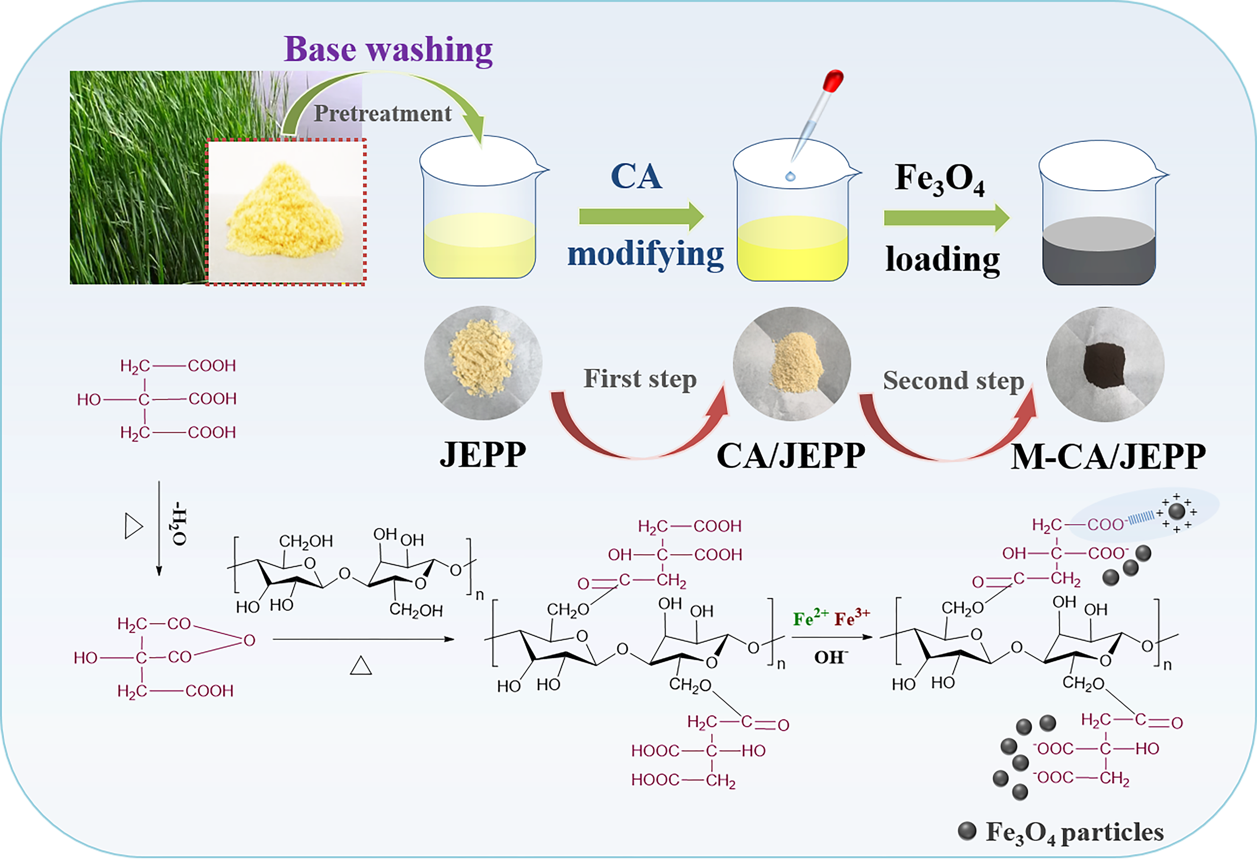
Chemical structure and morphology characterization of M-CA/JEPP
The functional groups of JEPP, CA/JEPP and M-CA/JEPP and their positions were determined by FTIR. As shown in Fig. 1a, a broad peak between 3000–3700 cm− 1 was linked to -OH stretching vibrations involved in the hydrogen bond interaction [43]. The absorption peaks observed at 2922 cm− 1 and 2848 cm− 1 corresponded to C-H stretching vibrations from CH and CH2 in cellulose, hemicellulose and lignin. Similarly, multiple peaks in the regions between 1000–1800 cm− 1 were related to the functional groups in cellulose, hemicellulose and lignin of the plant. Among them, the peak centered at 1736 cm− 1 was attributable to the C = O stretching of hemicelluloses, the peak located at 1513 cm− 1 was associated with C = C groups from lignin and the peak around 1250 cm− 1 came from C–O stretching in lignin and hemicelluloses [40, 44, 45]. The peaks of CA/JEPP near 1250 cm− 1 and 1513 cm− 1 were dramatically reduced, indicating a decrease in lignin and hemicellulose, which may be due to the alkali treatment, while a significant enhancement of the peak at 1736 cm− 1 could prove that citric acid have reacted with cellulose successfully. After magnetizing, the visible peak of M-CA/JEPP around 580 cm− 1 belonged to Fe-O bonds in Fe3O4, and the peak appeared at 1400 cm− 1 indicated the presence of COO−. The peak of C = O vibration from citric acid moved from 1736 cm− 1 to 1613 cm− 1 because of the influence of covalent bonds on the surface of Fe3O4 [46], implying the interaction existed in Fe3O4 and citric acid.
The X-ray diffraction patterns were conducted to identify the crystal structure of JEPP and M-CA/JEPP (Fig. 1b). Weak and broad peaks of the origin JEPP around 16.7° and 22.6° represented the amorphous region and crystalline area of cellulose [40]. The XRD pattern of M-CA/JEPP exhibited the typical diffraction peaks of trans spinel structure which was consistent with Fe3O4. The characteristic peaks at 30.2°, 35.5°, 43.2°, 53.7°, 57.1° and 62.7° were attributed to (220), (311), (400), (422), (511), (440) crystal planes, respectively [47].
To analyze the chemical composition and electronic states of various elements, XPS analysis was also performed and the results were compiled in Fig. 1c-f. The presence of carbon, oxygen, and iron peaks was confirmed by the XPS wide-scan survey of M-CA/JEPP (Fig. 1c). In the high-resolution XPS spectrum for C1s (Fig. 1d), there were three peaks located at 284.8 eV, 286.5 eV, and 288.1 eV attributing to C-C, C-O, and C = O/O = C-O [48], respectively. The high-resolution XPS spectrum for O1s (Fig. 1e) could be deconvoluted into four individual peaks around 530.0 eV, 531.3 eV, 532.8 eV and 533.1 eV which was corresponded to Fe-O, C = O/O = C-O, O-H, and C-O [48, 49], respectively. The Fe2p spectrum (Fig. 1f) showed two main peaks at 710.3 and 724.9 eV, which could be assigned as Fe2p3/2 and Fe2p1/2 from Fe3O4 [50] The deconvoluted peaks of Fe2p spectrum were linked to octahedral Fe3+ species, tetrahedral Fe3+ species, satellite peak of Fe3+ and Fe2+ ions, and octahedral Fe2+ species [51, 52]. Combined the results of FTIR, XRD and XPS, it could indicate that magnetic Fe3O4 was synthesized and incorporated into the CA-modified JEPP successfully.
The microstructure and surface morphology of initial JEPP and M-CA/JEPP were characterized by SEM. As shown in Fig. 2a, JEPP presented the shape of framing scaffold structure and smooth surface. Micro pores could be observed on the framing scaffold structure which were several microns in size. After modification and magnetizing, the surface of obtained M-CA/JEPP became extremely rough with the large pores disappeared, but some cracks and pits which might be conducive to adsorption were formed (Fig. 2b, d, e). The EDS spectra of M-CA/JEPP (Fig. 2c) only exhibited the peaks of C, O, and Fe, which were three major constituents of CA-modified fibers and magnetite, confirming there hasn’t impurities introduced during the synthesis process. Additionally, Fig. 2e showed there were plenty of magnetic nanoparticles covering on the surface of modified JEPP and the specific surface area was 34.15 m2/g (Fig. S2). Moreover, the EDS mapping were performed (Fig. 2f). The resulting patterns showed that C, O, Fe elements were uniformly distributed throughout the adsorbent powders, which was strong evidence for the combination of Fe3O4 and CA/JEPP.
The size and distribution of magnetic nanoparticles on adsorbents were shown in TEM images (Fig. S3a, b). The Fe3O4 nanoparticles were evenly distributed on the surface of the powders, whose average size was 6.60 nm (Fig. S3d). There was no serious aggregation between those nanoparticles. The ultrasonic assistance and citric acid modification in the fabrication process could reduce the possibility of aggregation and was beneficial for better dispersion [53]. As presented in high-resolution transmission electron microscopy (HRTEM) image (Fig. S3c), the lattice fringes of the samples displayed interplanar spacings of 0.254 nm and 0.302 nm in the nanoparticles, which matched well respectively with the (311) and the (220) characteristic lattice planes of Fe3O4 [54].
Adsorption properties of the adsorbent
Effects of initial pH on MB adsorption
The initial pH value of the dye solution is a significant influencing factor for adsorption performance. Alkaline conditions were conducive to the adsorption of MB dyes by M-CA/JEPP. It could be seen from Fig. 3a that the adsorption efficiency of MB was gradually enhanced with pH increasing from 4 to 11. At pH 4, the adsorption efficiency of MB was 77.61%, while the adsorption efficiency of MB increased to 98.62% at pH 11. Similarly, Fig. 3b implied that the adsorption capacity was increased from 155.22 mg/g to maximum value (197.25 mg/g) while the pH was adding up from 4 to 11. Therefore, pH = 11 was chosen to study the adsorption properties in other experiments.
The pH of the solution will affect the dissociation of functional groups on the surface of M-CA/JEPP, which has a corresponding effect on the adsorption behavior. Under the acidic condition with low pH, more free hydrogen ions (H+) in the solution competed with the cationic MB molecules to occupy the active site of the adsorbents, which inhibited the adsorption of MB [55]. As the pH rose from 4 to 11, the numbers of OH− ions in the solution increased, promoting the dissociation of H+ ions which came from -OH and -COOH functional groups on the surface of M-CA/JEPP. The electronegativity of the adsorbent enhanced, and the adsorption efficiency and adsorption capacity became higher owing to the electrostatic attraction between cationic dye and adsorbents.
Effects of contact time on MB adsorption
The contact time between adsorbents and dyes always affects the final result of the adsorption. Figure 3c presented rapid adsorption of dye in the first 10 minutes, for MB dye solution with initial concentrations of 50 mg/L, 100 mg/L, 150 mg/L, the adsorption efficiency could reach 98.34%, 95.21%, 88.93%. Thereafter, the adsorption efficiency increased gradually, and the adsorption quickly reached equilibrium in around 60 minutes. With increasing of initial dye concentration, the adsorption capacity could reach up to a higher equilibrium value with time increasing (Fig. 3d). But the increased in contact time has promoted the aggregation of dye molecules [56], which made it almost impossible to diffuse deeper into the adsorbent structure and hindered the unlimited increasing of adsorption capacity.
Effects of initial dye concentration on MB adsorption
The effect of initial dye concentration in the range of 10 to 500 mg/L was investigated and the results were shown in Fig. 3e, f. As the initial dye concentration increased, the adsorption efficiency declined (Fig. 3e). While the adsorption efficiency for MB was found to be 98.62% for 100 mg/L of initial concentration, the value was 28.82% with the initial dye concentration was 500 mg/L. As shown in Fig. 3f, the adsorption capacity increased and finally tend to be stable with the initial dye concentration increasing. Since high initial dye concentration might provide the high driving force for the mass transfer [57], the adsorption capacity increased from 197.25 mg/g to 272.08 mg/g with the initial dye concentration changed from 100 mg/L to 150 mg/L. However, as the adsorbent dose was fixed, which meant the available active sites were limited, so the adsorption capacity would become similar if the initial dye concentration was higher than 150 mg/L.
Magnetic property and reusability of M-CA/JEPP
The M-CA/JEPP showed powerful magnetic property. The magnetization curve in Fig. 4a revealed the ferromagnetic behavior of M-CA/JEPP, its saturation magnetization was 24.8 emu/g, which was significantly stronger than 3.6 emu/g [58] and 5.0 emu/g [59] in other magnetic cellulose-based adsorbent works. Therefore, this new type of adsorbent showed powerful magnetic responsivity, and it could be separated conveniently within ten seconds from the treated solution with the help of an external magnetic force (Fig. 4b). In other words, it can be used as a magnetic trigger to finish the separation and reuse process in wastewater treatment.
The cyclic adsorption experiment was performed to investigate the regeneration of the adsorbent (Fig. 4c). The adsorbent was regenerated by using 0.1 M HCl as desorption reagent after adsorption. The results showed that for 50 mg/L MB solution, the removal efficiency of the M-CA/JEPP towards MB could maintain a level above 98% after five cycles, which was hardly reduced, suggesting that the adsorbent had outstanding recyclability.
Fitting results of the adsorption process
Adsorption kinetics
Four typical kinetic models including pseudo-first-order, pseudo-second-order, Elovich and intraparticle diffusion were conducted to calculate the rate constants and other parameters of the M-CA/JEPP for MB adsorption according to Equation S1-S4. The fitting results and parameters were shown in Fig. 5 and Table 1.
The correlation coefficient (R2) was used to assess the degree of conformity between the experimental values and the theoretical values of the model. According to the fitting results of different concentrations of MB, the R2 values calculated by the pseudo-first-order dynamic model were between 0.969–0.989. The R2 values of the intraparticle diffusion model were between 0.577–0.977, and its fitted line were not zeroaxial, which indicated that intraparticle diffusion was not the rate-limiting step of the whole adsorption [34]. Elovich model was commonly used to deal with the chemical adsorption mechanism in nature, the R2 values of Elovich model were in the range of 0.935–0.975 [63]. The R2 values calculated by the pseudo-second-order dynamic model were above 0.996 and the obtained qe,cal values were also closest to the original experimental values. So pseudo-second-order model was more appropriate for defining this adsorption process, suggesting that the rate control step occurred during the entire adsorption process [64].
Table 1
Kinetic parameters for different initial concentrations of MB on M-CA/JEPP with various models
| Kinetics models | Parameters | Initial concentration |
| 50 mg/L | 100 mg/L | 150 mg/L |
| Pseudo-first-order | qe,exp (mg/g) | 98.837 | 197.247 | 276.450 |
| | qe,cal (mg/g) | 97.678 | 192.348 | 267.360 |
| k1 (min− 1) | 2.298 | 0.851 | 1.554 |
| R2 | 0.989 | 0.986 | 0.969 |
| Pseudo-second-order | qe,exp (mg/g) | 98.837 | 197.247 | 276.450 |
| | qe,cal (mg/g) | 99.783 | 200.526 | 275.755 |
| k2 [g/(mg min)] | 0.050 | 0.006 | 0.010 |
| R2 | 0.999 | 0.999 | 0.996 |
| Elovich | α [mg/g min] | 1.667E + 9 | 7.482E + 3 | 8.630E + 5 |
| | β [g/mg] | 0.237 | 0.049 | 0.053 |
| R2 | 0.974 | 0.935 | 0.975 |
| Intrapaticle diffusion | kp,1 [mg/g min− 0.5] | 29.089 | 95.758 | 86.421 |
| | C1 (mg/g) | 51.373 | 12.256 | 107.896 |
| R2 | 0.997 | 0.965 | 0.992 |
| kp,2 [mg/g min− 0.5] | 0.166 | 1.886 | 2.742 |
| C2 (mg/g) | 97.641 | 180.751 | 253.277 |
| R2 | 0.938 | 0.577 | 0.649 |
Adsorption isotherm
Langmuir, Freundlich and Temkin adsorption isotherms were used to explain the relationship between the adsorbent and adsorbate according to Equation S5-S7. The fitting results and parameters were shown in Fig. 6 and Table 2.
The results of the MB adsorption experiment at different temperatures indicated that the increase in temperature will make the adsorption capacity lower. Langmuir isotherm model was the most suitable model, whose R2 values were above 0.982. The maximum adsorption capacity of M-CA/JEPP for MB at 303 K was 293.132 mg/g, which surpass many magetic adsorbents derived from plants (Table 3). The Langmuir isotherm is often used to describe monolayer adsorption on uniform surfaces, which is also based on the assumption that the adsorption energy at each point of the surface is the same and there is no interaction between the adsorbate molecules attached to the surface. The Freundlich isotherm which suppose the energy on the surface is uneven is often used to describe multilayer adsorption. Furthermore, The Temkin isotherms assume that the adsorption heat of all molecules in the layer will decrease linearly with coverage rate due to the adsorbent/adsorbent interaction, which is commonly used to explain the interaction between adsorbent molecules adsorbed on the surface [65, 66]. The optimal adaptation of the Langmuir isotherm substantiated that the MB adsorption by M-CA/JEPP was monolayer adsorption rather than multilayer adsorption. In addition, one essential characteristic of the Langmuir isotherm could be expressed by the separation factor RL calculated from Equation S8. In the present study, RL values were between 0–1, indicating that the adsorption process under experimental conditions is conducive to proceeding [67].
Table 2
Isotherm parameters for MB on M-CA/JEPP with various models at different temperature
| Isotherm models | Parameters | Temperature |
| 303 K | 313 K | 323 K |
| Langmuir | qmax (mg/g) | 293.132 | 284.929 | 279.584 |
| | kl (L/mg) | 1.124 | 1.159 | 1.076 |
| R2 | 0.986 | 0.982 | 0.992 |
| Freundlich | 1/n | 0.154 | 0.154 | 0.156 |
| | kf [(mg/g)/(mg/L)1/n] | 133.957 | 130.151 | 126.474 |
| R2 | 0.782 | 0.787 | 0.813 |
| Temkin | B | 33.578 | 32.440 | 31.948 |
| | AT (L/g) | 46.887 | 48.667 | 47.059 |
| R2 | 0.876 | 0.876 | 0.903 |
Table 3
The M-CA/JEPP for MB compared with other reported magnetic adsorbents which derived from plants
| Adsorbents | qm (mg/g) | Reference |
| durian shell fiber-Fe3O4-MOF | 53.31 | [68] |
| magnetic rice husk ash | 150.5 | [69] |
| Fe3O4-loaded biochar from sorghum straw | 166.67 | [70] |
| Magnetic biochar nanocomposite from raw avocado peel | 62.1 | [71] |
| Seawater/K2FeO4-derived magnetic biochar from jackfruit peel | 129.61 | [72] |
| Magnetic biochar from agricultural waste | 55.0 | [73] |
| Fenton-modified biochar from rubber tree bark | 258.14 | [74] |
| Hydrochar magnetic adsorbents from Chinese medicine industry waste | 60 | [75] |
| Citric acid-modified Juncus effusus pith powders loaded with Fe3O4 nanoparticles | 293.132 | This work |
Adsorption thermodynamic.
After the experimental data fitted by Equation S9-S11, the thermodynamic parameters (Gibbs free energy change (ΔG), enthalpy change (ΔH), entropy change (ΔS) of this adsorption process could be calculated, the fitting lines and the calculated results were displayed in Fig. S4 and Table S2. Among the results, ΔG < 0 proved that the adsorption was a spontaneous process. The Negative ΔH indicated that these processes were exothermic, which further confirmed that temperature increasing was not beneficial for the adsorption. ΔS > 0 suggested that due to the increased randomness of the solution/solid interface, adsorbent/adsorbent composites are formed [76, 77]. Moreover, the absolute value of ΔH was between 0–84 kJ/mol, which meant this MB adsorption process could be mainly regarded as physical adsorption [78].