Materials
Kaempferol (KAE) and Dimethyl sulfoxide were provided by Sinopharm Chemical Reagent Co., Ltd. (Shanghai, China). D-α-Tocopherol polyethylene glycol 1000 succinate (TPGS), methanol, phosphoric acid, polyvinyl pyrrolidone (PVP), 1,1-dioctadecyl-3,3,3,3-tetramethylindotricarbocyaine iodide (DIR), Tween-20 (T-20), Tween-80 (T-80), Sodium dodecyl sulfate (SDS) and polyethylene glycol 2000 (PEG-2000) were obtained from Macklin Biological Technology Co., Ltd. (Shanghai, China). Methanol (HPLC grade) was ordered from Concord Technology Co., Ltd. (Tianjin, China). Chemical reagents were all of analytical grade or higher.
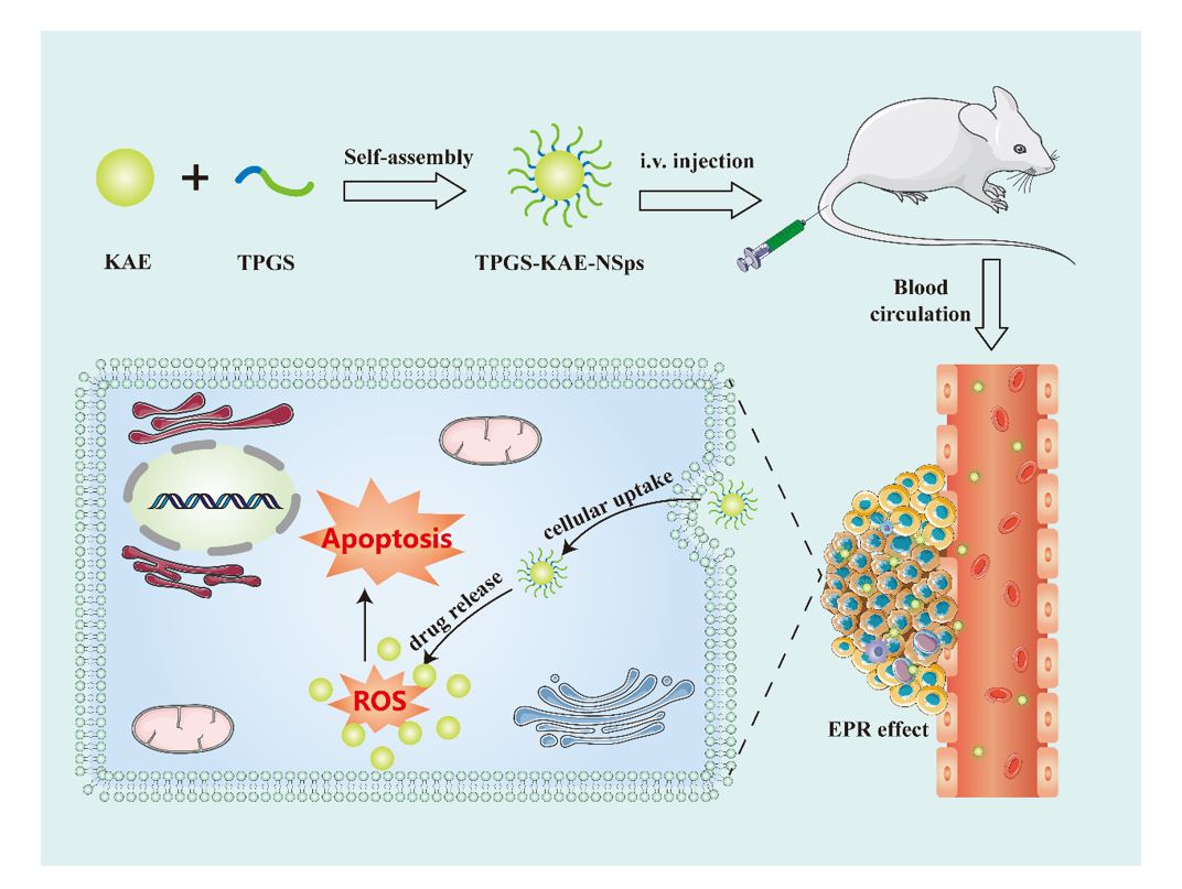
Preparation and Process optimization of nanosuspensions
Anti-solvent precipitation and high-pressure homogenization reported were the common methods of preparing nanosuspensions[35]. In this study, Kaempferol nano suspensions (KAE-NSps) were prepared by anti-solvent precipitation combined with high-pressure homogenization. Briefly, a certain amount of KAEs and stabilizers were dissolved in organic solvent together, the mixture was added to deionized water dropwise under ultrasonication at 45 ℃ with Ultrasonic instrument (Shanghai kedao Co., Ltd). Then the organic solvent was evaporated under vacuum at 50 ℃ by Rotary evaporator(Shanghai yarong Co., Ltd). Subsequently, the resulting solutions were subjected to homogenization (ATS Engineering Inc., Shanghai, China) to produce KAE-NSps. Finally, the lyophilization process of KAE-NSps was conducted by LD-A80-21L freeze dryer(Shanghai Binglin Electronic Technology Co., Ltd.) for the further characterize. There were many factors including different stabilizer, organic solvent, ultrasonic power, drug-load ratio, homogenization pressure, and homogenization times et al. affected the particle size of KAE-NSps during the preparation process. Here we took the particle size of KAE-NSps prepared as the final index as well as the method of Single factor analysis was applied to optimize the preparation process.
Physicochemical characterization of nanosuspensions
The method of Dynamic light scattering (DLS) was applied to determine the particle size, polydispersity index (PDI), and zeta potential (ZP) of TPGS-KAE-NSps prepared using Zetasizer Nano ZS90 (Malvern Instruments Ltd, Malvern, England). Diluting the specimens (60-fold) by deionized water before measurement to obtain the particle densities required for DLS and all the tests were repeated in triplicate under room temperature. JEM-2100F Transmission electron microscope (TEM) (JEOL, Japan) was used to observed the morphology of TPGS-KAE-NSps and TPGS, the sample dilution liquid of which was placed on a 200-mesh copper grid. The X-ray diffraction(XRD) of samples was detected using Ultima IV X-ray diffractometer (Rigaku, Japan) and scanned at a diffraction angle range of 5 ~ 60° using a Cu-Ka radiation generator set at 200 mA and 40 kV, with step length of 0.02 °and speed of 5 °/min. Fourier transform infrared spectroscopy (FTIR) spectra of the samples were detected using AVATAR370 spectrometer (Nicolet, USA) with the scanning wavelength set 4000 − 500 cm− 1. Thermogravimetric analysis (TG), Derivative thermogravimetric analysis (DTG) and Differential scanning calorimetry (DSC) of samples were detected by STA449F3 simultaneous thermal analyzer (NETZSCH, Germany). Approximately 10 mg of each sample placed and sealed in standard aluminum pan was detected at 10°C/min from 30°C to 600°C under nitrogen environment[36].
Stability of nanosuspensions
The stability of nanosuspensions mainly includes storage and various physiological media stability. The particle size and PDI of TPGS-KAE-NSps stored in a vial and placed at room temperature were tested on the 1st, 3rd, 5th, 7th, 10th, and 14th days to evaluate the storage stability. TPGS-KAE-NSps prepared was mixed with 2 × Phosphate buffer solution (PBS, pH7.4), 1.8% NaCl solution, 10% glucose solution respectively (1:1, v/v) to get an isotonic solution, while the nanosuspensions was also mixed with plasma (4:1, v/v), artificial intestinal fluid (in PBS (pH 6.8) with 1% trypsin) and artificial gastric fluid (in 1 mol/L diluted HCl with 1% pepsin). All the samples were placed in a water bath at 37 ℃ and the particle size was determined at 0, 2, 4, 8, and 12 h to evaluate the physiological media stability. Then the protein adsorption test was conducted to investigate whether the increased particle size of TPGS-KAE-NSps prepared in plasma was related to the adsorption of proteins.
Optimization of lyophilization for nanosuspensions
Three parts of TPGS-KAE-NSps freshly prepared were accurately absorbed into 10mL vials, as well as 2% w/v glucose, sucrose and PVP were added into for lyophilized protectors to obtain lyophilized powder by freeze-drying at -90 ℃ for 24h using the LD-A80-21L freeze dryer(Shanghai Binglin Electronic Technology Co., Ltd.). 0.5%, 1%, 2% and 5% w/v glucose were added into TPGS-KAE-NSps freshly prepared, lyophilized as before, respectively. All the lyophilized powder dissolved in 2 mL of deionized water was determined the particle size by DLS.
Hemolysis evaluation of nanosuspensions
The hemolysis experiment of TPGS-KAE-NSps was performed to evaluate the biocompatibility and whether it was applied to intravenous injection[37]. The whole blood was derived from Balb/c mice orbit. Whereafter, the supernatant plasma was removed by centrifugation at 5000 rpm to obtain 4% red blood cell suspension (v/v), which was mixed with 0.5 mL 1 × PBS, 0.5 mL deionized water and 0.5 mL TPGS-KAE-NSps of specific concentrations, respectively. All of samples were incubated in a water bath at 37 ◦C for 3 h and centrifuged at 4000 rpm for 10 min, then the supernatants of each were determined at 540 nm using microplate reader (Thermo, USA). The hemolysis ratio was obtained by the formula as follows:
Hemolysis ratio (%) = A-APBS/AH2O-APBS×100%
A, APBS and AH2O are the absorbances of the sample at 540 nm with TPGS-KAE-NSps, PBS and deionized water treatment, respectively.
High-performance liquid chromatography(HPLC) analysis
Kaempferol absorption wavelength was determined on ultraviolet spectrophotometer (U-3010, Hitachi, Japan). The quantification of kaempferol was performed using HPLC (Shimadzu, 20 AT, Japan), the chromatographic column of which was the C18 column (250×4.6 mm, 5 µm). The mobile phase consists of methanol and 0.4% phosphoric with a ratio of 70:30 (v/ v). The sample was under isokinetic elution conditions, the flow rate was 1 mL/min, the detection wavelength was 366 nm, the column temperature was 30°C, and the injection volume was 20 µL. The mass concentration(X) of kaempferol reference solution with the peak area(Y) was used to obtain the standard calibration curve.
Drug loading capacity (DLC)
TPGS-KAE-NSps lyophilized powder were completely destroyed in methanol and centrifuged at 12000 rpm for 10 min. Then the supernatants of each were passed through a 0.22µm filter membrane to obtain the filtrate, whose concentration was measured by HPLC. The DLC was obtained via the following formula:
DLC%=W1/W2×100%
W1 and W2 is the weight of drug in TPGS-KAE-NSps and the weight of TPGS-KAE-NSps, respectively.
Solubilization capacity of nanosuspensions
Respectively, 1×PBS (pH 7.4), PBS containing 0.5 and 1% SDS and PBS containing 0.5 and 1% T-80 were added with excessive TPGS-KAE-NSps and KAE in a vial. All the mixed samples, which were oscillated at 150 rpm for 72 h at 37°C, centrifuged at 12000 rpm for 10 min, then the supernatants were determined by HPLC to obtain the concentration of KAE.
In vitro drug release behavior of nanosuspensions
2 mL of TPGS-KAE-NSps were put into a dialysis membrane (cellulose ester, MWCO of 14 kDa), then immersed it in 250 mL of PBS (pH 7.4) containing 1% T-80 to dialysis under stirring at 37 ℃ (200 rpm). The dialysate was withdrawn regularly while an equal volume of fresh PBS solution was refilled, and change the release medium every 24h. Finally, the dialysate was quantified using HPLC for the concentration of KAE. Each time point was performed in triplicate.
Cell culture and cytotoxicity assay
Tumor cells ordered from the Chinese Academy of Sciences (Shanghai, China), including human hepatoma carcinoma cell line (HepG2 cells), glioma cell line (U251 cells), gastric cancer cell line (SGC-7901 cells) and mouse breast cancer cell line (4T1 cells). Fetal bovine serum(FBS), Roswell Park Memorial Institute-1640 (RPMI 1640) cell culture medium, Dulbecco’s modified Eagle’s medium (DMEM) cell culture medium and penicillin–streptomycin were supplied by Dingguo Biological Technology Co., Ltd. (Shanghai, China). Tumor cells were cultured in DMEM or RPMI 1640 cell culture medium containing 1% penicillin–streptomycin and 10% FBS at 37°C in a 5% CO2 atmosphere.
The cytotoxicity study of nanosuspensions was conducted using a CCK-8 Kit (DOJINDO Laboratories, Japan) in accordance with the manufacturer’s instructions. Briefly, 4T1, U251, SGC-7901 and HepG2 cells seeded evenly in 96-well plates (5 × 10³ cells per well) were incubated for 24 h and treated with TPGS-KAE-NSps and KAE at the set concentrations for another 24 h, and then, CCK-8 Kit was added to each well, the cell viability of which was determined using microplate reader (Thermo, USA) at 450 nm. Finally, the cell viability (CV, %) was obtained by the formula as follows:
CV = ODS-ODB/ODN-ODB×100%
ODS, ODB and ODN are the optical density (OD) values of the samples, blank control and negative control, respectively.
Cell scratch
The migration potential of cancer cells was evaluated by the scratch method[38]. 4T1 and U251 cells were seeded evenly in 6-well plates (2.5 × 10⁵ cells per well). After 24 hours of incubation, scraping each well with a 200 µL pipette tip while washing the wound with D-Hanks to remove loose and floating cells. Cells were then treated with specific concentrations of TPGS-KAE-NSps and KAE, as well as cells untreated served as controls. The conditional of wound closure was observed by an inverted microscope(XDS-1B, USA) and photographed using a camera 24 hours later. Images were analyzed using ImageJ software, and wound healing was calculated via the following formula:
Wound Closure = L0-L1/L0
L0 and L1 are Wound width at 0 h, 24 h.
Cellular uptake
A lipid-soluble infrared fluorescence probe DIR was added into kaempferol (1:40,w/w), and then the nanosuspensions was prepared by the same method to obtain TPGS-KAE-NSps labelled with DIR. 4T1 and U251 cells seeded evenly in confocal plates (2.5 × 10⁵ cells per well) were incubated for 24 h, then the TPGS-KAE-NSps labelled with DIR were added and incubated for 6 h. Subsequently, cells were subject to fixing with paraformaldehyde solution and staining with DAPI. Finally, the fluorescence intensity was observed by FV1000 confocal laser scanning microscopy (CLSM, Japan).
The detection of intracellular reactive oxygen species(ROS)
The intracellular ROS levels were quantified using the method of DCFH-DA. Briefly, 4T1 and U251 cells seeded evenly in confocal plates (2.5 × 10⁵ cells per well) were maintained for 24 h, then the cells were treated with TPGS-KAE-NSps and KAE, incubated for 24 h while cells untreated served as the control. Subsequently, the ROS assay kit (Beyotime Laboratories, China) was used in accordance with the instructions. 4T1 and U251 cells were dyed with DCFH-DA for 15 min and imaged by CLSM using the FITC channel[39].
Cell apoptosis
The test of cell apoptosis was conducted by Annexin V–FITC/PI Apoptosis Detection Kit (BD, USA). 4T1 and U251 cells seeded evenly in 6-well plates (2.5 × 10⁵ cells per well) were maintained for 24 h, and it was cultured with TPGS-KAE-NSps and KAE for another 24 h, then the cells were treated in accordance with the instructions and analyzed using Beckman Coulter flow cytometer (San Jose, CA). Each experiment was performed three times.
In vivo antitumor efficacy study
Female Balb/c mice were ordered from Shanghai Jiesijie Experimental Animal Co., Ltd. The establishment of the 4T1 tumor-bearing mice model was to inquire the in vivo antitumor efficacy. All mice were inoculated with 0.1mL of 4T1 cell suspension (1.0 × 10⁶ cells) subcutaneously to prepare tumor-bearing mice, which were divided into six groups randomly when the tumor volume grew to nearly 200 mm3 (n = 5): a) control saline, b) KAE, 15 mg·kg− 1, ig, c) TPGS-KAE-NSps, 15 mg·kg− 1, ig, d) TPGS-KAE-NSps, 5 mg·kg− 1, iv, e) TPGS-KAE-NSps, 10 mg·kg− 1, iv, f) TPGS-KAE-NSps, 15 mg·kg− 1, iv. The intravenous group was treated every other day, as well as the gavage group was daily for two weeks. The body weight of mice and tumor size were measured every second day. After treatment, the mice were sacrificed while the subcutaneous tumor and major organ including heart, liver, spleen, lung, kidney, and brain were dissected completely as well as blood was harvested. Each major organ was weighed and the blood was centrifuged for subsequent study. Tumor volume and the tumor inhibition rate (TIR) were obtained via the following formula:
Vtumor= LW2/2
TIR(%) = V0-V/V0
L and W is the tumor length and tumor width, while V0 and V is the tumor volume of control group and experimental group, respectively.
In vivo biodistribution study
The establishment of 4T1 tumor-bearing mice was the same as the antitumor efficacy study. TPGS-KAE-NSps labelled with DIR was injected intravenously when the tumor grew to a certain extent, and then, the mice were sacrificed at a specific time point set (0.5, 1, 2, 4, 8 and 24 h), the tumor and main organs including heart, liver, spleen, lung, kidney and brain were harvested for ex vivo imaging. The biodistribution of TPGS-KAE-NSps was monitored using UVP iBox® Explorer2TM in vivo imaging system (Analytik Jena, Germany).
In vivo safety evaluation study
In the antitumor efficacy study, primary tissues were gathered in sacrificed mice, and all the blood samples were centrifuged at 3000 rpm for 20 minutes to obtain serum, whose alkaline phosphatase (ALP), aspartate aminotransferase (AST), lactate dehydrogenase (LDH) and alanine aminotransferase (ALT) levels were measured to assess liver function, as well as serum creatinine (CRE) and urea nitrogen ( UREA) levels were determined to assess nephrotoxicity. All biochemical indicators of serum were measured by commercial kits (Biosino Biotechnolgy and Science inc., China) using microlab 300 (VITALAB, Netherlands) along with all experiments of histological were conducted by standard inspection procedure. Specimens were obtained by embedding primary organs fixed with 4% (v/v) paraformaldehyde in paraffin blocks, then sectioned at 5 µm thickness and mounted on glass slides. Sections were stained with hematoxylin and eosin (H&E) and examined under an upright microscope (Nikon Corporation, Japan).
Statistical analysis
All results were presented as mean ± standard deviation. T-test and one-way ANOVA were applied to establish the statistical differences between groups. The difference was considered significant at P < 0.05, and all statistical analyses were performed by the GraphPad Prism 8.0.