Animals and production system
Twenty naive zebu Gudali female and male calves born on the experimental animal farm of IRAD, Wakwa, located in the High Guinea Savannah (Adamawa plateau) of Cameroon were successively recruited into the study soon after birth (between October 2005 and May 2006) and were identified by ear tags. The animals were raised in a sedentary low input traditional cattle production system with grazing in fenced paddocks. Salt supplementation was all year round and cotton seed cake was fed only during the dry season. From birth, the experimental calves were left to graze with their mothers in the same paddocks in both pre- and post-weaning management. The calves were weaned at the age of nine months.
Local conditions including altitude, seasons, temperature and hygrometry
The experimental farm is located at E07", N013"35.895' and at an altitude of 1217 metres. The high altitude of this region (ranging between 1000 and 1300 meters) provides a relatively cool climate but about two decades ago the effect of climate change saw increasing temperatures ranging between 22–25°C and the average annual rainfall decreasing to between 900 mm to 1500 mm[13]. However, the farm's weather station recorded 1600 mm of rainfall during the year of the study. The climate is of the Sudanese tropical type with two seasons: the dry season that occurs from November to March, followed by the wet season (April to October). The area is covered by discontinuous wooded vegetation consisting of savanna grasses such as Hyparrheenia, Panicum and Sporobolus.
The Gudali breed
The Gudali is a short-horned and short-legged zebu cattle (Bos indicus) mainly occurring in Nigeria and Cameroon. A small population of the Gudali breed have also been seen in Burkina Faso (personal communication). They have a high potential for beef and consist of two major sub-types: the Sokoto and Adamawa Gudali. The latter comprises three regional variants, namely the Ngaoundere, Banyo and Yola Gudali [14]. Only the Ngaoundere Gudali were used without spraying with any acaricide during the entire study period.
Collection of blood and identification of ticks
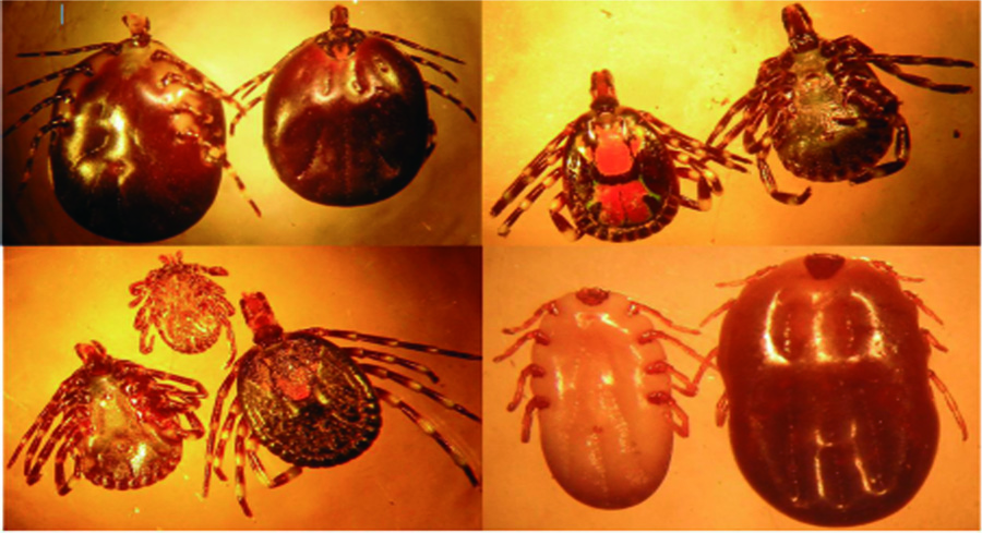
Once a week, the animals were restrained and all ticks were removed using a forceps. The ticks were stored in labelled vials containing 70% alcohol and as soon as possible they were counted and identified morphologically using a low-power stereomicroscope with the aid of standard identification keys [15]. All 16450 Amblyomma spp. collected, 1101 nymphs and larvae and 573 ticks of the Boophilus (Rhipicephalus) genera were examined for spp. identification (Figure 1).
Blood was collected from the jugular vein of each animal fortnightly for the first year and thereafter once a month until the animal attained the age of 18 months when it was removed from the study. The blood was used for measurement of packed cell volume (PCV) and DNA extracted from its buffy coat. Each buffy coat sample was stored at +4°C until it was used for DNA isolation.
DNA extraction, amplification and analysis
DNA extraction from the buffy coat was undertaken using the PureGene Genomic DNA Isolation kit (Biozym, Belgium) following the instructions of the manufacturer. The DNA samples were amplified by semi-nested polymerase chain reaction (PCR) and gel electrophoresis was used to visualise the amplicons. The analysis was based on the amplification of the 18S rRNA gene for the detection of all species of Babesia and was similar to that described by Devos & Geysen [16]. To amplify the 16S rRNA gene for the detection of Anaplasma spp. and Ehrlichia spp. the primers used were developed at IMT (Antwerp, Belgium).
The isolated DNA was used for species-specific detection of the pathogens by restriction fragment length polymorphism (RFLP).
Polymerase Chain Reactions
In each reaction microtube, 5 µl of template and 20 µl of the master mix were added. The latter contained 1µl of Yellow SubTM (Geneo BioTech Hamburg, Germany), 11.2 µl distilled water, 5µl of buffer (20 mMTris-HCl, pH 8.4; 100mM KCl), 1.6µl of MgCl (25 mM), 0.2µl of dNTP (100mM each), 0.4 µl each of Babesia, Theileria, Anaplasma and Ehrlichia primers (Table 1) (25µmol/µl) and 0.2µl of Taq polymerase (5U/µl). A thin layer (2 drops) of mineral oil was placed over the content of all microtubes. The whole tube was put in a preheated (84°C) thermocycler (PTC- 100 TM or T3 BiometraR, Westburg, Germany) programmed as follows: denaturing at 92°C for 30 seconds, annealing at 62°C for 45 seconds, extension at72°C for 1 minute and this whole cycle was undertaken 39 times. A last extension of 8 minutes was added before further analysis. Double distilled water was used as a negative control and a known Babesia bigemina, T.mutans, Anaplasma or Ehrlichia sample was used as a positive control in each case.
In the second round reactions, microtubes containing 24.5µl of the master mix consisting of 1µl ofYellow SubTM (Geneo BioTech Hamburg, Germany), 15.7µl of distilled water, 5µl of buffer solution, 1.7 µl of MgCl2 (25 mM), 0.4 µl of each primer, 0.13µl ofTaq polymerase and 0.5µl of the first round PCR product (template) were each covered with 2 drops of mineral oil. The negative and positive control samples were similarly prepared. The reaction tubes were then preheated and maintained at 84°Cand the thermocycler was programmed as follows: denaturation at 92°C for 30 seconds, annealing at 62°C for 45 seconds, extension at 72°C for 1 minute and this whole cycle was undertaken 24 times. A last extension of 8 minutes was added before further analysis. A total of 548 samples were examined using this nested PCR technique.
Five microliters of each PCR product was mixed with 2 µl of loading buffer and loaded into a 2% agarose gel wells and allowed to migrate in the agarose gel in TAE (0.5%) buffer at 100 Volts for 20 minutes. Thereafter, the gel was dipped in an ethidium bromide solution (1µg/ml) for 30 minutes to permit visualisation of the DNA using UV light. A photograph of each gel was made (Figure 2 and 3).
Table 1: Primers used in PCR for detection of Babesia/Theileria andAnaplasma/Ehrlichia groups.
|
Pathogen group
|
Name
|
Sequence
|
|
Babesia andTheileria
|
|
|
|
|
BabF3
|
ATGTCTAAGTACAAGCTTTTTACGGT
|
|
|
BabR2
|
TTGATTTCTCTCAAGGTGCTGAAGGAGTCG
|
|
|
BabR3
|
AAAGGCGACGACCTCCAATCCCTAGT
|
|
Anaplasma and Ehrlichia
|
|
|
|
|
EHR 16SD
|
GGTACCYACAGAAGAAGTCC***
|
|
|
EBR3
|
TTGTAGTCGCCATTGTAGCAC
|
|
|
EBR2
|
TGCTGACTTGACATCATCCC
|
Primer F: forward, R: reverse. *The primers are based on the amplification of the16S rRNA and 18S rRNA genes for the simultaneous detection of Anaplasma/Ehrlichia and Babesia/Theileria, respectively. That for the 16S rRNA were developed at ITM (Antwerp, Belgium).
Restriction Fragment Length Polymorphism (RFLP) and Sybr Green digestion by enzymes
Positive PCR samples were further analysed by RFLP to determine the pathogen species implicated. Four to six microliters of each positive PCR product was digested by restriction enzymes (Hind61 incubation temperature at 37°C and BseDI incubation temperature at 55°C overnight) in a mixture of RO-DI water, the enzyme and corresponding buffer in a final volume of 15 µl consisting of 11 µl of reaction mix and 4-6 µl of PCR product.
Four to 6µl of enzyme-digested product was mixed with 2µl of loading buffer and loaded into marked wells of a polyacrilamide (PAGE) gel that was totally immersed in TBE buffer. The power unit was set at 100Volts and allowed to run for 2hours 30 minutes. After migration, the gel was transferred into Sybr Green and maintained therein for 40 minutes. All positive PCR samples were submitted to RFLP where enzymatic digestion and their different fragment profiles were used to identify most of the parasite species.
Sequencing and Blast
On RFLP we could not distinguish the fragment profile of Theileria spp. from Ehrlichia spp. For that reason, we had to sequence our PCR products to confirm the diagnosis of infection by Theileria spp. and/or Ehrlichia spp. In each case one animal with a respective positive RFLP profile was randomly selected for sequencing.
Purification of the PCR products was done using QIAquick® purification kit (QIAGEN®) according to the manufacturer’s protocol. Cloning was then carried out using the TOPO TA Cloning® kits 2006 (Invitrogen™, California USA) according to the manufacturer’s protocol. Positive colonies were selected for sequencing and further analysed by comparing the sequences to the NCBI nucleotide database using Blast and Jalview 2.8.2 [17].
Analysis of collected data
Data was collected from each animal only during its first 18 months of life. Adjustments were made to permit data collected from animals with the same age and during the same season to be grouped together for analysis. Analysis of covariance to look for the effect of season and tick infestation rate on PCV was undertaken. A logistic regression was undertaken to determine the effect of the different tick genera infestation rates on animal blood parasite/bacteria incidence. The logit model employed the maximum likelihood function using the Newton-Raphson algorithm. Threshold significance value was 5%.