Study participants
The committees for ethical review of research involving human subjects at the Second Affiliated Hospital of Guangzhou Medical University (Guangzhou, China) approved this study. All patients provided written informed consent. Fourteen NMOSD patients who fulfilled the diagnostic criteria [12] and 14 non-autoimmune encephalitis neurological disease patients (Control) were enrolled in the study to estimate magnesium levels in the CSF.
Animals
Adult female C57/BL6 mice aged 6–8 weeks were purchased from Guangdong Medical Laboratory Animal Center. All mice were maintained on a standard 12-h light/dark cycle (8:00–20:00 light period) in a temperature-controlled room (21 ± 25°C) and were given free access to food and water, except for during MgT treatment. All animal experiments were conducted in accordance with the regulations of the Administration of Affairs Concerning Experimental Animals (China) and were approved by the Guangzhou Medical University Animal Ethics Committee.
Disease model induction
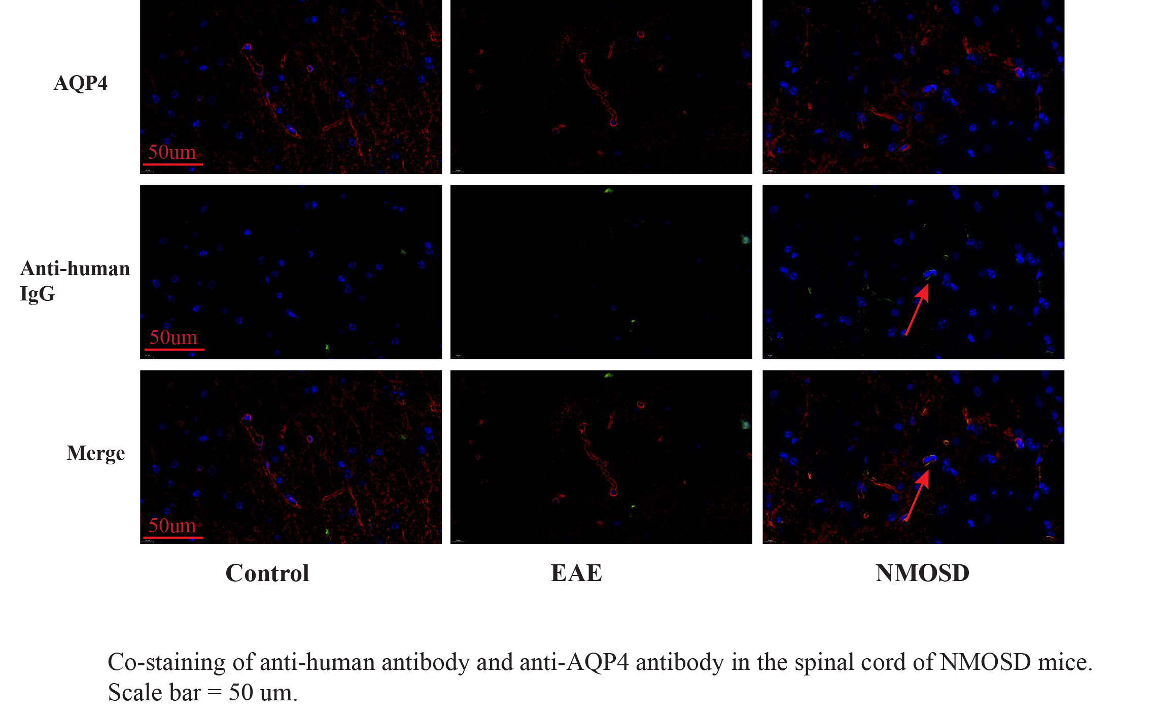
Experimental autoimmune encephalomyelitis (EAE) induction in mice was performed as described previously [13]. In brief, each mouse received 0.15 mL (total volume) of emulsified MOG35–55-CFA as three 0.05-mL injections subcutaneously on Day 0 and two 0.05-mL injections subcutaneously on Day 7. MOG35–55 and heat-killed H37Ra Mycobacterium tuberculosis (Difco, USA) were administered to each mouse at a dose of 200 µg in each injection. The ratio of (MOG35–55-DPBS) to (TB-CFA) was 1:1.3. Each mouse received 0.3 mL of 500 ng pertussis toxin intraperitoneally on Days 0, 1, and 7. To prepare an NMOSD mice model by induction [14], a series of 10 intraperitoneal injections of human IgG purified from 3 NMOSD serum samples were administered on Day 10 to a total volume of 12 mg in each mouse.
Tissue preparation and immunofluorescence assay
Adult female C57/BL6 mice were anesthetized with isoflurane and sacrificed by intracardial perfusion with heparinized saline (0.9% NaCl) followed by ice-cold 4% paraformaldehyde (PFA). The brain tissues were removed, post-fixed in 4% PFA, and embedded in paraffin. Subsequently, 5-µm thick adjacent serial sections were cut using a microtome. For immunohistochemistry, the sections were deparaffinized and rehydrated in xylene and ethanol. Endogenous peroxidase was quenched by incubation the sections in 3% hydrogen peroxide, and antigen retrieval was performed in 0.01 M citrate buffer. The sections were then blocked and incubated with primary antibodies, namely, AQP4 (1:200 dilution, 16473–1-AP, PTG) and glial fibrillary acidic protein (GFAP; 1:500 dilution, BA0056, Boster), overnight at 4°C. After washing, the sections were incubated at 4°C with biotinylated secondary antibodies. The sections were mounted on a slide with a Vector shield mounting medium containing DAPI (Vector Laboratories Inc.) and visualized using an automatic digital slide scanner (PANNORAMIC MIDI, 3DHISTECH).
Treatment with magnesium L-threonate
Magnesium L-threonate (Magceutics Inc., China) was orally administered to mice through drinking water at a dose of ~ 604 mg/kg/day (~ 50 mg/kg/day elemental magnesium) [15]. The average volume of drinking water per day was determined (~ 6 mL/day for each mouse), and the drug, at the required dose, was dissolved in daily drinking water. MgT treatment was started when the mice received subcutaneous injections of emulsified MOG35–55-CFA on Day 0 and lasted until the mice were sacrificed. All mice were maintained on standard food containing 0.15% elemental magnesium. Immortalized hCMEC/D3 was purchased from Meisen, Inc. (Pan’an, China) and cultured in an endothelial cell medium (ECM; ScienCell, USA). For MgT pre-treatment experiments, hCMEC/D3 were grown on 6-well plates and treated with a modified culture medium containing 10% NMOSD serum or healthy control (HC) serum with or without 10 mM MgT individually for 24 h. After incubating the cells for 24 h, the cell culture supernatants were aspirated, and the cells were collected and stored at − 80°C for western blot analysis or real-time reverse transcription–polymerase chain reaction (RT-PCR) assay.
Western blot analysis
After the last AQP4-IgG injection, mice were anesthetized with isoflurane, and brain tissues were removed. The prefrontal cortex and hippocampus tissues were rapidly dissected and frozen on dry ice. All tissue samples were sonicated in radioimmunoprecipitation assay buffer with a protein protease inhibitor cocktail, and the homogenate was centrifuged at 12,000× rpm for 30 min under 4°C. The supernatants were collected and mixed with 5× loading buffer. The proteins (30 µg sample) were separated on sodium dodecyl sulfate–10% polyacrylamide gels and transferred to polyvinylidene difluoride membranes (Merck Millipore, Germany) according to standard procedures. The blotted membranes were treated with blocking buffer (5% skimmed milk in 25 mM Tris-HCl, pH 7.6, 125 nM NaCl, 0.5% Tween 20) for 1 h at room temperature (RT) and incubated with antibodies specific to human Claudin-5 (1:3000 dilution, 352588, Invitrogen), ZO-1 (1:1000 dilution, 61-7300, Invitrogen), Occludin (1:1000 dilution, 40-4700, Invitrogen), TRPM7 (1:2000 dilution, ab245408, Abcam), GAPDH (1:3000 dilution, BM1623, Boster), and beta-actin (1:4000 dilution, ab8226, Abcam) for 15 h at 4°C. The membranes were incubated with secondary horseradish peroxidase–conjugated antibodies for 1 h at RT. After washing with Tris-buffered saline with Tween 20, the membranes were incubated with chemiluminescence detection solutions (PerkinElmer, USA). FluorChemTM SP software was used to measure band densitometry.
siRNA preparation and transfections
Small interference RNA (siRNA) was used to knockdown specific gene expression. Three pairs of siRNA targeting human TRPM7 were purchased from Shanghai GenePharma (Shanghai, China). The siRNA targeting human GAPDH was used as the positive control (PC), and the corresponding scrambled siRNA was used as negative control (NC). FAM(5-Carboxyfluorescein)-siRNA was used to illustrate transfection efficiency. Transient transfection of siRNA was performed using jetPRIME transfection reagent (Polyplus-transfection, France). One day before transfection, cells were trypsinized and seeded on a 6-well plate at a density of 1.5 × 10⁵ cells/well containing 2 mL of ECM. Subsequently, 2 µg of siRNA was diluted with 200 µL of jetPRIME buffer, and it was mixed by pipetting up and down. Then 4 µL of the jetPRIME reagent was added, and the mixture was vortexed for 10 s, which was centrifuged for a short time. The mixture was incubated for 10 min at RT, and the transfection mix was added to the cells in ECM. The cells were assayed or treated with NMOSD or HC serum with or without MgT for 24 h after siRNA transfection.
Real-time reverse transcription PCR
After different treatments, cells were harvested, and total RNA was extracted using the RNeasy Mini kit (Qiagen, US) and subsequently quantified using a spectrophotometer (NanoDrop 2000, Thermo Fisher Scientific, US). cDNAs were synthesized using a reverse transcription kit (Qiagen, US). Real-time RT-PCR was used to estimate the mRNA levels using a LightCycler 480 RT-PCR Detection System (Roche, US) with the following primers: GAPDH, F-5′-GGAGCGAGATCCCTCCAAAAT-3′, R-5′-GGCTGTTGTCATACTTCTCATGG-3′; Occludin, F-5′-GGAGCGAGATCCCTCCAAAAT-3′, R-5′-GGCTGTTGTCATACTTCTCATGG-3′; ZO-1, F-5′-AAGATCCAGCAATGAAGCC-3′, R-5′-TGAGAAGTGGGTTTGGGA-3′; Occludin, F-5′-TCCCTTCCATTTCTGACCT-3′, R-5′-GGGCATCACTTTATACGCA-3′; TRPM7, F-5′-ACTGGAGGAGTAAACACAGGT-3′, R-5′-TGGAGCTATTCCGATAGTGCAA-3′. The 2−ΔΔCt method was used to calculate gene fold changes. The level of specific mRNA was normalized according to GAPDH.
Transmission electron microscopy
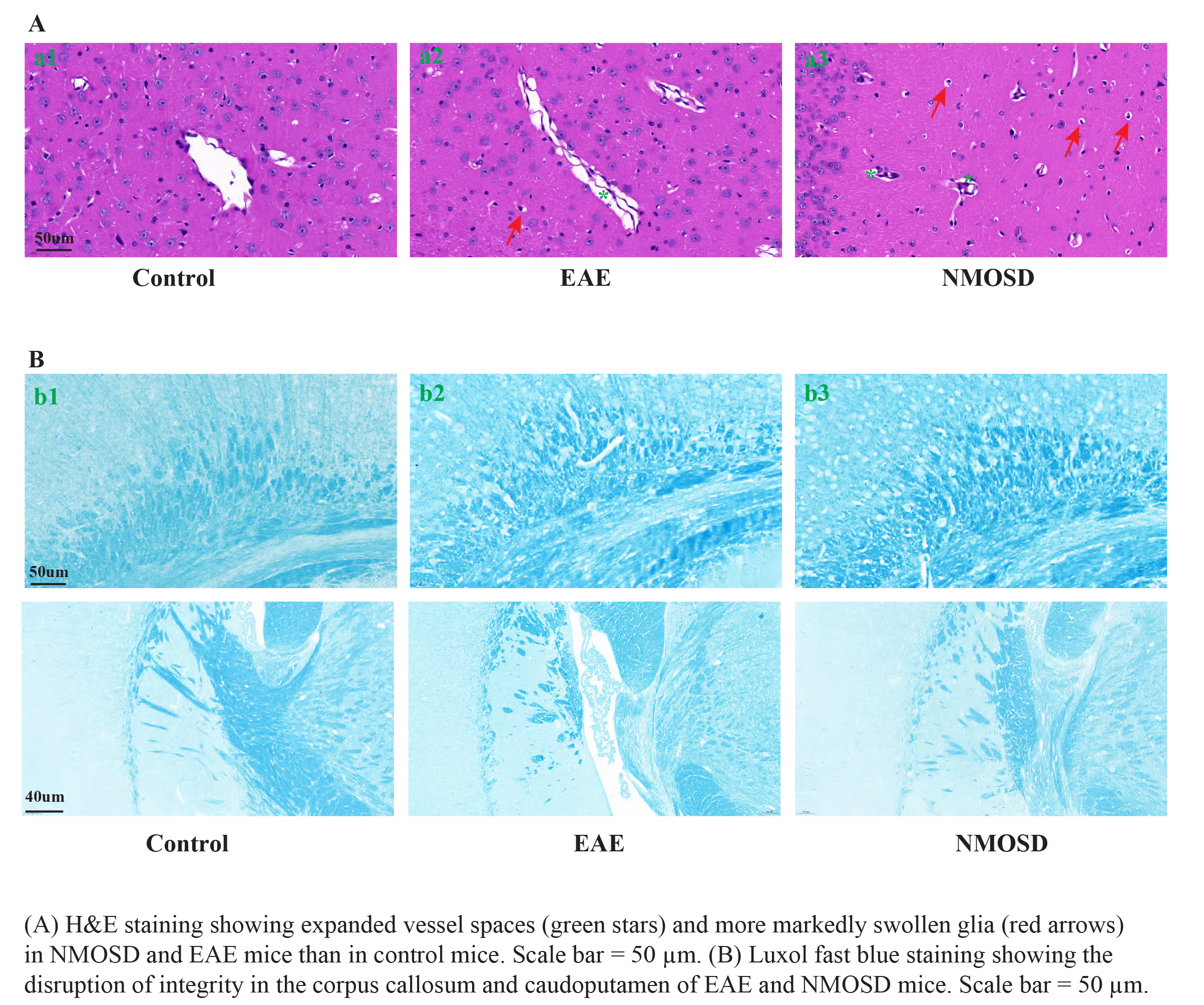
To observe the ultrastructure of BBB and myelin integrity, a transmission electron microscope (TEM) was used. Mice were anesthetized with isoflurane and sacrificed by intracardial perfusion with heparinized saline (0.9% NaCl), followed by ice-cold 2.5% glutaraldehyde. The brain and spinal cord tissues were removed, dissected to 1 mm3, and post-fixed in 2.5% glutaraldehyde (pH 7.4) for 2 h. After washing the tissues thrice with 0.1 M phosphate buffer (pH 7.2) and fixing them in 1% osmic acid at 4°C for 2 h, the samples were dehydrated with a graded series of ethanol. Subsequently, the samples were embedded in Epon-Araldite resin for penetration and placed in a model for polymerization. After a semi-thin section was used for positioning, an ultra-thin section was prepared for microstructure analysis, followed by counterstaining with 3% uranyl acetate and 2.7% lead citrate. The specimens were observed under a TEM (HT7800, Hitachi, Japan).
Measurement of CSF magnesium levels
Magnesium levels in the CSF from 14 GAFP-A patients and 14 non-autoimmune encephalitis control patients were measured using a commercially available kit (Abcam, ab102506).
Statistical analysis
Data are shown as mean ± standard deviation. Student’s t-test was used for comparisons between two groups, and one-way analysis of variance was used to compare three or more data sets, followed by Tukey's multiple comparisons test, using SPSS 20.0 software. Pearson’s correlation analysis was used for correlation analysis. A probability (p) value of < 0.05 was considered to indicate statistical significance.