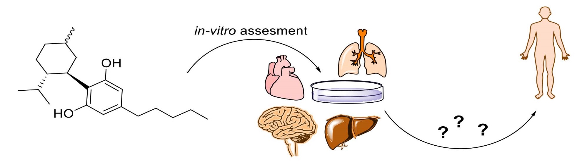
Lung Fibroblasts (NHLF) play a role in remodeling of the airway (alveolar structure) as well as airway inflammation, these cells are also commonly employed in lung cancer studies due to their prominence in disease commencement. Monitoring the NHLF, the number of viable cells in culture were based on quantitation of ATP present, an indicator of metabolically active cells. H4CBD was compared to a negative control (1% DMSO) and a positive control (1% SDS in media) with a concentration ranging from 1.56 to 50 µM. In figure 1 as shown below, the range of testing for NHLF and other cell lines aside from hERG, ranged from 24hr-72hr for detailed results. At the 24hr mark at the lowest concentration cell viability was high, with increasing concentrations, cell viability did decrease, although on the 48hr and 72hr mark, as shown below, at lower concentrations the cell viability remained close to the same as the 24hr mark. Sustained micromolar concentration over 24hrs is the only way to start inducing cytotoxic effects, effectively making low concentrations sustained safe, since the body metabolizes the cannabinoids efficiently sustaining concentrations would be improbable.
Hepatocytes play a huge role in a complex system of facilitating purification of blood components and external contaminants as well as endocytic uptake of nutrients and needed trophic agents9. The in-vitro assessment of H4CBD towards the toxicity of cultured hepatocytes was conducted, as shown in figure 2 below, H4CBD did not show significant drop in the cellular viability across varying concentrations at varying times, compared to the control. At the 24hr mark across multiple concentrations, cell viability remained constant, drops in cell viability were not noticed across concentrations until the 48hr and 72hr marks. At 6.25 µM and 25 µM at 48hr and 72hr respectively cell viability remained high, which remains of interest. Speculation for the sustained viability might be due to the adaptable function of hepatocytes to process compounds, although the SDS control did have sustained reduced viability, as well as the control, the cannabinoids through evidence does not pose cytotoxicity towards the hepatocytes.
Human neural progenitor cell (NPC) give rise to the glial and neuronal cells that populate the central nervous system (CNS) but does not assist in the generation of non-neural cells17. H4CBD did not exhibit low cell viability for NPC at the 24hr mark until the 6.25 µM concentration as shown in figure 3 below. Cell viability remained low as depicted at the 48hr mark across all concentration of H4CBD. Severe loss of cell viability is observed at the 72 hr period, with all concentrations exhibiting the same cellular viability characteristics. Observed half-life of cannabinoids18, fall below the 24hr mark of cell viability limitations. The sustained high concentrations contribute to the loss of cell viability, noting that continuous elevated concentrations would be improbable.
Most pharmaceuticals that undergo safety studies will either do a hERG assay or some type of myocyte safety test to determine the viability of myocytes and ensure that QT interval19 prolongation resulting in potentially fatal ventricular tachyarrhythmia called Torsade de Pointes does not occur. As well to evaluate anticipated cardiovascular effects, early evaluation of hERG toxicity has been strongly recommended for instance by the regulatory agencies such as U.S. Food and Drug Administration (FDA) and European Medicines Agency (EMA)20. The hERG study tested the effects of H4CBD on cloned hERG potassium channels (encoded by KCNH2 gene and expressed in HEK293 cells) was examined, as Cisapride21 the positive control. H4CBD was exposed to hERG at 0.0625, 0.125, 0.625, 1.25, 6.25, 12.5, 25, and 50 µM concentrations (n ≥ 3). The duration of exposure to each test concentration was a minimum of 3 minutes. The positive control data confirmed the sensitivity of the test systems to ion channel inhibition.
Results shown in table 1 below, suggests that H4CBD does not block the hERG-encoding channels that are expressed in the HEK293 cells until above the 1.25 µM concentrations. In order to determine if H4CBD effects other channels, Cav1.222 and Nav1.523 assays were conducted. The results showed H4CBD inhibited Cav1.2 calcium and Nav1.5 sodium channels with the same effect as the hERG potassium channels as shown in table 2 and table 3 below. This effect of the depolarization and repolarization by these channels results in a net zero inhibition24.
Table 1. IC50 value of H4CBD and Mean percentages of hERG inhibition
|
Test Article ID
|
IC50 (µM)
|
Conc. (μM)
|
Mean % hERG Inhibition
|
Standard Deviation
|
Standard Error
|
n
|
|
H4CBD A
|
<6.25
|
0.0625
|
7.1
|
4.3
|
1.6
|
7
|
|
0.125
|
15.5
|
7.0
|
4.0
|
3
|
|
0.625
|
38.0
|
5.9
|
3.4
|
3
|
|
1.25
|
64.7
|
9.7
|
3.4
|
8
|
|
6.25
|
100.0
|
0.3
|
0.1
|
9
|
|
12.5
|
99.6
|
1.0
|
0.3
|
9
|
|
25
|
100.0
|
0.2
|
0.1
|
5
|
|
50
|
98.1
|
3.7
|
1.6
|
5
|
|
Cisapride
(Positive control)
|
|
0.05
|
66.5
|
2.2
|
1.1
|
4
|
hERG in-vitro assay was conducted, and determined IC50 value, as well as mean inhibition of H4CBD at varying concentrations.
Table 2. Effects of H4CBD on Cav1.2 Ion channel current
|
Test Article ID
|
IC50 (µM)
|
Conc. (μM)
|
Mean % hCav1.2 Inhibition
|
Standard Deviation
|
Standard Error
|
n
|
|
H4CBD A
|
<6.25
|
0.0625
|
3.0
|
3.9
|
2.2
|
3
|
|
0.125
|
8.4
|
5.8
|
3.4
|
3
|
|
0.625
|
24.4
|
3.5
|
2.0
|
3
|
|
1.25
|
36.7
|
7.3
|
3.0
|
6
|
|
6.25
|
97.1
|
2.8
|
1.4
|
4
|
|
12.5
|
97.8
|
3.1
|
1.3
|
6
|
Cav1.2 assay was conducted to provide better evidence of the inhibition of the ion channel at specific concentrations.
Table 3. Effects of H4CBD on Nav1.5 Ion channel current
|
Test Article ID
|
IC50 (µM)
|
Conc (μM)
|
Mean % Late Nav1.5 Inhibition
|
Standard Deviation
|
Standard Error
|
n
|
|
H4CBD A
H4CBD A
|
<6.25
<6.25
|
0.0625
|
5.0
|
3.7
|
2.1
|
3
|
|
0.125
|
1.7
|
1.9
|
1.1
|
3
|
|
0.625
|
20.7
|
6.9
|
2.4
|
8
|
|
1.25
|
27.8
|
7.5
|
3.3
|
5
|
|
6.25
|
70.0
|
8.8
|
3.6
|
6
|
Nav1.5 assay was conducted to provide better evidence of the inhibition of the ion channel at specific concentrations.
Figure 4 below is data important to the AMES study done on mutagenesis of H4CBD to E.Coli.
Table 4. AMES test on various E. Coli. Strains, and their accompanying dose and mean revertants
|
Strain
|
Test Item
|
Dose level per plate (µg/plate)
|
Mean Revertants Per Plate
|
|
TA98
|
DMSO
|
-
|
14.0
|
|
|
H4CBD
|
1
|
15.0
|
|
|
|
5
|
8.5
|
|
|
|
10
|
9.0
|
|
|
|
50
|
-
|
|
|
2NF
|
2.5
|
663.5
|
|
TA100
|
DMSO
|
-
|
115.0
|
|
|
H4CBD
|
1
|
99.0
|
|
|
|
5
|
-
|
|
|
SA
|
1
|
472.0
|
|
TA1535
|
DMSO
|
-
|
10.0
|
|
|
H4CBD
|
1
|
9.0
|
|
|
|
5
|
-
|
|
|
SA
|
1
|
436.0
|
|
TA1537
|
DMSO
|
-
|
3.5
|
|
|
H4CBD
|
1
|
3.0
|
|
|
|
5
|
-
|
|
|
ICR
|
0.5
|
133.5
|
|
WP2uvrA
|
DMSO
|
-
|
46.5
|
|
|
H4CBD
|
1
|
52.0
|
|
|
|
5
|
49.0
|
|
|
|
10
|
48.0
|
|
|
|
50
|
43.5
|
|
|
|
100
|
42.0
|
|
|
|
500
|
25.5
|
|
|
|
1000
|
27.0
|
|
|
|
5000
|
14.0
|
|
|
NQNO
|
2
|
254.5
|
Vehicle and positive controls are, 2NF (2-Nitrofluorene), DMSO (Dimethylsulfoxide), ICR (Acridine), NQNO (4-nitroquinoline-N-oxide), SA (Sodium azide). Postfixes, P (precipitate), R (Reduced), SR (Slightly Reduced). Dose level per plate = µg/plate.
The conducted AMES test resulted in precipitation observed at very high concentration of drug (≥ 500 µg/plate) in strains TA98, TA100, TA1535, TA1537 with and without S9, and in strain WP2uvrA without S9; and at 5000 µg/plate in strain WP2uvrA with S9. Cytotoxicity was observed at ≥ 5 µg/plate in strains TA100, TA1535, TA1537 without S9; at ≥ 50 µg/plate in strain TA100, TA1537 with S9 and in strain TA98 without S9; at ≥ 100 µg/plate in strain TA1535 with S9; at ≥ 500 µg/plate in strain TA98 with S9; and at 5000 µg/plate in strain WP2uvrA without S9. The vehicle and positive controls responded appropriately. There were no increases in the mean number of revertant colonies as compared to the vehicle control in any strain. Thus, H4CBD is not mutagenic, up to cytotoxic concentrations, in strains TA98, TA100, TA1535, and TA1537 with and without S9, and in strain WP2uvrA with S9, and up to precipitating concentrations in strain WP2uvrA without S9.