3.1 Preparation, characterization and in vitro photo-thermal effect of HMON@CuS/Gd
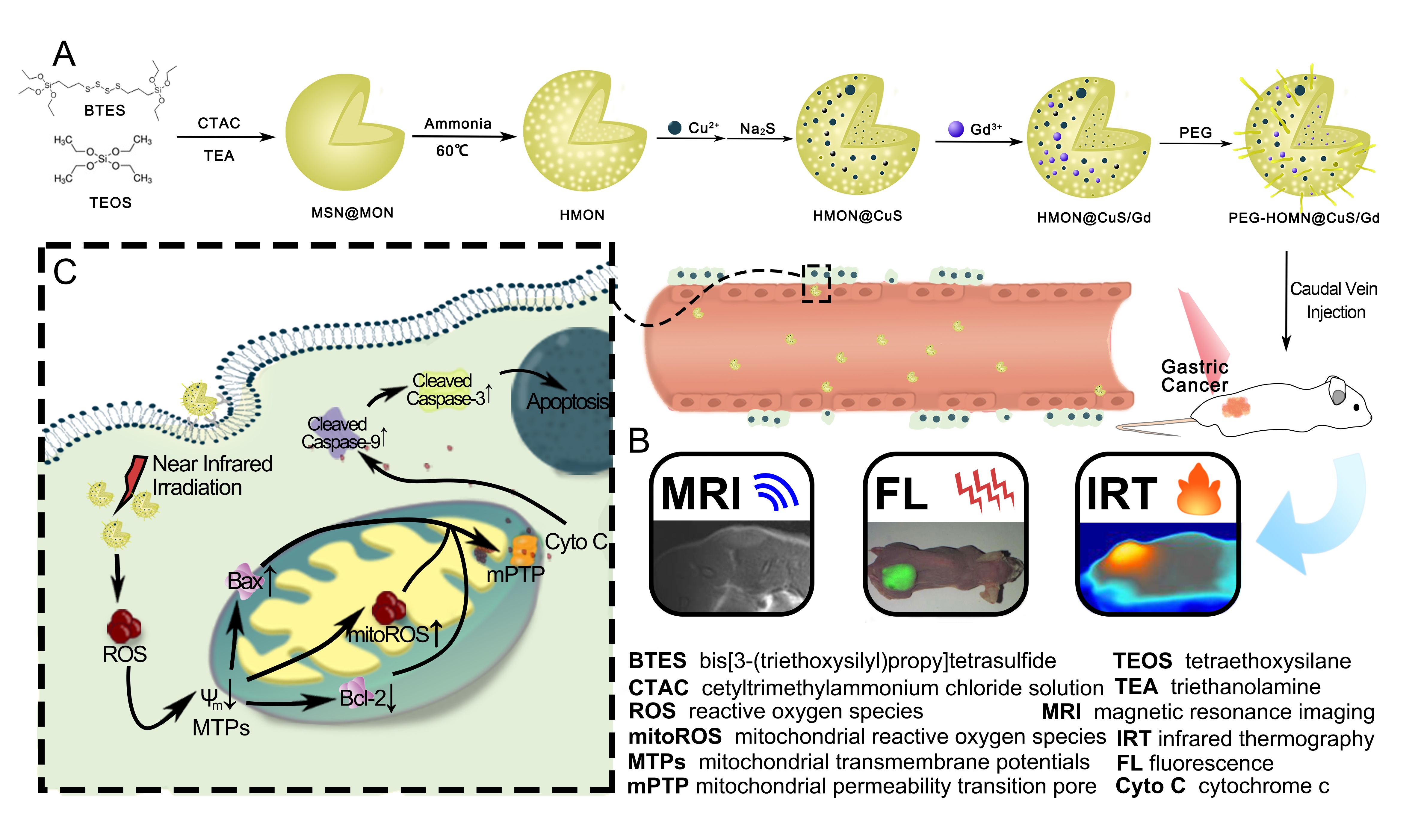
HMON were synthesized via a mild selective etching progress. In brief, mesoporous organosilica nanospheres with large pore were firstly synthesized by the dual hydrolysis and condensation of BTES and TEOS under the catalyzation of TEA, while CTAC was used for pore forming. As the dominant -Si-O- bonds were weaker than the –Si-C- bonds within the outer sites, the inner part of the mesoporous organosilica nanospheres were then etched using diluted ammonia to form the hollow mesoporous structure, bridged by –S-S- bonds [21]. Though it has been reported that CuS could be conjugated onto the surface of HMON through thiol groups, it required respective synthesis of HMON and CuS following the conjugation progress. The multi-steps protocol might be simplified as it has also been reported that Si-O- groups on the surface of the HMON have the potential to bond with metal ions (such as Fe3+, Au+, etc.) without the need of additional molecular chelator [27, 28]. In our previous work, CuS nanocrystals were in situ grown onto the surface of hollow mesoporous TaOx nanospheres without the help of thiol groups [19]. Herein, CuS nanocrystals were in situ grown onto the surface of the HMON similarly. Briefly, Cu2+ ions were added directly into the HMON solution and adsorbed onto the HMON, following the addition of Na2S to achieve the in situ nucleation and growth of CuS nanocrystals. Finally, the HMON@CuS/Gd NPs were constructed with the addition of GdCl3·6H2O. Then, the element mapping of HMON@CuS/Gd showed that the Cu2+ and Gd3+ ions were successfully jointed together onto the surface of HMON (Figure 1A). It is noticeable that the etching degree by diluted ammonia has great influence on the ability of the HMON to absorb Cu2+ ions. HMON with excessive etching leading to the poor conjugation with Cu2+ and Gd3+ ions due to the depletion of –Si-O- bonds. Eventually, C18PMH-mPEG was applied to improve the stability and dispersity of the HMON@CuS/Gd nanospheres.
The as-prepared HMON showed specific strong and broad peaks at 2927.32 cm-1 and 2855.28 cm-1 (attributed to -CH2) [29], which represented the covalent grafting of C18PMH-mPEG chains onto the surface of HMON, compared to non-PEGylation HMON. Meanwhile, new peak at 627.03 cm-1 (attributed to Cu-S) was also observed in HMON@CuS and HMON@CuS/Gd [30]. Therefore, the FI-TR results consistently confirmed the successful synthesis of HMON@CuS/Gd (Supplementary files, Figure S1). UV-vis spectra results revealed that HMON@CuS and HMON@CuS/Gd both have strong absorption in NIR region (Supplementary files, Figure S2), which is mainly contributed to the effective adherence of CuS nanocrystals on the surface of HMON [21]. XPS results demonstrated the presence of C (C 1s peaks at 284.95 eV), O (O 1s peaks at 531.99 eV), Si (Si 2p peaks at 102.53 eV), Cu (Cu 2p peaks at 933.27 eV), S (S 2p peaks at 163.36 eV) and Gd (Gd 4d peaks at 142.72 eV) (Supplementary files, Figure S3) [31]. From XRD results, HMON@CuS and HMON@CuS/Gd both showed specific characteristic diffraction peaks [30], which is indexed as a typical covellite crystalline phase of CuS (Supplementary files, Figure S4). It is noteworthy that CuS crystal was found in HMON@CuS and HMON@CuS/Gd, while no Gd-related crystal was observed in HMON@CuS/Gd, indicating that Gd was doped in HMON, but not as a crystal form. Taken together, the above results consistently confirmed the successful synthesis of HMON@CuS/Gd. In addition, the diameters of HMON was 115.5±1.46 nm, while CuS or/and Gd3+ loading led to a slight increase in the size of the HMON@CuS (116.7±1.51 nm) and HMON@CuS/Gd (117.1±1.71 nm), and all of them exhibited unimodal size distribution. Meanwhile, the data also indicated that both of HMON@CuS and HMON@CuS/Gd possessed negative net charge, which could help to improve their circulation stability (Supplementary files, Figure S5). Compared to these PEG modified HMON NPs, HMON-based materials without PEG modification also showed negative net charge, and slight minor diameters. However, the diameters’ SD value in HMON (108.2±4.54 nm), HMON@CuS (109.4±3.41 nm) and HMON@CuS/Gd (110.1±4.15 nm) NPs were larger than those in PEG-modified HMON NPs, indicating that C18PMH-mPEG could improve their stability (Supplementary files, Figure S6). Consequently, PEG modified HMON NPs were chosen for further experiments in our research. Due to the presence of the disulfide bonds in the framework of HMON, HMON@CuS/Gd exhibited time-dependent biodegradable behavior in the glutathione (GSH) solutions (Figure 1B), indicating that HMON@CuS/Gd NPs could be bio-degradable and eliminated through feces and urine, thus the drug accumulation leaded toxicity could be avoided.
It could also be expected that the as-prepared NPs possess the potential to be applied as MRI imaging contrast agent due to the addition of Gd [32, 33]. In vitro experiment demonstrated that T1 signal intensity was enhanced with the increasing concentration of HMON@CuS/Gd solutions. As calculated, significant increase of SNR (signal to noise ratio) could also be observed, indicating the outstanding potential for MRI imaging (Figure 1C&D). Besides the MRI imaging properties, we further investigate the photo-thermal conversion efficiency of HMON@CuS/Gd by monitoring the temperature changes under NIR laser irradiation in vitro using an infrared thermal imaging camera. After NIR laser irradiation (0.8 W/cm2) for 5 mins, dramatical temperature increase was observed in HMON@CuS/Gd and HMON@CuS groups, while no obvious temperature change was shown in PBS or HMON@Gd groups with the same laser irradiation. The maximum increased temperature (ΔTmax) of HMON@CuS/Gd and HMON@CuS groups (1000µg/mL) ∼38 °C, whereas the ΔTmax of other HMON@CuS/Gd and HMON@CuS groups (100µg/mL, 50µg/mL and 12.5µg/mL) increased to ∼16.0 °C, ∼8.0 °C and ∼6.0 °C respectively (Figure 1E&F; Supplementary files, Figure S7). It is worth pointing that HMON@CuS/Gd and HMON@CuS showed good temperature response under the NIR laser irradiation conditions, which demonstrating that the photo-thermal conversion efficiency was contributed to the presence of CuS. Moreover, HMON@CuS/Gd NPs showed an excellently stable photo-thermal performance, as the heat-cool curve of HMON@CuS/Gd NPs had no significant difference within five cycles of NIR laser irradiation (Supplementary files, Figure S8). According to the linear regression curve between the cooling stage and negative natural logarithm of driving force temperature of HMON@CuS/Gd, the photo-thermal conversion efficiency of HMON@CuS/Gd NPs was calculated to be 82.4% (Figure 1G). In addition, to determine whether HMON@CuS/Gd NPs could produce ROS under NIR laser irradiation, a singlet oxygen sensor was applied to detect ROS levels. As shown in Figure 1H, HMON@CuS/Gd NPs exhibited concentration-dependent production of ROS in deionized water, indicating that HMON@CuS/Gd NPs possessed excellent PDT ability. Moreover, HMON@CuS showed similar ROS generation ability in the presence of NIR, while no excessive singlet oxygen was observed in HMON@Gd NPs, with or without NIR treatment (Supplementary files, Figure S9), which demonstrating that the ROS generation capacity of HMON@CuS/Gd NPs were contributed to the presence of CuS. Therefore, HMON@CuS/Gd would be promising PTT, PDT and functional imaging NPs for the treatment of GC
3.2 In vitro photo-therapeutic effect of HMON@CuS/Gd nanoparticles in GC cells
Firstly, we tested the in vitro photo-therapeutic effects of HMON@CuS/Gd on HGC-27 cells. Briefly, HGC-27 cells were first incubated with HMON@CuS/Gd at different concentrations for 4 h and then were irradiated with an 808 nm laser at a power density of 0.8 W/cm2 for 5 mins. After 24 h, CCK-8 assay was applied to evaluate the antitumor therapeutic efficacies of HMON@CuS/Gd. As shown in Figure 2A, free HMON@CuS/Gd and single NIR laser treatment did not induce significant changes in cell death, since the half maximal inhibitory concentration (IC50) of HMON@CuS/Gd was as high as 570.01µg/mL. Furthermore, dramatical decreased cell viabilities were observed in HMON@CuS/Gd plus NIR group (IC50=50.51µg/mL), indicating that HMON@CuS/Gd exhibited an effective photo-therapy effect with NIR irradiation. In the other hand, HMON@CuS exhibited similar anti-tumor effect with NIR irradiation, compared to HMON@CuS/Gd plus NIR irradiation group, indicating that photo-therapy effect was contributed to the addition of CuS, while Gd showed negligible cell toxicity (Supplementary files, Figure S10). Consistently, LDH assay revealed that HMON@CuS/Gd plus NIR induced higher levels of LDH leakage than blank group, while HMON@CuS/Gd only and single NIR laser did not induce any LDH changes (Figure 2B). These results suggested that HMON@CuS/Gd could provide a promising killing effect to GC cells, with the irradiation of NIR.
As it has been reported, several CuS-based NPs had been applied for PDT&PTT synergistic therapy [34, 35]. However, the therapeutic effect of HMON@CuS-induced PTT and PDT at mild temperature, has not been fully discussed. It is still wondered whether the therapeutic effect was achieved by PTT ablation alone, or PDT also played some certain role in the therapeutic progress. In order to evaluate the PDT effect, HGC-27 cells were placed on an ice box while being irradiated with NIR light and the temperature was controlled below 10°C to minimize the effect of NIR-induced PTT ablation. To our surprise, significant cell death was still observed in this condition, though not as obvious as HMON@CuS/Gd plus NIR treatment, as IC50 raised to 122.4 µg/mL (Figure 2C). Furthermore, evident LDH leakage was also found, shown in Figure 2D.
When referring to PTT effect, clinicians have pointed out that tumor cells might occur apoptosis under 43-45°C, while normal cells are generally tolerant to this temperature condition [36]. In another word, PTT effect refers to generating external 45 °C heating at cancer region, which is supposed to have a direct cytotoxic effect on tumor cells, and enhance the efficacy of chemotherapy and radiotherapy, improve the body's immunity, and thus suppress tumor progression [18, 37]. Coincidentally, it could be observed by the IR camera that when HGC-27 cells were treated with HMON@CuS/Gd (50 µg/mL) plus NIR irradiation (0.8 W/cm2, 5 mins), the temperature could raise up to around 45 °C at 37 °C temperature condition. Consequently, HMON@CuS/Gd with 50 µg/mL concentration were chosen for the following cellular experiments. Simulated 45 °C external temperature condition using a cell culture incubator was set as control. It could be seen that slight cell death and LDH leakage were observed in the external heating group, comparing with NIR irradiation group, indicating hyperthermia might not be the only factor (Figure 3C&D). Taken together, it could be concluded that HMON@CuS/Gd could induce combined PDT and PTT effect, while this obvious anti-tumor cell death was contributed to their PDT&PTT synergetic effect.
To address biocompatibility issues, in vitro experiments were performed by incubating HMON@CuS/Gd NPs with normal GES-1 cells. CCK-8 assay revealed above 85% cell viability rates for GES-1 as the concentration ranging from 0-512 µg/mL, while the IC50 of HMON@CuS/Gd was as high as 957 µg/mL to GES-1 cells (Figure 2E). However, when treated with non-PEG modified HMON@CuS/Gd NPs, cell viability rates for GES-1 cells showed significant decrease (IC50=312 µg/mL; Supplementary files, Figure S11), indicating the addition of PEG could improve their bio-safety, and PEG modified HMON@CuS/Gd were consequently chosen for further biological experiments in our research. Surprisingly it could be observed that IC50 to GES-1 cells was much higher than HGC-27 GC cells, indicating that the as-prepared HMON@CuS/Gd showed specific toxicity to GC cell. Furthermore, HMON@CuS/Gd (50 µg/mL) were also co-incubated with RAW264.7 murine macrophage-like cells. This immunotoxicity experiment revealed that HMON@CuS/Gd treatment did not elicit any inflammatory response at the cellular level (Figure 2F). These results strongly indicated that HMON@CuS/Gd NPs have good in vitro biocompatibilities, highlighting their value for clinical translation as drug carriers.
3.3 In vitro cellular uptake and anti-GC effect of HMON@CuS/Gd nanoparticles
Cellular uptake of HMON@CuS/Gd were then investigated by flow cytometry and CLSM assays, while FITC were firstly encapsulated into HMON@CuS/Gd NPs. After incubating with HMON@CuS/Gd (50 µg/mL) for 1-4 h, the FITC fluorescence intensities increased significantly as time passed by (Figure 3A&B), indicating the excellent cellular uptake efficiency of HMON@CuS/Gd. Since HMON@CuS/Gd could efficiently enter GC cells, we further detected the apoptosis rates determined by flow cytometry, which demonstrated that HMON@CuS/Gd plus NIR laser irradiation induced 2-fold higher levels of total apoptosis (14%) than blank group (5%), while free HMON@CuS/Gd and single NIR laser did not induce any apoptotic change (Figure 3C). EdU dye is a kind of thymidine nucleoside analogues, which could specifically insert into DNA molecules of rapid proliferation cells, and higher EdU-positive cell rates usually demonstrate better cell growth abilities [38]. In our research, after conjugated reactions with EdU, we found that HMON@CuS/Gd plus NIR laser irradiation induced 2-fold lower levels of EdU-positive rates (15%) than blank group (40%), while free HMON@CuS/Gd NPs and single NIR laser did not induce any EdU-positive rates change (Figure 3D). These results suggested that HMON@CuS/Gd provided a promising anti-proliferation and promoting apoptotic effects to GC cells, with the irradiation of NIR.
3.4 Anti-tumor mechanism of photo-therapeutic effect induced by HMON@CuS/Gd
Though excellent anti-tumor effect has been proved, their relevant photo-therapeutic mechanism remained unknown. To examine the cancer-killing mechanism of HMON@CuS/Gd, we incubated HGC-27 cells with HMON@CuS/Gd (50 µg/mL) for 4 h, irradiated with an 808 nm laser (0.8 W/cm2, 5 mins), then observed by TEM. Surprisingly, dozens of mitochondria were shown in non-treated HGC-27 cells, while no mitochondria were observed in treated HGC-27 cells (Figure 4A). Meanwhile, it could be proved that MTPs of HGC-27 cells was reduced by HMON@CuS/Gd NPs with the assistance of NIR irradiation, further indicating the damage of mitochondria (Figure 4B). Consequently, we assumed that HMON@CuS/Gd induced PTT&PDT probably resulted in mitochondrial damage to exert its function.
Mitochondria are central organelles for the regulation of cancer cell life and death, to which the damage can directly activate the intrinsic apoptosis pathway. When cells receive certain external stimuli, the mitochondrial electron transport is blocked, MTPs changes, the maintain of mPTP is disrupted and Cyto c release, followed by activates caspase-depended apoptosis pathway [39]. Actually, the mitochondrial-dependent damage could be triggered via a range of exogenous and endogenous stimuli, such as oxidative stress, ischemia and DNA damage [40, 41]. As it is well-known, ROS are inevitable products of cell metabolism, while high levels of intracellular ROS could attack mitochondria and cause mitochondrial-damage-dependent apoptosis [42]. Accumulating studies had proved that NIR-mediated PTT&PDT could induce the outbreak of ROS, following by activating oxidative stress and mediating mitochondrial damage in cancer cells [15, 16, 43]. As it has been demonstrated in the CCK-8 results, HMON@CuS/Gd did induce obvious anti-tumor cell death, partly contributed by their PDT effect. In order to clarify the ROS generation, DCFH-DA was applied as biological probe to monitor the intracellular level of ROS [44]. Increasing ROS generation was found to be significantly increased in HMON@CuS/Gd plus NIR treated HGC-27 cells (Figure 4C). To further identify the PDT effect, HGC-27 cells were placed on an ice box while being irradiated with NIR light, and the temperature was controlled below 10°C to minimize the effect of NIR-induced PTT ablation. Interestingly, dramatical increase of ROS was still observed in this condition, though not as obvious as HMON@CuS/Gd plus NIR treatment. Meanwhile, our results showed that slight ROS was produced in the external heating group (Supplementary files, Figure S12). Consequently, it could be concluded that HMON@CuS/Gd could induce ROS generation, was also mainly contributed to their PDT effect.
Meanwhile, in order to clarify whether the generated ROS could cause mitochondrial dysfunction, MitoSOX Red was used as an indicator to discern the superoxide in mitochondria through flow cytometry analysis (FACS) in this study [45]. Notably, a sharp increase in red fluorescence (refer to mitochondrial ROS, also called mitoROS) was detected in HMON@CuS/Gd plus NIR group (Figure 4D). Taken together, we assumed that photo-therapeutic effect induced by HMON@CuS/Gd NPs might promote intracellular ROS level and induce mitochondrial dysfunction to some extent. The formation of much more amount of mitoROS species was also discovered, which in turn aggravate the damage of mitochondria and might activate the Caspase-depended apoptosis pathway. To verify whether the Caspase-depended apoptosis pathway was activated, the expression of pro-apoptotic protein (Bax) and anti-apoptotic protein (Bcl-2) was detected. It could be seen that pro-apoptotic protein (Bax) dramatically elevated and anti-apoptotic protein (Bcl-2) decreased (Figure 4E; Supplementary files, Figure S13). As Bax was a pro-apoptotic protein with multiple Bcl-2 homology domains, this alteration could alter mPTP [46] (Figure 4F; Supplementary files, Figure S14). MPTP opening is the primary event of the mitochondrial intrinsic apoptosis pathway, could be referred as sudden mitochondrial permeability transition and loss of inner mitochondrial potential, leading to the increase of cytochrome c (Cyto C) (Figure 4E; Supplementary files, Figure S13). Considering Cyto c release from mitochondria to the cytoplasm has been verified as the most important event in caspase depended mitochondrial mediated apoptosis signal transduction pathway, the expression of apoptotic proteins in treated cells were further detected. From the western blot results, the scale of the expressed well-defined apoptosis protein markers (cleaved caspase-9/caspase-9, cleaved caspase-3/caspase-3) was markedly increased in HMON@CuS/Gd plus NIR group, compared with other three groups (Figure 4G; Supplementary files, Figure S15). Through the involvement of caspase-9, Caspase-3 is subsequently cleaved, which could activate DNA fragmentation, thereby inducing cell apoptosis [47, 48]. Taken together, our research demonstrates that HMON@CuS/Gd plus NIR treatment did induce mitochondrial damage to trigger GC cells’ caspase-dependent apoptosis pathway.
In order to further confirm whether the mitochondrial-damage-dependent apoptosis pathway contributed by HMON@CuS/Gd induced PDT effect, N-Acetyl-L-cysteine (NAC, an anti-oxidant containing sulfhydryl group) was applied for the rescue experiments [49]. With the addition of NAC, intracellular ROS level in HMON@CuS/Gd plus NIR treated HGC-27 cells was dramatically decreased (Figure 5A). Interestingly, MTPs, mitoROS and mPTP in treated HGC-27 cells was consistently reversed (Figure 5B&C&E; Supplementary files, Figure S16). Furthermore, we also found the expression of apoptosis protein markers (Bax) and Cyto C was significantly decreased, while the anti-apoptotic protein (Bcl-2) increased (Figure 5D; Supplementary files, Figure S17), which both indicated that mitochondrial damage was partly rescued. Since Cyto C has been reported to be one of the main activators during the caspase-dependent cell death, caspase-pathway would be inhibited if Cyto C’s release was decreased. As expected, the expression of apoptosis protein markers (cleaved caspase-9/caspase-9, cleaved caspase-3/caspase-3) were significantly decreased (Figure 5F; Supplementary files, Figure S18). Furthermore, cell viabilities, LDH leakage, cell proliferation and apoptotic levels were consistently reversed, when ROS was suppressed by NAC (Figure 5G&H&I&J). In all, these results indicated that HMON@CuS/Gd plus NIR could exert anti-proliferation effect through activating the ROS/mitochondria-damage/caspase pathway.
3.6 In vivo multi-mode imaging behaviors of HMON@CuS/Gd nanoparticles
Before moving forward to study in vivo tumor photo-therapy, in vivo biodistribution behaviors of such agent in HGC-27 tumor-bearing mice were studied. DIR, as a near-infrared lipophilic carbocyanine dye, has been used for many cell biology applications [50, 51]. DIR were first encapsulated into HMON@CuS/Gd NPs (HMON@CuS/Gd-DIR). Following that, tumor-bearing mice were treated with HMON@CuS/Gd-DIR via tail vein injections. After 0, 3, 6, 12, 18 and 24 hours, the mice were imaged using a small animal in vivo fluorescence imaging system (DIGITAL FPRCISION MEDICINE Company, Beijing, China). The in vivo fluorescence images indicate that HMON@CuS/Gd could access the cancer region in 3 hours, then gradually enriched in the tumor site from 3h to 24h (Figure 6A; Supplementary files, Figure S19). As shown in Figure 6A, a significant fluorescence signal accumulated in the tumor in the HMON@CuS/Gd-treated mice and peaked at 24h, which provided an optimal therapeutic time window for subsequent therapy in vivo. Meanwhile, tumor-bearing mice were sacrificed 24h post injection, and major organs were collected for fluorescence imaging. Ex vivo imaging also showed that HMON@CuS/Gd NPs were mainly accumulated in the tumor region (Figure 6B), which further confirming that their passive tumor-targeting abilities through enhanced permeability and retention (ERP) effect [52, 53]. Since HMON@CuS/Gd NPs were accumulated in lung, spleen and liver, we have already carried out CCK-8 assays to further test the biocompatibility of HMON@CuS/Gd NPs, by incubating HMON@CuS/Gd with lung cells (BEAS-2B), primary spleen cells of mice (spleen cells) and liver cells (LO2). From the in vitro results, all the cells showed above 85% cell viabilities rates, which indeed confirm the biosafety of HMON@CuS/Gd NPs (Supplementary files, Figure S20). Moreover, to further demonstrate the enhancement of MRI, HGC-27 tumor-bearing mice were also detected using a 3.0 T MAGNETOM Skyra MRI scanner, as it has been verified that HMON@CuS/Gd could be used as MRI T1 contrast agent in vitro. HMON@CuS/Gd solution (6 mg kg−1) was tail-intravenous injected, and then MRI images of the tumor were obtained after 24h. Figure 6C&D displayed that HMON@CuS/Gd could significantly enhanced T1-weighted signal after tail-intravenous administered, and these results not only highlighted the potential of HMON@CuS/Gd for MRI, but also again confirmed that HMON@CuS/Gd could gradually access the cancer region within 24 hours, and 24h could serve as an optimal therapeutic time window for subsequent therapy in vivo. Subsequently, the IRT imaging abilities of HMON@CuS/Gd in vivo were further investigated. HGC-27 tumors on mice that were exposed to NIR irradiation (0.8 W/cm2, 8 mins), after tail-intravenous injected with HMON@CuS/Gd for 24 hours. As shown in Figure 6E&F, the tumor temperature of tumors increased from ∼36 °C to ∼44 °C within 3 mins, while maintained at 43°C∼45 °C in the next 5 mins. This temperature was much greater than that (36 °C) experienced by mice administered saline plus NIR irradiation (0.8 W/cm2, 8 mins), indicating that HMON@CuS/Gd did exhibit mild PTT effect (43-45 °C) under NIR irradiation (under 0.8 W/cm2) , which would be much more attractive for clinical photo-therapeutic treatment.
Development of multifunctional nanoplatforms integrating both diagnostics and treatment functions for cancer nano-theranostics have attracted widespread research interest in nanobiotechnology [54, 55]. However, it is still worth to construct theranostic nano-systems with multi-modal imaging to guide therapy. Our results revealed that HMON@CuS/Gd did exhibit good FL, MRI and IRT imaging capacities. Furthermore, HMON@CuS/Gd NPs could selectively cause PT at cancer region, guided by FL/MRI/ IRT imaging. Taken together, HMON@CuS/Gd NPs might be a promising treatment for solid tumors with precise photo-therapeutic efficiency.
3.7 In vivo photo-therapeutic treatments of HMON@CuS/Gd nanoparticles in GC
To further analyze the antitumor effects of HMON@CuS/Gd NPs in vivo, we constructed tumor-bearing mice. Then, we randomly divided these mice into four groups, treating them with saline (negative control), saline plus NIR irradiation, HMON@CuS/Gd and HMON@CuS/Gd plus NIR irradiation. Fourteen-days’ post-treatment, we found that the relative tumor volume showed no significant difference between the saline group and saline plus NIR group, which meant that NIR itself could not inhibit tumor growth (Figure 7A&B&C&D). Furthermore, inhibited tumor growth rate in the HMON@CuS/Gd plus NIR irradiation group was approximately 80% compared to control group, while free HMON@CuS/Gd induced no significant changes (Figure 7A&B&C&D). We also observed the body-weight-change in these four groups, while no body-weight-loss was found (Figure 7E). The blood serum samples were sent for the biochemical analysis of clinically relevant indicators (aspartate aminotransferase and alanine aminotransferase) for the liver and (blood urea nitrogen and creatinine) for the kidneys [47]. No significant differences were observed in free NIR, free HMON@CuS/Gd and HMON@CuS/Gd plus NIR groups, compared to saline control groups, indicating that no damage happened in livers or kidneys (Figure 7F). Therefore, these results indicated that the photo-thermal therapy induced by HMON@CuS/Gd NPs could efficiently kill tumor cells with less side effect, indicating HMON@CuS/Gd NPs have a promising application for GC treatment.
The isolated issues were further sent for histological detection, while H&E staining (Figure 7G) further confirmed the collected specimen were tumors. TUNEL termed as “Terminal deoxynucleotidyl transferase dUTP nick end labeling”, which is a classic marker to detect DNA fragmentation in apoptotic tumor cells [56]. Interestingly, we found that expression of TUNEL was upregulated in HMON@CuS/Gd plus NIR group, while no evident changes were found in other groups, compared with saline group (Figure 7G; Supplementary files, Figure S21). Consequently, the results of IHC consistently highlight the superiority of HMON@CuS/Gd plus NIR irradiation on anti-tumor proliferation, since this group showed highest expression of TUNEL.