Materials
The PCL (CAPA 6800; MW 80,000) used in the present work was obtained from Solvay Chemicals Co., Ltd. (Green River, WY, USA). Tris buffer, fetal calf serum (FCS), dexamethasone, 3-(4,5-dimethylthiazol-2-yl)-2,5-diphenyltetrazolelium bromide (MTT), minimum essential medium (MEM), Dulbecco's modified Eagle's medium (DMEM), dimethyl sulfoxide (DMSO), phosphate-buffered saline (PBS), and streptomycin were obtained from Gibco® (Waltham, MA, USA). SIGMAFAST pNPP, Alizarin Red S, paraformaldehyde, formalin, β-glycerol, ascorbic acid-2-phosphate, KCl, K2HPO4•3H2O, MgCl2•6H2O, and Na2SO4 were purchased from Sigma–Aldrich Co. (St Louis, MO, USA). BCIP/NBT stain (SK-540) was obtained from Vector Laboratories, Inc. (Burlingame, CA, USA).
Fabrication And Treatment Of Hydroxyapatite And Calcium Hydroxide
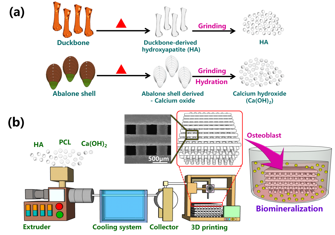
Discarded DBs and fish (Abalone) shells were collected from restaurants in Kaohsiung, washed with clean water, and treated with hot water (100°C). After removing the residual duck meat and fish flesh, the bones were placed in a 105°C vacuum drying oven. The dried DBs and fish shells were crushed into fragments, placed in a 1,000°C high-temperature calcinator for 3 h, and mechanically ground into powder after cooling to room temperature. The 10–30 nm particle size fraction was isolated using a cyclone separator. This fraction was then dried in the 105°C vacuum drying oven to yield samples of natural HA and CaO powder. Hydration of CaO yielded Ca(OH)2, which was then dried in vacuo until the moisture content was < 0.01%. The preparation procedure is illustrated in Scheme 1(a).
Fabrication Of The Composite And 3d Printing Filament:
Table S1 shows the composition of the various PCL/HA and PCL/MHA composites. The PCL, HA, and TAS samples were dried in the 50°C vacuum oven for 12 h prior to preparation of their composites. Different weight ratios were mixed in a TUS-194T extruder (Atlas Electric Devices Company, Chicago, IL, USA) at a screw speed and temperature of 20 rpm and 75–85°C, respectively. The reeler speed was adjusted to yield a filament of diameter 1.75 ± 0.05 mm suitable for 3D printing.
Scheme 1(b) illustrates the bone scaffolds constructed from the PCL/HA and PCL/MHA composite filaments using a 3D Cuby DW-300 printer (Dimension Way Inc., Taichung, Taiwan). The filament was fed into the 3D printer operating at 80°C and 5 rpm with x- and z-direction printing speeds of 130 and 2.5 mm/s, respectively. The z-direction thickness was about 0.3 mm. All of the printing processes were performed in an aseptic environment. Two porous scaffolds were designed: one with a diameter of 11 mm and thickness of 3 mm, and the other with a diameter of 28 mm and thickness of 3 mm. Both designs had a pore size of 0.30–0.35 mm. These samples were used in biocompatibility and biomineralization tests.
Instrumentation
X-ray diffraction (XRD) patterns of the PCL/HA and PCL/MHA composites were determined using the D2-PHASER instrument (Bruker, Karlsruhe, Germany) [25] operating with Cu Kα X-rays at a working voltage of 40 kV and current of 20 mA. Measurements were recorded at 2θ = 10°–60° at a scanning speed of 2°/min. Samples were gold-plated for 120 s before the surface morphology was examined using scanning electron microscopy (SEM; S-3000N; Hitachi, Tokyo, Japan) operating at 15 kV. Elemental compositions were determined by quantitative energy-dispersive X-ray spectroscopy (EDS) [26]. The water absorption of a scaffold was assessed by immersing it in distilled water for different time periods. The percentage weight increase due to water absorption, P, was calculated according to the following equation:
P = (Wa−Wb)/(Wb) × 100%
where Wb is the dry weight of the scaffold and Wa is the weight of the treated scaffold sample after removing excess water.
Cytocompatibility And Bioactivity
NIH3T3 fibroblasts (BCRC 60008; Bioresource Research Centre, Hsinchu, Taiwan) were cultured in growth medium (90% DMEM, 4 mM L-glutamine, 1.5 g/L sodium bicarbonate, 4.5 g/L glucose, and 10% FCS) until the cells were stable. Then, the growth medium was removed, osteogenic medium (90% DMEM/F12 medium, 10% FCS, 39.3 ng/mL dexamethasone, 50 g/mL ascorbic acid-2-phosphate, 10 µL/mL penicillin/streptomycin, and 756 g/mL β-glycerol) was added to the cells for osteogenic differentiation, and the NIH 3T3 derived osteoblast cells were obtained after the NIH fibroblast cells were continuously cultured to confluence for several weeks. The 3D-printed scaffolds of various compositions were then treated with these osteoblasts.
Osteoblast Viability
The PCL/HA and PCL/MHA scaffolds were loaded into 24-well plates, and the wells were filled with 2 ×10⁴ osteoblasts and cultured for 2 days. After removing the culture medium, the remnant was cleaned once with PBS. It was then mixed with trypsin-EDTA and placed undisturbed in the incubator for 7 min. This allowed the cells growing in the scaffold to suspend in the liquid. The scaffold was rinsed twice with PBS before removal. The suspending liquid was then centrifuged to remove the supernatant liquid. Then, cells mixed with MEM were injected into a cell culture box and placed in the incubator for 8 h. The osteoblasts grown on the PCL/HA and PCL/MHA scaffolds of different compositions were observed and analyzed using an inverted microscope.
A proliferation test was performed to establish the viability of osteoblasts on the PCL/HA and PCL/MHA scaffolds over different time periods. osteoblasts (2 × 10⁴) were placed into each well of a 24-well plate with a scaffold. They were cultured in the incubator, removed after 1, 3, and 5 days and mixed with the MTT reagent. They were left undisturbed for 3 h to allow the formation of Formazan violet crystals, which were then dissolved by the addition of DMSO. Cell viability was determined according to ELISA at 570 nm.
The cell cycle distribution pattern of osteoblasts implanted with the PCL/HA and PCL/MHA scaffolds was examined. Osteoblasts (2 × 10⁵) were implanted into each well of a 24-well plate with PCL/HA or PCL/MHA scaffolds, cultured for 2 days, trypsinized, washed with PBS, and centrifuged (to isolate the cells). The cells were then fixed with cold alcohol (–20°C) and centrifuged to remove the supernatant. Propidium iodide (PI) and Triton X-100 were added, and flow cytometry (FL2 PMT; BD, Franklin Lakes, NJ, USA) was performed. WinMDI 2.9 software was used for statistical analysis.
Changes in the quantities of normal, apoptotic, and necrotic osteoblasts cultured on the PCL/HA and PCL/MHA scaffolds were subjected to cell apoptosis analysis. Osteoblasts (5 × 10⁵) were implanted into each well of a 24-well plate with PCL/HA or PCL/MHA scaffolds and cultured for 1 day. The supernatant was then removed, and annexin V–FITC (fluorescein isothiocyanate) and PI staining solution were added. The cells were incubated in a lightproof environment at room temperature for 20 min and finally subjected to flow cytometry, with WinMDI 2.9 software used for statistical analysis.
Osteoblast Mineralization
Calcium ion deposition
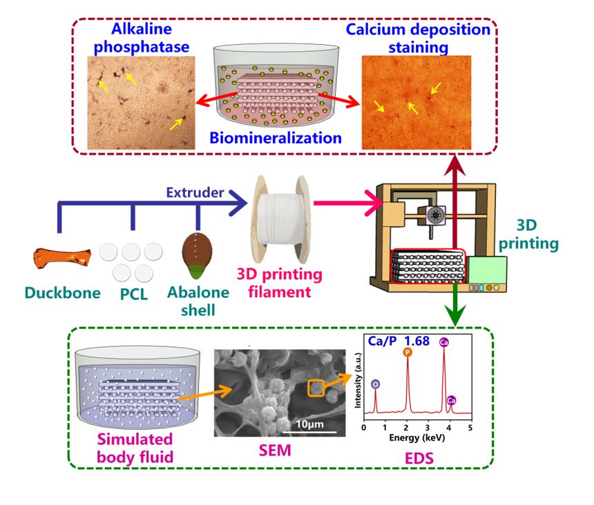
A scaffold sample was soaked in 75% ethanol for 1 min, exposed to ultraviolet (UV) light for 30 min, and placed at the bottom of a 3-cm-diameter culture dish. Osteoblasts (1 × 10⁵) were implanted into the scaffold sample. After 7 and 14 days of cultivation, the culture medium was removed, and the scaffold sample was cleaned once with PBS. After the addition of trypsin, the scaffold sample was left undisturbed for 5 min to suspend the cells. The scaffold sample was then removed, cleaned three times with PBS, and centrifuged at 2,048 rpm to remove the supernatant and obtain the osteoblasts. Then, the osteoblasts were cultured with fresh culture medium for 1 day. After removing the culture medium, the osteoblasts were cleaned three times with PBS and mixed with a 4% formalin solution for 20 min to immobilize the cells; 0.5% Alizarin Red S solution was then added. The sample was left undisturbed for 30 min and cleaned with deionized water (DIW), and the morphology of calcium ion (Ca2+) deposited on the osteoblasts was observed using a microscope. The osteoblasts were also dissolved in 10% acetic acid for 30 min, and the absorbance was measured at 415 nm by the ELISA reader to quantify Ca2+ deposition on the osteoblasts.
Alkaline Phosphatase Activity
Osteoblasts (2 × 10⁴) were implanted on the scaffold. After 2 days of cultivation, the culture medium was removed and the scaffold was cleaned once with PBS. After the addition of trypsin, the scaffold was left undisturbed for 5 min to suspend the cells. The scaffold was then removed, cleaned three times with PBS, and finally centrifuged at 2,048 rpm to remove the supernatant and obtain the osteoblasts. Then, the osteoblasts were cultured with fresh culture medium for 1 day. After removing the culture medium, the morphology and phosphorus ion deposition were examined. For morphological analysis, 2% paraformaldehyde was added to the cells for 5 min to immobilize them, and they were then stained with BCIP/NBT, left undisturbed for 4 h, and imaged microscopically. Phosphorus ion deposition on the osteoblasts was determined as follows. The cells were added to cell lysis and pNPP solutions, and incubated at 37°C for 30 min to transform p-NPP into p-nitrophenol; 3 M NaOH was added to stop the reaction, and the absorbance at 405 nm was measured by the ELISA reader to quantify the phosphorus ion deposition.
In vitro degradation and analysis of simulated body fluids
In vitro degradation
The scaffold samples were dried in a vacuum drying oven at 45 ± 2°C until the water content was < 0.01%, placed in beakers containing 1 × PBS, covered with aluminum foil, and maintained at room temperature. They were then removed on day 7, 14, 21, or 28; PBS was then rinsed from the surface of the samples, which were dried to a constant weight. The weights reflected the in vitro degradability.
In vitro apatite formation in simulated body fluid
A scaffold sample was placed in a container containing SBF (8.035 g NaCl, 0.355 g NaHCO3, 0.255 g KCl, 0.231 g K2HPO4•3H2O, 0.311 g MgCl2•6H2O, 0.292 g CaCl2, 0.072 g Na2SO4, 39 mL of 1 M HCl, and 6.18 mL of trihydroxymethylaminomethane/1 L DIW) at 37 ± 1.5°C. The SBF was replaced every 10 days until day 90. The sample residues were then removed by treatment with chromic acid (200 g/L) and distilled water, and dried in the oven at 50°C. Apatite formation was examined by SEM at various intervals.
Antibacterial Activity
Pseudomonas aeruginosa (BCRC 10303) and staphylococcus aureus (BCRC 10451) obtained from the Taiwan Bioresources Research Center (Hsinchu, Taiwan) were activated and cultured in liquid medium for 1 day, and then collected and uniformly smeared on a culture dish. PCL/HA and PCL/MHA scaffold samples (~ 1.10 cm in diameter and 0.3 cm thick) sterilized by alcohol and UV light were attached to the culture dish containing agar medium smeared with P. aeruginosa and S. aureus, sealed with film, and cultured for 1 day. The areas of inhibition of P. aeruginosa and S. aureus were measured.
The quantitative test for antibacterial activity was carried out as follows. PCL/HA and PCL/MHA scaffold samples (1 g) with different compositions were placed in culture bottles containing culture medium and 2 × 10⁵ CFU /mL of bacteria, and cultured for 20 h and counted the bacteria..