Understanding the neuropathological changes underlying intellectual disability and neurological features in individuals with DS is critical. Although these changes in the brain begin from the fetal period (17), neuroimaging studies, especially in early childhood, include a small patient cohort. Our study performed statistical shape analysis for CCs in DS cases during infancy and early childhood that had grossly normal CC morphology. This is the first modern morphometric shape analysis study to evaluate CC in DS patients in this age group.
The corpus callosum development commences with the development of the genu at about the 12th weeks of the intrauterine period, followed by the isthmus and splenium, and finally, the rostrum around 18-20th weeks of gestation (18, 19). The maximum number of fibers passing through the corpus callosum is reached in the intrauterine period, and myelination of these fibers continues in the postnatal period (20). In the 3rd month of the newborn period, the genu and the splenium are myelinated and thickened in the 4th -6th months (12, 13), and this causes the CC to increase in size with age. When the size of the corpus callosum was examined, we found a statistically significant increase, as expected, with increase in age in the healthy group. However, we could not find a relationship between age and CC size in the DS group. In the literature, the first sign of neuroanatomical abnormality is a characteristic reduction in brain size and an increase in progression in the last three months of pregnancy are observed in fetuses with DS (21, 22). Parallel to this, in our study, CC did not show the expected development with age in patients with DS in the postnatal period and fell behind.
Studies in the literature on children and youth with DS, in which MRI evaluated structural changes in the brain assessed primarily focused on volume analysis. In these studies, total brain volume reductions (23–26), cerebellar (23–25, 27) and hippocampal (24, 26, 27) volumes decrease, and also frontal and temporal lobes (25, 26) regional reductions were reported, whereas parietal lobe gray matter, thalamus, and basal ganglia volumes (24) were maintained. However, there is no evaluation of CC in these studies. In the pediatric age group, only in the study of Gunbey et al. (7), CC volume decrease in individuals with early childhood DS was reported, which reveals the loss of neocortical neuronal projections involved in the maintenance of higher cognitive processes. In our study, unlike volume studies, we aimed to determine whether the shape of CCs, which were evaluated to have qualitatively normal morphology, is also quantitatively normal; and if there is any abnormality and its location. We found a statistically significant difference between the healthy and patient groups regarding the corpus callosum shape. A shape difference was quite evident, especially at the splenium level. The splenium is the bulbous-shaped part of the CC at the most posterior part. The fibers in the splenium are projections from the occipital-parietal and temporal cortex (28). Splenial and hippocampal fibers cross the midline together and connect the cortices of these lobes (28). Although the function of the splenium remains unclear, an increase in the size of the splenium during adolescence could facilitate the maturation of multiple higher functions such as language, reading, computational skills, intelligence quotient (IQ), behavior, and consciousness, which require visuospatial information transfer; and can allow these functions to develop (29–31). The retardation in these functions, which may occur in individuals with Down Syndrome starting from the neonatal period, might be due to dysmorphic changes that are more clearly detected in the splenium part of the CC. In Gunbey et al’s. (7) study, while there was no statistically significant decrease in total and segmental brain volumes, hippocampal volume, and white matter volumes; a significant decrease in CC volume may be a finding that may support this observation. That is, while findings such as a decrease in total brain volume, especially diffuse cortical atrophy including parietal lobes, symmetrically sulci, and hippocampus in adults with DS (32, 33), are not yet seen in early childhood, the first deteriorations in the brain may start from the CC and especially from the splenium part of the CC. A theory that could explain this deformity might be that myelination deficiency or disorders in axons can be detected earlier in the CC, which has a more compact structure than the white matter and especially in the splenium, which is the thickest part of the CC.
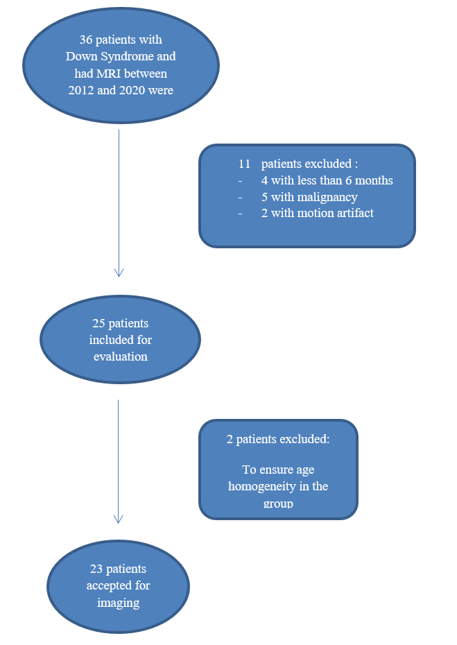
The study's limitations include the small patient cohort and, recruitment from a single center. Also, a technical limitation was performing morphometric analysis on a single plane (sagittal) T2-weighted images. The CC's landmarks and actual size could be better evaluated with a three-dimensional (3D) sequence. However, this was not possible due to the retrospective nature of the study.
To the best of our knowledge, this is the first statistical shape analysis study to evaluate CC in individuals with DS in early childhood. We think that the results of our study and other findings in the literature may contribute to the elucidation of how neurocognitive disorders seen in individuals with DS cause changes in the brain in the early stages. Besides, the results we obtained can be a crucial reference source for creating a database of artificial intelligence programs that we will be using more soon. However, the findings should be supported by other studies that are more comprehensive and include a larger number of patients. Also, more comprehensive studies that include other anatomical reference points in the brain will contribute to the field.