2.1. Sample capture.
All Pelvic floor vaginal anterior wall prolapse samples (n = 8, age from 50–75) were obtained from surgical patients at the department of obstetrics and gynecology department, West China Second University Hospital (Chengdu, China). All patients received written informed consent, which was approved by the Ethics Committee of Sichuan University. The patient was clinically diagnosed with serous III to IV degree of vaginal anterior wall prolapse and did not receive any other treatment 6 months prior to harvesting the sample.
2.2. Pelvic floor fibroblasts isolation and culture.
The obtained surgical sample was immediately cut into small pieces, digested with type-I collagenase at a concentration of 1 mg/ml, placed in a 37°C water bath shaker at 150 rpm for 2 hours, and then passed through 70µm and 40µm cell filters filtration in sequence, the obtained single cell suspension were cultured in DMEM medium (Hyclone) supplemented with 15% fetal bovine serum (Gibco) and 1% penicillin-streptomycin (Hyclone), and all cell cultured in a humidified incubator (Heraeus) with 5% CO2 saturation at 37°C, the medium was changed for every 3 day, the first passage cell confluence at 80% takes about 15 days and can be sub-cultured for further test, and the cells used in our experiment in the third or fourth passage.
2.3. Immunohistochemistry.
Immunocytochemistry was used to characterize fibroblasts specific surface markers, the cells were digested by trypsin-EDTA and seeded into 6 cell plates at the concentration of 1×10⁵ cells/well, and incubated in a 5% CO2 saturation at 37°C. When the cell confluence at 80%-90%, washed with PBS and fixed in 4.00% paraformaldehyde for 15 minutes, then, 0.50% Triton X-100 and 5.00% normal goat serum was used to permeate and block for 5 minutes, respectively. After that, the cells were incubated with the primary antibodies including the use of anti-Cytokeratin, anti-α-SMA and anti-Vimentin (1:200, Abcam) antibodies at 4°C overnight, and the second antibody was incubated at room temperature for 1 hours. Finally, the cells were counterstained with haematoxylin and visualized with the DAB.
2.4. Administration.
The effect of different concentration (range from 10− 5-10− 10 mol/L) of E2 on pelvic floor fibroblasts proliferation were tested by CCK-8 assay at 12h, 24h and 48h, After obtain the best administration concentration, cells are administrated with E2 (with the best concentration) and rapamycin (with the concentration of 10− 8 mol/L) for further detected, besides, cells are treated with lysosomotropic reagents chloroquine (with the concentration of 40 µmol/L for measuring "autophagy flux",(36) and all drugs are purchased from solarbio, China.
2.5. Proliferative assay.
Colorimetric Cell Counting Kit (CCK8, Beyotime, China) was used to test the cell proliferative capacity. The cells were seeded at a density of 1×10⁴/well into the 96-well plate, 10 µL CCK-8 reaction solution was added to each well after administration for 12h, 24h and 48h and incubated for 2 h. The microplate reader was used to measure the absorbance at 450 nm. The final absorbance value is calculated from the absorbance value of the test well minus the absorbance value of the reagent background.
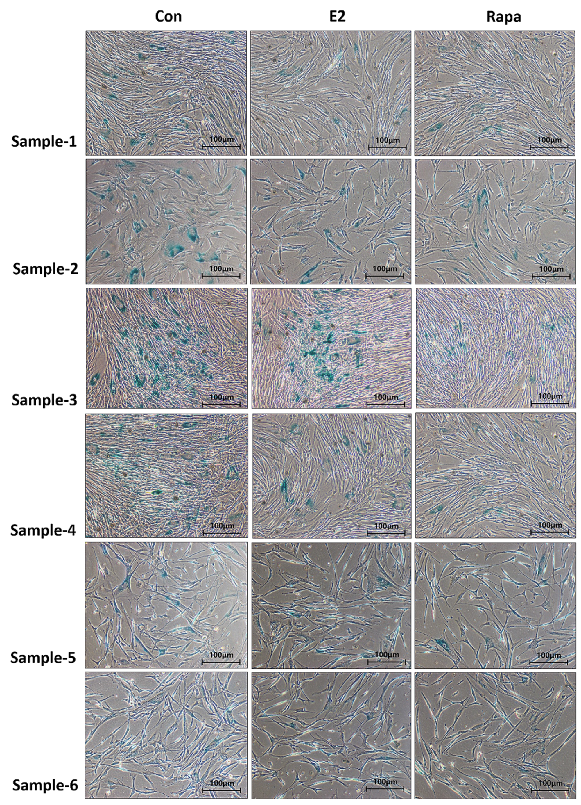
2.6. Senescence-Associated β-Galactosidase (SA-β Gal) Staining.
The senescence of the pelvic floor fibroblasts was evaluated by SA-β gal staining kit (Beyotime, China). The cells were seeded at a density of 1×10⁵ cells/well on coverslip, when the cell confluence at 80%-90%, fixed with 4% paraformaldehyde for 15 min and washed three times with PBS, finally, incubated the cells with SA-β-gal reaction solution at 37° C for 12 h according to the manufacturer’s protocol, then washed three times with double-steaming water, SA-β gal positive cells were counted under the optical microscope.
2.7. Western blotting analysis.
Western blots were used to measure protein expression. The concentration of protein was determined by the BCA Kit (Beyotime, China). The total protein equivalent (30 µg) of each sample was separated by 10% sodium dodecyl sulphate-polyacrylamide gel (SDSPAGE) and polyvinylidene fluoride (PVDF) membranes (Invitrogen Life Technologies, Inc) in the membrane transfer system. Then, the membrane was placed in 5% fatfree milk and sealed at room temperature for 2 hours. After washing with TBST (Tris Buffer Saline-Tween 20), Followed by incubated with primary antibodies against p16INK4A, p21, p-53 sirt-1(Cell Signaling Technology, Inc), COL-I & COL-III (Proteintech, Inc), LC3-I/II and GAPDH (Signalway Antibody, Inc) (1:1000 dilution) at 4 ℃ overnight, and then incubated with secondary antibody (anti-rabbit or anti-mouse IgG-conjugated with HRP) (1:3000 dilution) at room temperature for 1 hours. the membranes were visualized in Molecular Image® ChemiDocTM XRS + system (Bio-Rad Inc., USA) with Image LabTM Software and analyzed by Image J 1.44p software (National Institutes of Health, USA).
2.8. Ultrastructure of fibroblasts observed under transmission electron microscope (TEM).
Take the logarithmic growth phase fibroblasts, expand culture and collect cells, so that the total number of cells reaches 2×10¹⁰, washing with PBS and centrifuge at 1,000 rpm for 5 minutes, then resuspend the cells with 1:6 diluted fixating solution (3% glutaraldehyde: PBS) and stand at 4 ℃ for 5 min, after that, centrifuge at 10000 rpm for 15 min, discard the supernatant, and then fix with 3% glutaraldehyde at 4 ℃ for 2 h. after dehydration, immersion, embedding, slicing and staining, the cells are observed under transmission electron microscope (TEM).
2.9. Statistical analysis.
Results were analyzed by GraphPad Prism 9.0 Software (GraphPad, San Diego, CA, USA). The values are expressed as mean ± SEM at least three independent experiments performed. The data were analyzed using one-way analysis of variance (ANOVA) and post-hoc analysis, Pvalues < 0.05 were considered to indicate a statistically significant difference, the significance was calculated by comparing the controls with experimental groups.