3.1 Characteristics of PS-NPs
The results of the IR spectrum related to polystyrene nanoplastics are shown (Fig. 1-a). According to the results obtained from FTIR analysis, it can be seen that the broad absorption peak at 3386 cm− 1 is related to the stretching bond of the first type of amine N-H and the bending peak at 1580–1650 cm− 1 is related to the amine vibration of N-H. The appearance of medium and sharp peaks in the region of 2082–3026 cm− 1 is characteristic of C-H aromatic stretching vibrations.
The absorption band observed at 730–765 cm− 1 indicates the presence of a strong C = C bending and alkene bond. The broad and strong peak at 3400 cm-1 indicates the existence of O-H bond (He et al., 2020). According to the FESEM microscope images, PS-NPs have been synthesized in a spherical shape with a uniform and regular appearance. These images were taken with a magnification of 500 nm. (Fig. 1-b). The higher the value (positive or negative), the more stable the colloidal dispersion. After examining the zeta potential, PS-NH2 nanoparticles were found to have an electrical charge of 17.3–15.9 mV, where the zeta potential is close to zero (that is, the sample is approximately at the isoelectric point) and has a neutral electrical charge. Therefore, according to the obtained results, PS-NH2 nanoparticles, due to their surface charge range, cause the instability of the particles and this provides the possibility for flocculation and non-separation of the particles (Fig .1-c)(17)
3 − 2 Effect of PS-NPs on MDA-MB231, HFF-2 cell viability
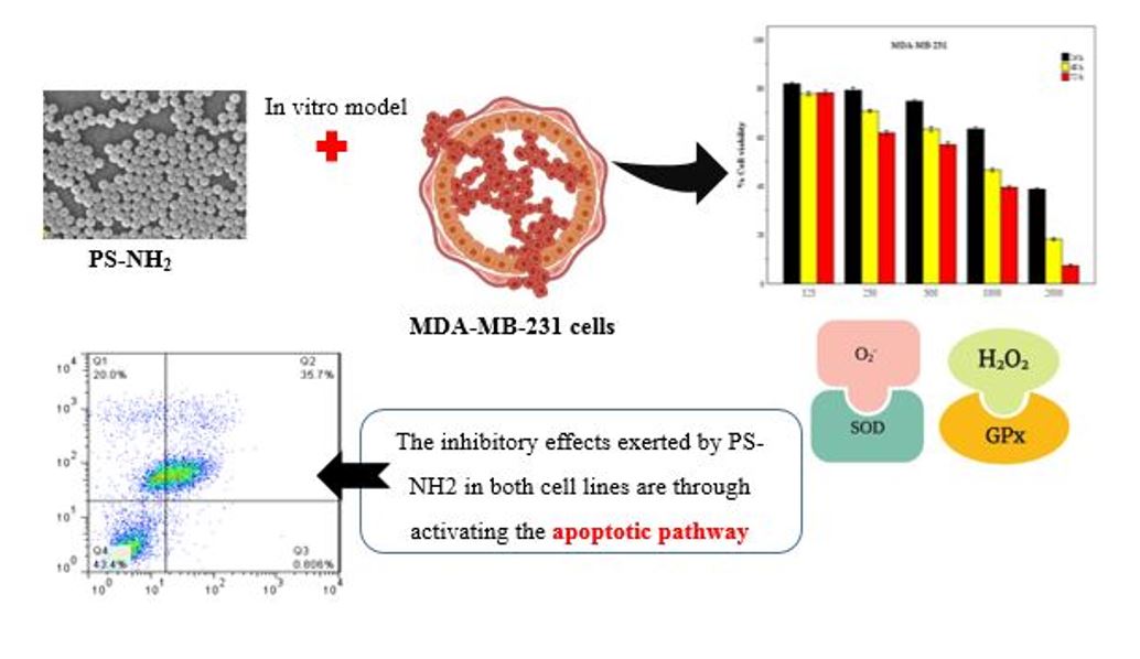
The effect of different concentrations of PS-NH2 on MDA-MB-231 and HFF-2 cell lines was investigated by cell culture(Fig. 2,3). According to the results obtained in this study in both MDA-MB-231 and HFF-2 cell lines, it was observed that the toxicity effect of PS-NH2 nanoparticle increases with increasing concentration and passage of time, while the smaller the size of the nanoparticle, more toxicity effect was observed. (12)
One of the goals of this study is to investigate the anti-tumor effect and apoptosis induction of a combination of nanoplastic polystyrene. The intensity of the color created in the MTT method is proportional to the cells with active mitochondria. In other words, this method is used to determine the rate of proliferation and survival of cells after exposure to cytotoxic substances. (12)
The results of two-way analysis of variance (ANOVA) show that the effect of time, concentration and size of PS-NH2 nanoparticles on the survival of breast cancer cells is significant (p < 0.001). (Table 2),(12). Independent T-test was used to compare the viability of MDA-MB-231 cells with HFF-2 cells. As independent T results show, in general, the average survival of MDA-MB-231 cells is not significantly different from HFF-2 cells, Therefore, it can be said that the viability of both cells is generally the same (Fig. 2,3).
The results of MTT show that the survival of MDA-MB-231 and HFF-2 cells depends on the concentration, size and time of exposure to polystyrene nanoplastic. In this research, it was observed that higher concentrations and smaller sizes of PS-NH2 have more cytotoxic effects than lower concentrations and larger sizes of this compound, so that it is accompanied by morphological changes such as reduction in cell volume and rounding was accompanied Also, PS-NH2 nanoplastic significantly reduced cell viability with increasing concentration (Fig. 4,5). The results of the MTT test at the concentration of 10, 50, 100 µg/ml clearly showed that PS-NH2 in the sizes used in this study did not have specific cytotoxic effects in vitro (He et al., 2020). By increasing the concentration of PS-NH2 above the concentration of 125 µg/ml, the life of cells decreases logarithmically (Chan et al., 2007).
The value IC50 for 90, 200, 300 nm value was about 500 µg/ml for 72 hours and 250 µg/ml for 24 hours. The obtained results suggest that polystyrene nanoplastic has cytotoxicity potential on MDA-MB-231 and HFF-2 cell lines. The most toxic was observed for the size of 90 nm with a concentration of 2000 µg/ml in 72 hours, because it has a smaller size than all the particles, so its ability to penetrate the cell is greater and its lethality is much higher, as a result, the number of living cells in these wells is less.
3–4 Antibacterial effect PS: Agar Dilution
Polystyrene nanoparticles in all three sizes of 90, 200, and 300 nm were investigated in order to determine their antimicrobial properties against Gram-positive S. aureus and Gram-negative bacteria.
The antibiotic streptomycin was used as a positive control to determine the sensitivity of the species (Fig. 6-g), and the culture medium with nanoparticles was used as a negative control (Fig. 6-h). Comparing the results of nanoparticles with the control showed that these particles in concentrations of 500, 1000, 1500, 2000 ug/ml had no antimicrobial effect on these two bacteria and no growth halo was formed (Fig. 6). In addition, the MIC results confirm this, and the combination of PS-NH2 did not prevent the growth of bacteria in any of the wells. Therefore, PS-NH2 has no antimicrobial effect on S. aureus and E. coli.
Based on the research results of Bamre et al. (2020), it has been reported that no microbial decomposition is observed in polystyrene sheets buried in the soil after 32 years (18). Hydrophobic groups in thermoplastics have made them resistant to hydrolysis (19). The molecular compounds of plastic affect the hydrophobicity of the polymer surface and cause the easy connection of microorganisms to each other on the plastic surface (20). The high molecular weight and low solubility of polystyrene in water have prevented it. These polymers pass through the membrane of microorganisms and intracellular decomposition takes place on them. The biological process of decomposing these compounds starts with the release of extracellular enzymes from the decomposing microorganisms (20). According to the results of studies by Lithner and his colleagues (2011), most plastics are very resistant to microbial degradation in everyday use (21)(Lithner et al., 2011)(21).
3–5 Antibacterial effect of PS: minimum inhibitory concentration (MIC)
Polystyrene nanoparticles in the sizes of 90, 200, 300 nm were investigated in order to determine the antimicrobial properties for the gram-positive bacteria S. aureus and the gram-negative bacteria E. coli. Mueller Hinton Broth culture medium with Neem-McFarland bacteria was used as a negative control (Fig. 7-b) and streptomycin antibiotic was used as a positive control (Fig. 7-a). After analysis by ELISA reader, no antibacterial effect was observed after treatment with PS-NH2. As in the antimicrobial test of diologen agar, in terms of quality, no growth halo was observed due to the use of PS-NH2, and the results of the MIC and diologen agar tests were also confirmatory and proved that PS-NH2 nanoparticles have an antibacterial effect on bacteria. It does not contain S. aureus and E. coli (Fig. 7),(18).
3–6 Apoptosis
To determine the apoptotic or necrotic effect of PS-NH2 on MDA-MB-231 and HFF-2 cell lines, we used the flow cytometry technique and compared the obtained results with the control. In MDA-MB-231 and HFF-2 cells, it was observed that with increasing concentration of PS-NH2 nanoparticles, the percentage of living cells decreased and they entered the phase of apoptosis and necrosis. Also, with the increase in nanoparticle size, its effect decreased in both cells. Statistically, samples treated with PS-NH2 nanoparticles in both MDA-MB-231 and HFF-2 cell lines have a significant difference with the control sample(Fig. 8,9a-g), (P < 0.001)
In this research, flow cytometry analysis was used for further studies of cell death method (apoptosis or necrosis). Our results showed that the percentage of apoptotic cells (annexin positive and PI negative) increases with the passage of time and increasing concentration and decreasing size of PS-NH2. According to the obtained results, by increasing the concentration from 500 µg/ml to 2000 µg/ml and reducing the size of PS-NH2 to 90 nm, the percentage of apoptosis increased significantly (P < 0.001).
The highest percentage of apoptosis induced in the concentration of 500 µg/ml of 90 nm size PS-NH2 after 48 hours of treatment was 50% in HFF-2 cells (Fig. 9-a). Apoptosis is defined as programmed cell death. If this process is disturbed, it causes pathological conditions, such as cancer, autoimmune disorders (22).Unlike apoptosis, necrosis is the pathological death of cells and this type of cell death occurs during severe damage to cells such as hypoxia, hyperthermia and external toxins. Radiotherapy, chemotherapy and hormone therapy methods all induce apoptosis in cancer cells. Of course, using higher doses of these compounds causes cancer cell death through other methods (22).
PS-NH2 in HFF-2 cells induced the peak of phosphatidylserine (one of the reliable indicators of apoptosis) in the flow cytometry histogram compared to the control group. And statistically, it has a significant difference with the control group (P < 0.001).
The general conclusion that can be drawn is that polystyrene nanoplastics in smaller sizes and higher concentrations have had more toxic properties over time. In this research, it was observed that the toxic effect of PS-NH2 on fibroblast cells is greater than on cancer cells, and this will be a warning sign for the high use of products containing this nanoparticle (Fig. 8,9a-g).
3–7 cell cycle
In the cell cycle, a regular and organized path of events is followed until replication takes place and it is divided into two general stages, interphase and mitosis (Futreal et al., 2005). According to the obtained results, MDA-MB-231 and HFF-2 cells are mostly stopped in G1 and Sub-G1 phase (Table 1). This result indicates the occurrence of apoptosis as a result of treatment with PS-NH2 nanoparticles. It was observed that the percentage of apoptosis induced in HFF-2 cells after treatment was higher than that of breast cancer cells. Statistically, both investigated cell lines have a significant difference with the control sample (p < 0.001).
Examining the results of the cell cycle in the samples treated with PS-NH2 in the present study shows the role of this compound in creating a break in the G1 phase of the cell cycle in MDA-MB231 and HFF-2 cells. The induction of cell accumulation in G1 phase by PS-NH2 is shown by increasing the concentration of this compound (Table 1).
The increase in the number of cells in G1 phase due to treatment with PS-NH2 in size 200, 90 and 300 nm in concentrations of 500 and 2000 mg/ml was significant compared to the control (p < 0.001). The effect of different concentrations of PS-NH2 is to cause an interruption in the G1 phase of the cell cycle and increase the accumulation of MDA-MB-231 and HFF-2 cells in this area, and it has increased compared to the control (P < 0.001), (Table 1).
In HFF-2 cells, G1 percentage is lower than MDA-MB-231 at the same concentration (Fig. 11). Although this treatment in HFF-2 cells was significant in the number of cells in the G1 and Sub-G1 phase compared to the control and caused an interruption in the G1 phase, which led to the induction of apoptosis in these cells. (p < 0.001). The percentage of cells present in G1 phase in 90 nm size is less than 200 nm and in these sizes, with increasing concentration, we will see a decrease in the amount of cells in G1 phase. In the concentration of 2000 µg/ml of 300 nm, the percentage of G1 cells is more than 500 µg/ml (Fig. 11a-g). In MDA-MB-231 cells, at 90 and 200 nm, the amount of S-phase cells decreases with decreasing concentration, but at 300 nm, with decreasing concentration, we see an increase in the amount of S-phase cells (Fig. 10a-g). In HFF-2 cells, the percentage of S-phase cells increases with the decrease of PS-NH2 concentration. Also, with the increase of PS-NH2 size, the percentage of S-phase cells increases. The percentage of cells in G2 phase in the cell cycle of MDA-MB-231 increases from 2000 to 500 µg/ml, while this amount decreases in HFF-2 cells. With the increase in the size of HFF-2 cells, the amount of G2 increases. In HFF-2 cells, the level of G2 is higher than in MDA-MB-231 cells (Table 1).
Therefore, the results of the cell cycle showed that MDA-MB-231 cells treated with amine polystyrene in all concentrations show a significant arrest in the G1 phase with the passage of time compared to the control cells and the increase in concentration The treatment causes a slight decrease in the percentage of cells in the S and G2 phase. In HFF-2 cells, it was observed that increasing the concentration of treatment causes a slight increase in the percentage of cells in the G1, S, and G2 phase .
In HFF-2 cells, we see a significant increase in the amount of Sub-G1. The cell population before the G1 phase indicates the occurrence of apoptosis. In MDA-MB-231 cells, this increase occurred only in the size of 90 nm.
The average results and standard deviation showed that the results were statistically significant compared to the control (P < 0.001). In addition, the percentage of cells entering the Sub-G1 stage, which indicates apoptotic cells, has also increased significantly, depending on the dose and size of nanoparticles compared to the control in both cells (Table 1).
According to the obtained results, it can be concluded that the inhibitory effects exerted by PS-NH2 on MDA-MB-231 and HFF-2 cell lines are probably through the activation of the apoptotic pathway (Fig. 10,11).
Hu et al. (2021) in a study investigating PS-NPs on zebrafish larvae and macrophage cells, according to their results, showed that the percentage of apoptotic cells in the early stages in the PS NPs group was higher than the control group. The percentage of apoptotic cells increased in a concentration-dependent manner with a maximum increase of about 10%. They also identified caspase-3 protease activity in zebrafish larvae, which played a central role in the execution of apoptosis. Their results showed that PS NPs treatments caused a certain degree of apoptosis (23).
In a study of the effect of microplastic polystyrene (PS-MPs) on granulosa cells (GCs) in rat ovaries, An et al. (2021) observed that treatment with this compound causes apoptosis. Their study showed that the apoptosis rate of granulosa cells significantly increased after treatment with PS-MPs. These results showed that PS-MPs cause apoptosis of GCs through oxidative stress (24).
3–8 Activity of antioxidant enzymes
GPx enzyme activity was measured in MDA-MB-231 cells compared to the control (Fig. 12). According to the obtained results, after the cells were exposed to PS-NH2 nanoparticles with a size of 90 nm at a concentration of 500 µg/ml, the enzyme activity increased about 2.3 times compared to the control. At the concentration of 1000 µg/ml, the enzyme activity decreased by 6.5 times compared to the control. Between the tested groups, with a decrease in the concentration of PS-NH2, there was a 13-fold increase in enzyme activity, and there was a statistically significant difference (P < 0.001).
GPx enzyme activity was measured in MDA-MB-231 cells compared to the control. According to the obtained results, after the cells were exposed to PS-NH2 nanoparticles with a size of 200 nm and a concentration of 500 µg/ml, the enzyme activity was about 1.5 times that of the control, and at a concentration of 1000 µg/ml, the enzyme activity was halved compared to the control. Between the tested groups, with a decrease in the concentration of PS-NH2, there was a 2.5-fold increase in enzyme activity, and there was a statistically significant difference (P < 0.001).
GPx enzyme activity was measured in MDA-MB-231 cells compared to the control. According to the obtained results, after the cells were exposed to PS-NH2 nanoparticles with a size of 300 nm, a concentration of 500 µg/ml, the enzyme activity decreased by 0.24 times than the control, and at a concentration of 1000 µg/ml, the enzyme activity was halved compared to the control. Between the tested groups, with the decrease in PS-NH2 concentration, there was a 1.5-fold increase in enzyme activity, and there was a statistically significant difference (P < 0.001). Thus, according to the results of the present study, the lower the concentration of this nanoparticle, the higher the activity of the GPx enzyme.
GPx enzyme activity was measured in HFF-2 cells compared to the control (Fig. 12). According to the obtained results, after the cells were exposed to PS-NH2 nanoparticles with a size of 90 nm, the concentration of 500 µg/ml, the enzyme activity was about 1.3 times, and at the concentration of 1000 µg/ml, the enzyme activity was doubled compared to the control. Between the tested groups, with the increase in PS-NH2 concentration, enzyme activity decreased about 0.68 times, and there was a statistically significant difference (P < 0.001).
GPx enzyme activity was measured in HFF-2 cells compared to the control. According to the obtained results, after the cells were exposed to PS-NH2 nanoparticles with a size of 200 nm and a concentration of 500 µg/ml, the enzyme activity increased by about 2.2 times and at a concentration of 1000 µg/ml, it increased by 1.3 times compared to the control. Between the tested groups, with the increase in PS-NH2 concentration, enzyme activity decreased about 0.68 times, and there was a statistically significant difference (P < 0.001).
GPx enzyme activity was measured in HFF-2 cells compared to the control. According to the obtained results, after the cells were exposed to PS-NH2 nanoparticles with a size of 300 nm and a concentration of 500 µg/ml, the enzyme activity increased by 1.5 times and at a concentration of 1000 µg/ml, it increased by 1.2 times compared to the control. Between the tested groups, with the increase in PS-NH2 concentration, enzyme activity decreased about 0.68 times, and there was a statistically significant difference (P < 0.001). Thus, according to the results of the present study, with the increase in PS-NH2 concentration, the level of GPx enzyme activity in HFF-2 cells decreases.
Glutathione is the most important intracellular non-protein antioxidant. This substance reduces the free radicals in the cell, so reducing the level of this substance increases the amount of free radicals in the cell and induces apoptosis in cancer cells. The results of measuring the activity of GPx enzyme in our research showed that the combination of PS-NH2 in sizes of 90, 200, 300 nm on HFF-2 cells caused an increase of 3, 2.2 and 1.2 in intracellular glutathione, respectively, compared to the control group. In HFF-2 cell line, and in MDA-MB-231 cells, this nanoparticle in the size of 90 and 200 nm increases the amount of intracellular glutathione by 2.2 and 1.1 times, respectively, compared to the control group and in the size of 300 nm (Fig. 12). Enzyme activity is halved compared to the control. In this cell, with the increase in nanoparticle concentration, we see a 13-fold decrease in the activity of GPx enzyme, which prevents the induction of apoptosis in these cells by increasing the level of glutathione. In both cells, a statistically significant difference was observed with the control group (p < 0.001)
The glutathione system plays a vital role in protecting the body from oxidative stress. This system converts hydrogen peroxides resulting from the activity of superoxide dismutase enzyme on superoxide ion, which is a dangerous reactive oxygen species (ROS), into water. Glutathione peroxidase and glutathione reductase are enzymes that play a vital role in this cycle, and the expression of their genes increases when ROS increase and the body is in a state of oxidative stress. Oxidative stress induced by reactive oxygen species causes apoptosis in cancerous and non-cancerous cells and their destruction.
GSH acts as a strong scavenger of free radicals, it is also responsible for maintaining the cellular redox state and protecting cells against oxidative stress (25). The content of GSH when HFF-2 cells were exposed to PS-NH2 was significantly increased compared to the control group (P < 0.001), indicating a strong induction of oxidative stress, and exposure to 500 µg/ml PS-NH2 in 90 nm size resulted in maximum increase in GSH content. In HFF-2 cells treated with PS-NH2 nanoparticles, with increasing size, we saw less GPx activity, and in 90 nm size, the activity of GPx enzyme increased about 3 times compared to the control, and in 200 and 300 nm sizes, the level of GPx enzyme activity increased, respectively. It increases 2 and 1 times compared to the control. In MDA-MB-231 cells, considering that the cell is cancerous and the cell has suffered more damage, the level of GPx enzyme activity was lower than that of the control group, and with the increase in the size of the nanoparticles, we saw a decrease in the activity of the enzyme. The level of GPx enzyme in MDA-MB-231 cells is lower than the level of HFF-2 and it has a statistically significant difference compared to the control group (Fig. 12) (p < 0.001).
SOD is the cell's first line of defense against ROS. Usually, superoxide radical is the first free radical that is produced during stress, and SOD quickly converts superoxide radical into hydrogen peroxide and molecular oxygen, and by removing superoxide, it plays a more vital role in the antioxidant system than enzymes. Catalase and peroxidase play a role, so it increases the resistance of cells to environmental stresses (26–28). In most studies, the level of SOD activity is variable. This is partly due to the difference in the tested factors such as cell type, type of treatment, concentration of treatment and duration of treatment (29). The increase in SOD activity is due to combating oxidative stress and protecting the cell's defense system against oxidative damage. Also, the increase in enzyme activity is due to the increase in superoxide, which probably superoxide ions increase the expression of superoxide dismutase genes through a signaling role (Fig. 13),(30). by examining the activity of superoxide dismutase enzyme in MDA-MB-231 and HFF-2 cells, which were treated for 48 hours with concentrations of 500, 1000 µg/ml of PS-NH2 combination. had been treated, they showed that the enzyme activity increased at low treatment concentrations, but the enzyme activity decreased with increasing treatment concentration. The decrease in enzyme activity at high concentrations is probably due to the increase in the production of H2O2 in the cell and the inactivation of the enzyme by H2O2 or the binding of PS-NH2 to the active site of the enzyme, and as a result, the enzyme activity decreases (31).
Statistical analysis of the data obtained from the measurement of superoxide dismutase enzyme activity using one-way ANOVA showed that the enzyme activity in MDA-MB-231 and HFF-2 treated with PS-NH2 was significant compared to the control. is (P < 0.001). Comparison of the average activity of superoxide dismutase enzyme in HFF-2 cells showed that treatment with concentrations of 500, 1000 µg/ml of PS-NH2 increased the activity of this enzyme compared to the control, but with increasing concentration in this cell, the activity of the enzyme decreased. We were in control. In MDA-MB-231 cells compared to the control group, the activity of SOD enzyme decreases, while increasing the concentration of nanoparticles also decreases the activity of this enzyme. The highest activity of superoxide enzyme was at a concentration of 500 µg/ml in HFF-2 cells, which showed an increase in SOD enzyme activity compared to the control group (Fig. 13). In normal and oxidative stress conditions, there are advanced antioxidant defense systems to eliminate excess ROS in cells. For example, it is known that SOD converts superoxide into H2O2(Nordberg & Arner, 2001). Environmental pollutants can induce ROS production and subsequent oxidative stress. In general, SOD is responsible for maintaining the dynamic balance between ROS production and scavenging under normal conditions. Therefore, SOD activity indicates the antioxidant capacity of cells.
The increase in SOD activity and GSH content in HFF-2 and MDA-MB-231 cells at higher doses of PS-NH2 indicates the impairment of antioxidant capacities, which play a fundamental role in eliminating superoxide anion (O2•-) and H2O2. Imbalanced antioxidant capacity against oxidative stress causes disturbance in antioxidant structures and as a result leads to cell death.
In the electron transfer chain, O2•- is produced by decreasing the valency of oxygen molecules, along with the leakage of electrons. After production, it can be spontaneously catalyzed to H2O2 by SOD. In addition, H2O2 can be reduced by enzymatic antioxidants such as glutathione peroxidase (GPx), where GSH acts as an exclusive electron donor (32).This is consistent with previous studies that the cytotoxicity of nanoparticles is dependent on surface charge (33) and concentration (34).
PS-NH2 significantly decreased SOD activity and prevented the increase in GSH content. Previous studies have reported that inhibition of SOD activity leads to the accumulation of O2•- (35) and GSH is necessary to reduce H2O2-induced oxidative damage (36).
During a study of the effect of PS-NH2 nanoparticles on HepG2 cells, He et al. (2020) showed that SOD enzyme activity decreases with increasing concentration after treatment with these particles. Our results in this research are in accordance with their results and we have seen a decrease in enzyme activity with increasing treatment concentration in both MDA-MB-231 and HFF-2 cells (12).
An et al. (2021) showed the effect of polystyrene microplastics (PS-MPs) on granulosa cells (GCs) in rat ovaries that PS-MPs lead to an increase in MDA enzyme and a decrease in GSH-PX, CAT and SOD in ovarian tissue. became. And furthermore, the level of ROS was significantly higher in GCs treated with PS-MPs. Our results showed that PS-NH2 nanoparticles decrease SOD and GPx enzymes compared to the control group and were in accordance with the results obtained from the study of An and his colleagues who reported that PS-MPs cause oxidative stress in the ovaries of rats. desert (24)