Synthesis and Characterization of Polymers and Hydrogels
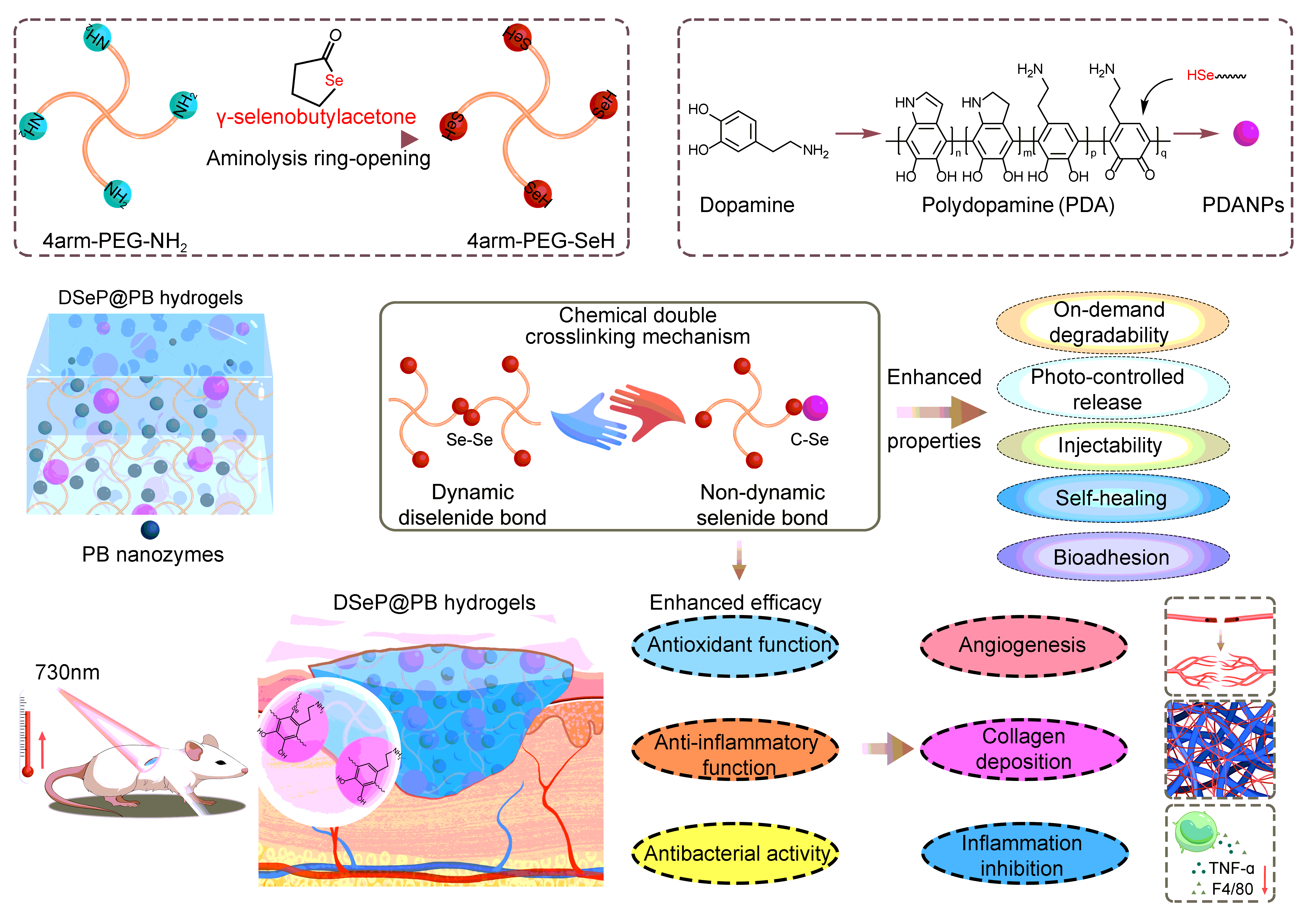
Scheme 1 shows a schematic of the fabrication process for obtaining different hydrogels. First, 4-arm-PEG-SeH (4-arm polyethylene glycol (PEG) with selenol) was synthesized by linking ring-opening γ-selenobutylacetone to 4-arm-PEG-NH2. PEG-Se2 (diselenide-containing polyethylene glycol hydrogels) was then produced by oxidizing 4-arm-PEG-SeH. DSeP (PEG-Se2 loaded with PDANPs) and DSeP@PB (PEG-Se2 loaded with PDANPs and PB nanozymes) were produced by mixing 4-arm-PEG-SeH and PDANPs or 4-arm-PEG-SeH, PDANPs and PB nanozymes, respectively.
PEG was selected as the key component of hydrogels owing to its high hydrophilicity, low immunogenicity, tunable physicochemical properties and ease of use39. 4-arm-PEG-NH2 is a multiarm PEG derivative that has amine groups attached to the terminals of each arm and is connected to one pentaerythritol core.25 To endow 4-arm-PEG-NH2 with on-demand degradable properties, the γ-selenobutylacetone (γ-SBL) ring was opened and linked to 4-arm-PEG-NH2 via a nucleophilic addition reaction to synthesize 4-arm-PEG-SeH (Fig S1A).24 1H NMR analysis confirmed the successful synthesis of 4-arm-PEG-SeH (Fig. 1A). Signals that correspond to the protons of the -CH2-labeled terminal amino group shifted from approximately 2.82 ppm to 2.91 ppm, whereas peaks that were consistent with the protons of the ring-opened SBL presented at approximately 1.95 and 2.35 ppm, indicating the successful selenol modification of 4-arm-PEG-NH2. The calculated conversion rate of the protons of the -CH2-labeled terminal amino group was approximately 89%. Therefore, we successfully synthesized 4-arm-PEG-SeH with a one-pot procedure without using any additives or tedious steps. PEG-Se2 was then produced by oxidizing 4-arm-PEG-SeH with air (O2) in a stoichiometric ratio of 1:1 at ambient temperature, and the gelation of hydrogels was demonstrated by the inverted tube approach (Fig S1B and C).
We previously reported that diselenide bonds are too sensitive to maintain the rigid structure of hydrogels, and the mechanical strength of diselenide-crosslinked hydrogels can sharply decline in an aquatic environment.13 Furthermore, the low adhesion property of PEG hydrogels is not conducive to wound healing. Hybrid hydrogels that integrate nanoparticles into their hydrogel networks through chemical interactions can be tailored to promote the mechanical and other desirable properties of PEG hydrogels.25 PDANPs known to have enhanced hydrophilicity, anti-erosion capacity, and superior biochemical characteristics were incorporated into 4-arm-PEG-SeH to develop DSeP by means of Michael addition and crosslinking of 4-arm-PEG-SeH with PDANPs (Fig S1D-F). Simultaneously, the remaining selenol can be gradually oxidized under the action of oxygen to form diselenide. DSeP (0.01), DSeP (0.1) and DSeP (1) were defined as DSeP containing 0.01, 0.1 and 1 wt% PDANPs, respectively. The FTIR spectrum of DSeP exhibited enhanced peaks at 3460, 2860 and 1645 cm-1 corresponding to NH2, C-H and C = O, respectively, compared with the FTIR spectrum of PEG-Se2 formed through the oxidation of 4-arm-PEG-SeH without PDANPs (Fig. 1B). Compared with the XPS spectra of PEG-Se2, DSeP showed enhanced typical peaks corresponding to O and N (Fig. 1C). It is noteworthy that the peak corresponding to Se was at approximately 56 eV, and a sharp peak was detected in PEG-Se2, indicating that there is only one type of Se in the polymer. After the Michael addition reaction of the selenol of 4-arm-PEG-SeH with PDANPs, a mellow peak was detected in DSeP, signifying the incorporation of Se in the hydrogels. Altogether, the results verified the presence of PDANPs in DSeP.
With the aid of scanning electron microscopy (SEM), the surface morphology of the hydrogels was analyzed. SEM showed homogeneous pores with round shapes in all the hydrogels (Fig. 1D). The pore size of the hydrogels increased with the addition of PDANPs. In addition, the strong structural stability of the hydrogels was indicated by the pore structure integrity as well as its interconnectedness with the rest of the structure. When calculating the amount of time required for gelation, the point at which the curves representing storage modulus (G') and loss modulus (G") generated from the rheological test intersected was considered the gelation point. It was discovered that the time required for gelation is proportional to the weight ratio of PDANPs present in this system (Fig. 1E). The gelation time was the longest (6.3 ± 0.3 h) when 0.01 wt% PDANPs were incorporated into the hydrogels and gradually decreased to 4.6 ± 0.7 and 3.7 ± 0.1 h after the weight ratio of PDANPs was elevated to 0.1 and 1 wt%, respectively. The mechanical strength of DSeP was improved in a PDANP dose-dependent manner, suggesting that increasing PDANP concentrations in the hydrogel network might enhance gelation efficiency (Fig S2).
We speculate that PDANPs at a low concentration (0.01%) may hinder the oxidative coupling of diselenide in the hydrogels, thus prolonging the gelation time. However, when the concentration of PDANPs increases in DSeP, the possibility of reactivity between selenol and PDANPs increases, resulting in a shorter gelation time. The G' and G" curves of the hydrogels were recorded over time to analyze the effect of varying PDANP weight ratios on the rheological characteristics of the hydrogels. The G´ of DSeP (0.1) (1500 Pa) was higher than that of DSeP (0.01) (220 Pa), DSeP (1) (750 Pa) and PEG-Se2 (190 Pa), suggesting that introducing a sizable quantity of PDANPs into the system may enhance the crosslinking density, thus increasing the G' of hydrogels (Fig. 1F). The G' and G'' curves of DSeP over frequency and strain were also recorded (Fig S3). These results indicate that the hydrogels can remain stable under high frequency and high strain conditions.
To determine the mechanical properties of the hydrogels, rotational stress‒strain measurements were taken. As depicted in Fig. 1G, the incorporation of PDANPs into PEG-Se2 effectively enhanced the mechanical properties of the hydrogels, and DSeP (0.1) exhibited the highest stress at the same strain. In particular, the increase in stress ranged from 400 Pa to 2000 Pa at a strain of approximately 250% along with an increase in the PDANP weight ratio from 0 to 0.01, 0.1 and 1 wt%. Applying a strain of 100% throughout 50 loading‒unloading rotating test cycles facilitated the assessment of the recovery and resilience of the DSeP (Fig S4). The recovered hydrogels showed stress‒strain curves that were similar to those of the original hydrogels following 50 loading‒unloading cycles, indicating that the hydrogels exhibited high fatigue resistance. Consequently, hydrogels with excellent mechanical properties can withstand stress without breaking, thus protecting surrounding tissues from damage.
Hydrogels are made of a network of polymers that can absorb large amounts of water and have a three-dimensional structure.1 Because hydrogels may reduce wound effluence and lower the risk of infection, they are useful for accelerating the healing process. The swelling behavior of the hydrogels is illustrated in Fig. 1H. PEG-Se2 continuously absorbs water and gradually disintegrates into small fragments, making the swelling process of hydrogels difficult to measure owing to the weak dynamic covalent bonds of Se–Se in the hydrogels.13 After 16 h, all hydrogels absorbed water to the maximum capacity, which varied between 1,550 and 2,000% of their starting weight. The lowest water absorption of approximately 1500% was observed in DSeP (0.01), followed by DSeP (0.1) and DSeP (1) (1900% and 2000%, respectively), demonstrating that the swelling rate increased when swelling equilibrium was reached as the concentration of PDANPs increased. After the addition of PDANPs, the hydrogels reached swelling equilibrium as a result of the reaction between selenol and PDANPs to generate a nondynamic monoselenide bond.25 Therefore, the hydrogels can absorb water as much as 15–20 times their weight, which might aid in the removal of fluid from the wound area, thus speeding up the healing process.
Hydrogel disintegration at a rate that is suitable for biological uses is crucial.40 The degradation rates of hydrogels were discovered to be adjustable in this study by changing the concentration of PDANPs (Fig. 1I). After approximately 1 week, PEG-Se2 showed the fastest degradation rate of 100%. With increasing concentrations of PDANPs, the weight ratios of DSeP (0.01), DSeP (0.1) and DSeP (1) were 22.3%, 24.3% and 50.0%, respectively. These findings suggest that increasing the mass ratio of PDANPs in the hydrogel network enhances the cross-linking efficiency and stability. Overall, the hydrogel DSeP(0.1) with the best mechanical properties and comprehensive properties was selected for subsequent experiments.
The injectability of hydrogels is critical for wound healing because injectable hydrogels can easily reach deep defects through a simple and minimally invasive procedure.41 In this study, the hydrogels showed good stability over a range of frequencies from 1 Hz to 10 Hz and shear thinning properties (Fig. 2A), indicating the remarkable injectable property of the hydrogels. Injectability was also evaluated via simulation experiments, demonstrating that the hydrogels were readily injectable, could flow easily through a needle and could be recovered as stable hydrogels (Fig. 2B). Self-healing capability is another important property of hydrogels, and hydrogels with self-healing properties may repair themselves and return to their former form.42 A rheology recovery test under various stresses was performed to examine the hydrogels' self-healing abilities. The hydrogel network failed at a strain of approximately 800%, as determined by a strain amplitude sweep of DSeP. Subsequently, the rheological recovery tendency of the hydrogels was examined using a continuous step strain test (Fig. 2C). The recovered hydrogels experienced a dramatic drop in G' from 1500 to 110 Pa after being subjected to a first high strain of 1000%. Moreover, G'' was greater than G', suggesting that the hydrogel network had collapsed. In the presence of a low strain (1%), the G' of hydrogels rebounded to 1500 Pa, implying the partial restoration of the cross-linking. After another cycle of the test, the healed hydrogels displayed almost the same values of G' and G'' as the second cycle, demonstrating the self-healing of DSeP. The hydrogels also exhibited excellent self-healing ability after cutting and attaching. Altogether, these results suggest that, owing to chemical double cross-linking mechanisms (diselenide and selenide bonds), DSeP represents an ideal wound dressing capable of adapting to various irregularly shaped wounds.
An effective skin wound dressing requires both high adhesion performance and wound-repairing properties of the bioactive constituents of hydrogels.30 The adhesive potential of the hydrogels was estimated using the tensile lap-shear test (Fig. 2D and E). The adhesive strength of the hydrogels markedly increased as the weight ratio of PDANPs increased. All hydrogels showed good adhesive strength between 20 and 80 kPa, which is considerably better than that of commercial dressings (approximately 5 kPa).43 These results indicate that DSeP has potential applications in wound protection. Possible mechanisms for hydrogel adhesion include catechol and quinone groups on DSeP reacting with amino or thiol groups on the protein through imide formation or Michael-type reactions.28
Diselenide bonds are responsive to redox conditions.13 Using this redox-dependent response, we observed the on-demand degradability of DSeP(0.1) under oxidative (H2O2) or reducing conditions (glutathione, GSH). As depicted in Fig. 2F, DSeP(0.1) could be dissolved in 15 min in the presence of H2O2 (3 wt%) or GSH (1 wt%). A gauze soaked in hydrogen peroxide (H2O2) was placed on half of the hydrogel covering a patch of porcine skin to simulate a dressing change. Complete dissolution of DSeP(0.1) occurred after 20 min of contact with the gauze. Similarly, the hydrogels were completely dissolved under reducing conditions (GSH), indicating that the hydrogels can be removed without causing any tissue damage. These results suggest that DSeP(0.1) has on-demand degradability and can be removed without damaging the tissue.
Selenium-containing hydrogels with on-demand degradation as attractive minimally invasive tools have a wide range of potential clinical applications 44. However, typical selenium-containing hydrogels are usually mechanically weak. Unfortunately, the mechanical properties of hydrogels applied in wounds or tissue engineering are crucial because hydrogels inevitably tolerate mechanical forces through in vitro culture and in vivo healing45. In this work, reinforcement by PDANPs in the hydrogels provides mechanical strength and structural integrity, whereas the diselenide bonds help to maintain the dynamic characteristics. The idea of chemical double cross-linking mechanisms (diselenide and selenide bonds) may provide a general strategy to realize on-demand degradation and robust mechanical strength.
Synthesis And Characterization Of Dsep@pb
PB nanozymes can degrade H2O2 and scavenge superoxides, thus exhibiting potent anti-inflammatory activity.34, 35, 46 Under red light irradiation, PB nanozymes exhibit excellent photothermal properties, leading to the effective and rapid killing of bacteria.47, 48 In this study, PB nanozymes were synthesized as described previously.37 PB nanozymes had an average particle size of approximately 70 nm, with a small polydispersity index (PDI) of 0.2 and a zeta potential of -15.6 mV (Fig S5A). Transmission electron microscopy (TEM) was used to examine PB nanozymes and determine their morphology, revealing that the nanozymes had a cubical shape with a diameter of 30 nm (Fig S5B). The discrepancy between DLS and TEM measurements is attributed to the following reason: DLS represents the hydrodynamic size, whereas TEM indicates the dried size of nanoparticles. The FTIR (Fig S5C) and UV‒vis absorbance (Fig S5D) spectra revealed the typical characteristic peaks of PB nanozymes, indicating the successful synthesis of PB nanozymes.
Furthermore, PB nanozymes were embedded in DSeP to fabricate hybrid hydrogels termed DSeP@PB. Compared with DSeP, the FTIR spectra of DSeP@PB (Fig S6A) showed the typical peak corresponding to the CN group at 2090 cm-1. Compared with DSeP, the XPS spectra of DSeP@PB (Fig S6B) showed that the peak corresponding to Se was wider, indicating the interaction of selenium with PB nanozymes. Energy dispersion spectroscopy (EDS) was used to characterize the elements on the surface of hydrogels (Fig S6C–D). As depicted in Fig S6E–F, iron and selenium were distributed in a uniform manner on the surface of the hydrogels, suggesting that PB nanozymes were uniformly incorporated into the hydrogels. Altogether, these results verified the presence of PB nanozymes in DSeP@PB.
Photothermal Properties And Controllable Release Of Hydrogels
Owing to the presence of PB nanozymes in DSeP@PB, we speculated that the hydrogels can exhibit photothermal properties under 730-nm red light irradiation (Fig. 2G).48 As expected, the temperature of PEG-Se2 only showed a moderate increase under 730-nm red light irradiation (Fig. 2H), whereas that of DSeP eventually increased to 25°C after 20 min. Notably, after the incorporation of PB nanozymes, DSeP@PB exhibited better photothermal properties, and their temperature significantly increased to 35°C after irradiation for 12 min. These findings illustrated that the introduction of PDANPs and PB nanozymes synergistically enhanced the photothermal activity of PEG-Se2. To simulate in vivo conditions, the photothermic property of DSeP@PB was evaluated on 5-mm-thick porcine skin. The temperature of the hydrogels increased to 17°C and achieved equilibrium. At this temperature, the antibacterial activity could be maintained without harming normal body tissues.48 Furthermore, the photothermal stability of the hydrogels was evaluated through light switch experiments and several photothermal cycles (Fig. 2I). After four ON/OFF laser cycles, there was almost no change in photothermal performance, indicating the high photothermal stability of DSeP@PB.
Numerous stimuli-responsive hydrogels designed for controlled drug release are excellent platforms because they can recognize stimuli such as light or temperature and can be triggered to release drugs.16, 49 Considering that light's wavelength, intensity, exposure area, and time could be remotely tuned for "on-demand" regulated drug delivery, using light as an external stimulus could provide precise spatiotemporal regulation of drug release.13 We determined whether light could trigger the release of PB nanozymes from DSeP@PB. As illustrated in Fig. 2J, in the absence of light irradiation, PB nanozymes were released slowly from DSeP@PB, whereas constant light irradiation triggered a faster release of PB nanozymes. Importantly, the release of PB nanozymes from DSeP@PB under 2-h light/dark cycles showed a light-triggered pattern. The release rate of PB nanozymes was significantly higher in the 2-h light irradiation cycle than in the 2-h dark cycle. We speculated that the light-controllable release of hydrogels was attributed to the light-triggered dynamic exchange of diselenide bonds and photothermal effect-induced enhanced drug release.23, 50 Altogether, these findings highlighted the light-triggered sustained release of PB nanozymes from DSeP.
Antibacterial Activity of DSeP@PB In Vivo and In Vitro
Colony formation assays in spread plates were conducted to assess the capacities of hydrogels and PB nanozymes as antibacterial agents against S. aureus and E. coli (Fig. 3A–D). Under conditions devoid of red light, the antibacterial efficiency of DSeP, PB nanozymes and DSeP@PB against E. coli was 23% ± 2%, 1% ± 2% and 22% ± 3%, respectively, and that against S. aureus was 25% ± 3%, 13% ± 2% and 26% ± 4%, respectively, indicating their weak antibacterial ability against S. aureus and E. coli. Following 15 min of 730-nm red light irradiation, the antibacterial efficiency of DSeP, PB nanozymes and DSeP@PB against E. coli was significantly increased, reaching up to 63% ± 7%, 98% ± 1% and 98% ± 1%, respectively. In addition, the antibacterial efficiency of DSeP, PB nanozymes and DSeP@PB against S. aureus was 12% ± 3%, 98% ± 1% and 99% ± 1%, respectively. These findings illustrate that both DSeP@PB and PB nanozymes can effectively fight against both S. aureus and E. coli when subjected to red light irradiation.
SEM was used to evaluate changes in bacterial structure and integrity following treatment with hydrogels (Fig. 3E). After being exposed to red light for 15 min, both S. aureus and E. coli retained their normal shapes (the control group). After treatment with DSeP, the morphology of S. aureus and E. coli was also hardly altered, with only minor structural damage in the body's periphery. Following treatment with PB nanozymes and DSeP@PB, both E. coli and S. aureus showed serious deformations and shrinking, indicating that the bacteria were damaged after light irradiation. Based on the promising antibacterial efficacy of PB nanozymes and DSeP@PB in vitro, their in vivo therapeutic efficacy was evaluated in wound infections caused by S. aureus. A majority of wound infections are caused by the gram-positive bacterium S. aureus; therefore, a mouse model of wound infection caused by S. aureus was established.51, 52 Each mouse had a wound 15 mm in diameter carved off its back. Wounds then received various posttreatment care after bacterial solution was added to the wounds. Bacterial counts in S. aureus-infected wounds were evaluated using the colony formation-based spread plate method. As depicted in Fig. 3F and G, no significant differences were observed in S. aureus counts (P > 0.05) among all the groups without light irradiation. After light irradiation, the DSeP@PB and PB groups showed excellent antibacterial efficiency (DSeP@PB: 99% ± 1%, P < 0.0001; PB nanozymes: 99% ± 1%, P < 0.0001), which is consistent with their superior antibacterial activity observed in vitro. Altogether, these results demonstrate that both DSeP@PB and PB nanozymes exhibit superior antibacterial performance under 730-nm light irradiation.
Biocompatibility And Antioxidant Efficacy Of Hydrogels And Pb Nanozymes
Via distinct redox pathways, PB nanozymes replicate the actions of catalase, peroxidase, and superoxide dismutase (SOD).36, 38 Consequently, the xylenol orange test was used to analyze the PB nanozyme capacity for the total degradation of H2O2. The results illustrated that PB nanozymes could degrade H2O2 in a time- and concentration-dependent manner (Fig S7A). Although a low concentration of PB nanozymes (25 µg·mL-1) had minimal effects on the degradation rate of H2O2, PB nanozymes at higher concentrations led to a significant enhancement of the degradation rate. In the presence of 50 µg·mL-1 PB nanozymes, approximately 23% ± 9% of H2O2 was degraded within 60 min, and by the 120-min mark, the rate of degradation had reached 53% ± 3%. In the presence of 100 µg·mL-1 PB nanozymes, the degradation rate of H2O2 was closer to 100% after 60 min. Furthermore, potent superoxide-scavenging capacity was shown in PB nanozymes in a dose- and time-dependent manner (Fig S7B). PB nanozymes (10 µg·mL-1) scavenged 3% ± 1% and 5% ± 2% superoxide radicals within 30 and 60 min, respectively. At a dosage of 100 µg·mL-1, PB nanozymes showed the strongest superoxide-scavenging capability, with removal rates of 26% ± 4%, 33% ± 5% and 43% ± 3% at 30 min, 60 min and 120 min, respectively. Altogether, these findings proved that PB nanozymes are effective ROS scavengers.
Furthermore, the biocompatibility of DSeP@PB and PB nanozymes was evaluated using NIH-3T3 fibroblasts. As shown in Fig. 4A, live/dead staining with calcein-AM/PI did not show a significant loss of cell viability or changes in cell morphology in cells incubated with PB nanozyme and DSeP@PB (Fig. 4B). These results demonstrated that the hydrogels and PB nanozymes had a negligible impact on cell viability, which is in line with the results of the research by Han et al.37 We speculate that when suspended in growth media, the hydrogels and PB nanozymes did not damage the cells. Even after being subjected to 730-nm red light, the cells may have been damaged but could quickly recover to normal growth over time.
The capacity of PB nanozymes and hydrogels to scavenge ROS was verified by measuring the levels of intracellular ROS using a fluorescent probe named dihydrofluorescein diacetate (DCFH-DA) (Fig. 4C). As anticipated, treatment with H2O2 resulted in a considerable increase in the intracellular fluorescence signal, which indicated a remarkable increase in ROS generation in the treated cells. The level of intracellular ROS was reduced in the DSeP-treated group, indicating that DSeP had certain antioxidant properties. Notably, in the presence of PB nanozymes and DSeP@PB, the fluorescence signal dramatically decreased, suggesting that PB nanozymes and DSeP@PB had efficient ROS-scavenging activity. The in vitro cytoprotective effects of PB nanozymes and DSeP@PB were examined in the presence of H2O2 (Fig. 4D). As expected, cell viability significantly decreased to 35% ± 3% after H2O2 treatment. However, DSeP did not affect cell viability, implying minimal cytoprotective effects. Furthermore, after the addition of PB nanozymes, cell viability markedly increased to 60% ± 5%. Although 730-nm light irradiation did not affect the cytoprotective effects of PB nanozymes and DSeP, it significantly promoted the cytoprotective effects of DSeP@PB (P < 0.05), suggesting that light-triggered release of PB nanozymes contributes to the cytoprotective effects of DSeP@PB + RL (DSeP@PB combined with red light irradiation). This confirmed our hypothesis that DSeP@PB + RL showed improved resistance to oxidative stress by scavenging exogenous oxidants.
In Vivo Wound Healing in a Diabetic Mouse Model
To assess the in vivo toxicity of the hydrogels and PB nanozymes, the body weight, blood biochemical parameters and histological characteristics were investigated in healthy ICR mice. The major organs of all treated mice exhibited no pathological changes, demonstrating that the hydrogels and PB nanozymes had no systemic toxicity (Fig S8A). In addition, the biochemical analysis showed that each index was within the normal range, indicating that the hydrogels and PB nanozymes had no systemic toxicity in vivo (Fig S8B). Administration of the hydrogels and PB nanozymes had a negligible influence on the body weight of mice, which increased steadily during the 21-day follow-up (Fig S8C). These results indicate that the hydrogels and PB nanozymes serve as an effective and safe wound dressing to promote wound healing.
The in vitro studies demonstrated that DSeP@PB is a promising candidate as a wound dressing for chronic diabetic wounds. Therefore, a diabetic rat model was used to assess the healing-acceleration performance of the hydrogels (Fig. 5A). The wound area in all seven groups decreased with an increase in postoperative time (Fig. 5B–D). DSeP, PB nanozymes and DSeP@PB exhibited accelerated wound closure rates in the presence of 730-nm light irradiation compared with the absence of irradiation, indicating that 730-nm light irradiation could promote wound healing. Compared with DSeP and PB nanozymes, DSeP@PB exhibited significant improvement in wound closure, indicating that DSeP and PB nanozymes have synergistic effects on wound healing. In the control group, wounds were still open on day 14 but were completely closed in the DSeP@PB + RL group. The quantitative examination of the wound site illustrated that wounds treated with DSeP@PB + RL had healing ratios of 99% ± 1%, which differed substantially from that of the control group (73% ± 1%) (P < 0.05), indicating that DSeP@PB and red light irradiation synergistically accelerated wound healing. Altogether, the results demonstrate that DSeP@PB + RL has superior wound healing ability compared to the other groups.
Histomorphological Evaluation
In diabetic wounds, histological examination was carried out to analyze the level of regenerated skin quality (Fig. 6A and B). The wound site was stained with H&E and Masson for the purpose of determining the extent of granulation tissue, the thickness of the epidermis, and the number of blood vessels, along with characterizing the regenerated skin layers. As depicted in Fig. 6C, wound length was assessed in the treatment groups on days 7 and 14. All treatment groups had shorter wounds than the control group (2.7 µm ± 0.1 µm), with the DSeP@PB + RL (0.2 µm ± 0.1 µm) group having the shortest wounds (P < 0.05).
In injuries accompanied by inflammation, parenchymal cells cannot complete the healing process on their own.53 For wounds of this kind, granulation tissue is a crucial component of the healing process. It proliferates at first, then degrades and absorbs foreign bodies and necrotic tissues to fill up the wound, and finally transforms into scar tissue to seal it.6 Therefore, the development of denser granulation tissue throughout the healing process is a crucial sign for gauging the success of wound repair. As depicted in Fig. 6B and D, all treated groups had denser granulation tissue than the control group on day 14 (P < 0.05), with the tissue being thickest in the DSeP@PB + RL group. These results indicated that DSeP@PB + RL irradiation had the best wound-healing effects.
Successful wound healing relies on the deposition of collagen. As the fibrin-fibronectin clot that forms at the initial stage of wound healing is gradually replaced by collagen, the wound becomes stronger and more resistant to further damage.7 The cells that contribute to angiogenesis and connective tissue development use collagen as a scaffold upon which to adhere, proliferate, and differentiate.54 Fig. 6B and E show the deposition of newly produced collagen in wounds. On day 14, the control group demonstrated sparse and disorganized low-level collagen deposition; however, the treated groups, especially the DSeP@PB + RL group, exhibited considerably increased deposition of collagens compared with the control group. Wounds subjected to DSeP@PB + RL showed satisfactory healing, as determined by H&E and Masson staining, with a quick healing rate and collagen synthesis at the wound site.
Furthermore, the effects of hydrogels and PB nanozymes on angiogenesis were investigated in vivo (Fig. 7A). To detect the presence of new blood vessels, the vessel walls were stained with immunofluorescence antibodies against CD31 (a marker unique to the vascular endothelium) and α-SMA (a marker specific to vascular smooth muscle cells). As illustrated in Fig. 7B and C, the expression of CD31 and α-SMA was highest in the DSeP@PB + RL group, followed by the DSeP@PB group. These findings suggest that DSeP@PB and red-light irradiation may function together to generate robust angiogenesis and neovascularization, thus promoting skin re-epithelialization, collagen deposition, and cutaneous maturation, all of which are necessary for wound closure.
We speculated that topical administration of DSeP@PB may reduce the recruitment of macrophages at the wound site since these hydrogels can serve as both ROS scavengers and anti-inflammatory mediators in vitro. Immunostaining for F4/80 glycoprotein, which is produced by macrophages and is often used to detect tissue macrophages, facilitated us in evaluating the magnitude of macrophage recruitment.35 As depicted in Fig. 7D, DSeP showed a minimal effect on the proportion of F4/80-positive cells, whereas DSeP + RL substantially reduced the proportion of F4/80-positive cells, suggesting that 730-nm light irradiation could reduce macrophage infiltration in the DSeP group. The decrease observed in macrophage infiltration in the PB, PB + RL and DSeP@PB groups may result from the potential anti-inflammatory action of nanozymes. Importantly, the proportion of infiltrating macrophages in the DSeP@PB + RL group was the lowest, being almost 8 times lower than that in the control group. Immunostaining was performed to evaluate the expression of TNF-α, which is implicated in the first stages of inflammatory processes and is released by activated macrophages as a proinflammatory cytokine.55, 56 As displayed in Fig. 7E, the DSeP + RL group had considerably lower TNF-α expression levels than the DSeP group, suggesting that 730 -nm light irradiation could reduce the expression of TNF-α in the DSeP group. Similarly, PB, PB + RL, and DSeP@PB all showed reduced TNF-α expression, which may be attributable to the potential anti-inflammatory effect of nanozymes. The expression of TNF-α in the DSeP@PB + RL group was the lowest, being almost 7 times lower than that in the control group. Therefore, DSeP@PB + RL showed the best in vivo anti-inflammatory effects to reduce the macrophage burden and prevent the secretion of proinflammatory cytokines in diabetic wounds.
Gene Expression Analysis
RNA-seq was performed to analyze gene expression in tissues surrounding diabetic wounds, using the untreated group as the control. Using the empirical Bayes method (fold change ≥ 4; q-value < 0.05), 1446 significant differentially expressed genes (DEGs) were discovered in the DSeP@PB + RL group, with 691 upregulated and 755 downregulated genes. The distribution of these DEGs was visualized on volcano plots (Fig. 8A). A list of the 30 most upregulated genes is presented in Fig. 8B.
When comparing the DSeP@PB + RL and control groups, KEGG pathway enrichment analysis revealed that a substantial number of DEGs in the DSeP@PB + RL group were enriched in pathways related to inflammatory reactions, including the NF-kappa B, IL-17, and TNF signaling pathways, as well as cytokine‒cytokine receptor interactions (Fig. 8C). Subsequently, Gene Ontology (GO) analysis was performed on DEGs, and the results were significantly different between the DSeP@PB + RL and control groups (Fig. 8D). More genes in the DSeP@PB + RL group than in the control group were found to be associated with molecular functions (MFs), cell components (CCs), and biological processes (BPs), such as inflammatory responses, angiogenesis, positive modulation of cell proliferation, epidermis development, cell migration, and growth factor activity, which are related to skin regeneration (Fig. 8E). The expression of inflammatory factors, including IL-β, Ccl3 and Nos2, which are detrimental for skin regeneration, was decreased in the DSeP@PB + RL group compared with the control group. The findings herein are in line with those of in vivo immunofluorescence studies (Fig. 8F). Genes involved in the positive control of cell proliferation, such as Agt, Bambi, Bmp4, Ednrb, Efemp1, and Fgf5, were more highly expressed in the DSeP@PB + RL group. In addition, genes involved in the positive modulation of angiogenesis, such as Cyp1b1, Foxc1, Notch1, and Sema3e, were expressed at higher levels in the DSeP@PB + RL group, whereas genes involved in the negative modulation of angiogenesis, such as Col4a2, Ephb2, Robo4, and Vash1, were expressed at lower levels.
Taken together, these findings illustrated that DSeP@PB combined with red light irradiation offers a promising strategy for diabetic wound healing. Our data help to elucidate the mechanism underlying the wound healing-promoting efficacy of DSeP@PB. After DSeP@PB is injected into diabetic wounds, PB nanozymes are triggered by RL to be released from the hydrogels under red light irradiation, exerting potent anti-inflammatory and antioxidant effects by scavenging exogenous oxidants. Simultaneously, RL irradiation remarkably augmented the anti-inflammatory and antioxidant effects of PB nanozymes. Finally, DSeP@PB + RL showed the most efficient wound healing activity by inhibiting inflammation and stimulating angiogenesis and collagen deposition.