Chemicals
CPO was purchased from Sigma-Aldrich Chemical Company (Gillingham, United Kingdom) and prepared at 50 μg/ml in dimethyl sulfoxide (DMSO 0.3%), which was purchased from ICI-UK. Local suppliers provided additional lab chemicals.
Cell culture
The cell culture division at VACSERA in Egypt provided the A549 human lung cancer cell line. A549 cells in the form of frozen ampoules bearing the reference number HTB-22 were imported from the American Type Culture Collection. Gibco® provided the RPMI-1640, foetal bovine serum (FBS), and penicillin/streptomycin (Invitrogen, Grand Island, NY, USA). A549 cells were grown in RPMI-1640 containing 10% FBS, 100 units/mL of penicillin/streptomycin, and 37 °C in an incubator with 95% humidity and 5% CO2. To maintain exponential growth, the cells were isolated using trypsin-EDTA and passaged every two to three days.
Cytotoxicity test
A 3-(4,5-dimethylthiazol-2-yl)-2,5-diphenyl tetrazolium bromide (MTT) assay was used to evaluate cytotoxicity, as previously described [18]. Cell viability after CPO treatment was determined using the MTT assay. Initially seeded in 96-well plates (TPP, Swiss), cells (2 × 10⁵ cells/ml) were then exposed to 2-fold serially diluted concentrations of CPO (up to 4,000 g/ml). An inverted microscope was used to find the cytotoxic effect (Hund, Germany). The media containing the drugs were taken out after 24 hours and medium containing MTT stain (Sigma, M5655-1G; 0.5 mg/ml) was added. The formed formazan crystals were solubilized with DMSO after four hours of incubation at 37°C. A microplate reader, the ELx 800, was used to measure the absorbance at each well at 570 nm (Biotek, Winooski, USA).
Evaluation of DNA damage
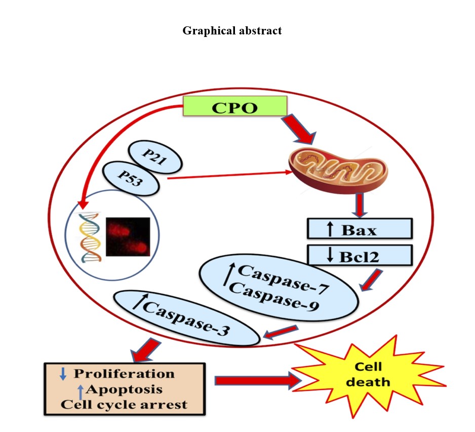
Evaluation of DNA damage was performed using single-cell gel electrophoresis [19] on control and CPO-treated A549 cells. To achieve a density of 2.5 × 10⁴/ml, cells were combined in a 1:1 ratio with 1% low melting point agarose after being washed twice with phosphate buffered saline (PBS). The slides were then covered with a thin layer of 300 µl of cells in agarose. The slides were electrophoresed at 25 V for 30 min after being incubated for one hour in lysis buffer (60 mmol/l NaOH, 1 mol/l NaCl, 0.5% (w/v) N-lauryl sarcosine, pH 12.5) and another hour in DNA unwinding solution (40 mmol/l NaOH, 2 mmol/l EDTA, pH 13). Comets were stained with ethidium bromide and then examined with fluorescence inverted microscopy (Olympus CKX41) at a magnification of 40x while using a green filter (Excitation filter BP480-550C). The pictures were then taken with a C-mount camera (Optika pro5 CCD camera). CASP software was used to directly calculate the percentage of the tail moment, the amount of DNA in the tail, and the length of the tail when quantifying DNA damage in the obtained images.
Flow cytometric analysis of the cell cycle
A FACS Caliber Flow Cytometer (USA, CA, Sunnyvale, Becton Dickinson) that features a small, air-cooled, low-ion laser beam (488 nm) with 15 mW argon was used to conduct the flow cytometry. Cell cycle analysis was carried out as previously mentioned [20]. The cells were incubated in a solution (200 L) containing 200 g/ml of RNase A (Invitrogen Biotechnology, Carlsbad, CA, USA), 20 g/ml of propidium iodide (PI) (Sigma, St. Louis, MO, USA), and 0.1% Triton v/v in PBS after the sample was prepared. The samples were subjected to flow cytometric analysis following a 30-minute dark incubation period. A percentage of cells in the G2/M, G0/G1, or S phases is displayed as a result.
Apoptosis detection
With the aid of an Annexin V-FITC Apoptosis Detection Kit and a BD FACS Calibur Flow Cytometer (BD Biosciences, CA, USA), lung cancer cells' apoptosis was discovered (Biovision, USA). The emission wavelengths used to gather data were 530 nm for fluorescein isothiocyanate (FITC) and 670 nm for PI. The argon laser's excitation wavelength was 488 nm. Glutathione peroxidase (GPx) and glutathione (GSH) were measured using ELISA analysis, as well as 4-hydroxynonenal (4-HNE) and proliferation markers KI67 and PCNA. GSH, GPx, Ki-67, and PCNA were quantified using a quantitative sandwich immunoassay. Using Biodiagnostic's kits, the concentrations of GSH and GPx activities were estimated (Giza, Egypt). Ki-67 and PCNA were measured using rat Ki-67 ELISA Kits from Biorbyt in the United Kingdom (cat. no. orb410642) and rat PCNA ELISA Kits from MyBioSource.com in California (cat. no. MBS2515480), respectively. According to the manufacturer's instructions (FineTest, Wuhan, China), lipid peroxidation was calculated by estimating the concentration of 4-HNE using a competitive inhibition enzyme immunoassay technique with a detection range of (31.25-2,000 pg/ml). ELISA analysis software was used to measure all markers.
Flowcytometric analysis of p53, p21, caspases (3, 7, and 9), Bax, and B-cl2
Santa Cruz Biotechnology provided the p53 (cat. no. sc-7480), p21 (cat. no. sc-6246), caspase-3 (cat. no. sc-271759), caspase-7 (cat. no. sc-56063), caspase-9 (cat. no. sc-56076), Bcl-2 (cat. no. sc-7382), and Bax (cat. no. sc-7480) (Santa Cruz, CA, USA). The right way to prepare cells was done so in accordance with the manufacturer's instructions. PBS/BSA buffer (1% BSA) was used to bring the cell suspension's concentration to 1×10⁶ cells/ml. Cell suspension was pipetted into test tubes. Fluorescein (FITC) conjugated antibodies that were appropriately labelled were added to the recommended dilution, thoroughly mixed, and incubated at room temperature for 30 min. After centrifuging the cells at 400 g for 5 minutes after being washed with 2 ml of PBS/BSA, the supernatant was discarded. Cells were suspended in 0.2 ml of either 0.5% paraformaldehyde in PBS/BSA or 0.2 ml of PBS/BSA. Data was collected using flow cytometry (Becton Dickinson, CA, USA). Cell Quest software (Accuri C6) was used to collect a total of 20,000 cells for analysis, and a histogram plot of conjugate fluorescence (x-axis) versus counts (y-axis) was made in logarithmic fluorescence intensity.
Statistical analysis
The one-way ANOVA POST HOC (Tukey's and Dunnett's) test was used in the statistical analysis, which was carried out using the GraphPad Prism software. Results were presented as the mean ± standard deviation (SD) from three independent experiments, and statistical significance was determined by P values of < 0.05 or < 0.001.