3.1 1H NMR analysis of the phenyl silicone oil standard
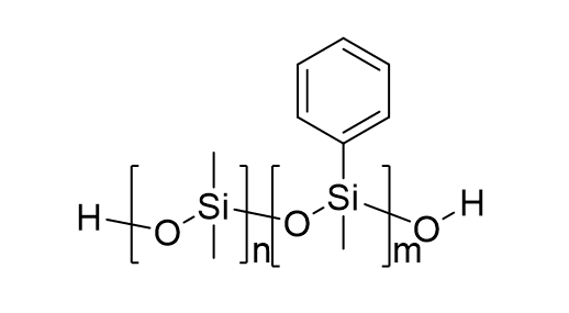
A silicone oil with high phenyl content was selected as the phenyl silicone oil standard, and its phenyl content was measured by 1H NMR. Figure 1 shows a 1H NMR spectrum of a CDCl3 solution of 38.1 mg phenyl silicone oil standard and 12.3 mg 1,4-dioxane. In the 1H NMR spectrum, two peaks at a low field of 7.2 ~ 7.6 ppm correspond to the protons on the phenyl ring, a signal at 3.64 ppm to methylene protons in the structure of 1,4-dioxane, and that at about 0.0 ppm to methyl proton in phenyl silicone oil. Thus, the phenyl content in the phenyl silicone oil standard was calculated to be 2.53 mmol/g, according to the peak area (5.22) of phenyl proton and that (16.11) of methylene proton.
3.2 Method development for the determination of the phenyl group
Toluene and THF (THF) are the common eluents for GPC analysis, and THF was selected as the eluent in this work due to its relatively low UV absorption. The UV absorption curve of the phenyl silicone oil standard solution was obtained in the wavelength range of 200–400 nm (the supplementary Fig. S1). As can be seen, a maximum absorption at 265 nm was observed, and thus the detection wavelength was set as 265 nm in the following experiments.
Under the optimized conditions described above, GPC-UV analysis was performed for a series of phenyl silicone oil standard solutions at the mass concentration of 5, 10, 50, 100, 500, and 1000 µg/mL (corresponding to phenyl concentrations of 0.972, 1.94, 9.72, 19.44, 97.2, 194.4 µg/mL, respectively). As shown in Fig. 2, a broad peak at tR 7.50 min, corresponding to the polymer of phenyl silicone oil, was observed in the GPC-UV chromatography.
A calibration curve was constructed by plotting peak area vs concentration (the supplementary Fig. S2). Good linearity was achieved in the range studied with the correlation coefficient (R2) at 0.9979. The limit of detection (LOD, S/N = 3) was 0.778 µg/mL, and the limit of quantification (LOQ, S/N = 10) was measured to be 0.972 µg/mL. Intraday reproducibility (RSD, n = 3) was determined to be 0.6%, and the interday reproducibility (RSD, n = 3) was 3.5% .
3.3 Evaluation of the anti-interference performance of the method
High selectivity is a key factor for a good analytical method. Potential interference factors for the determination of the phenyl group include the raw material (methylphenylcyclosiloxane), toluene solvent, and other types of silicone oil. Herein, the anti-interference performance of the established method on the presence of the above interfering substance.
Methylphenylcyclosiloxane is the raw material, and thus every phenyl silicone oil products contain some methylphenylcyclosiloxane, which can not be differentiated from phenyl silicone oil in 1H NMR analysis. To our interest, methylphenylcyclosiloxane is of small molecular weight, and can be easily separated from the polymer of phenyl silicone oil on a GPC column (Fig. 3). As shown in Fig. 3 and the supplementary Fig. S3, the peak at 7.52 min is attributed to phenyl silicone oil, and that at 10.52 min corresponds to methylphenylcyclosiloxane. The resolution (R) of the two peaks is calculated using the equation of \(\text{R}=\frac{2[{\text{t}}_{\text{R}\left(\text{B}\right)}-{\text{t}}_{\text{R}\left(\text{A}\right)}]}{1.699[{\text{W}}_{1/2\left(\text{B}\right)}+{\text{W}}_{1/2\left(\text{A}\right)}]}\), in which tR(A) and tR(B) stand for the retention time of phenyl silicone oil and methylphenylcyclosiloxane, and W1/2(A) (3.41 min) and W1/2(B) (2.00 min) refer to their peak width at half height. Thus, the calculated resolution is 1.52 for the two peaks, indicating a complete separation. By comparing the GPC-UV chromatogram of samples of phenyl silicone oil mixed with a different mass ratios of methylphenylcyclosiloxane in Fig. 3, the peak area of the signal at 10.51min rises significantly with increasing the ratio of methylphenylcyclosiloxane, while that of the signal at 7.52 min keeps almost constant with RSD at 1.8%. Therefore, the interference of methylphenylcyclosiloxane can be excluded due to the separation on a GPC column.
Toluene and xylene are common solvents for phenyl silicone oil products, which can not be differentiated from phenyl silicone oil in 1H NMR analysis, either. Similarly, toluene and xylene are also small molecular, and can be easily separated from the polymer of phenyl silicone oil on a GPC column (the supplementary Fig. S4 and Fig. S5). The R is 2.21 and 2.16, respectively, which indicated that the solvent peak can be completely separated from the peak of phenyl silicone oil. As can be seen, the peak area of the signal at 12.62 min rises with increasing the content of toluene, the peak areas of toluene as solvent and THF as solvent were 3411 and 3499, respectively, and the peak area of the signal at 12.52 min rises with increasing the content of xylene, the peak areas of xylene as solvent and THF as solvent were 3411 and 3446, respectively. Therefore, the interference of toluene and xylene can be excluded due to the separation on a GPC column.
However, various silicone oils, including methyl silicone oil, hydroxyl methylsilicone oil and vinyl silicone oil, are also polymers, which are co-eluted with phenylsilicon oil on a GPC column. Although these silicone oils show almost no absorption at 265 nm (the supplementary Fig. S6),, their co-elution with phenylsilicon oil results in a different matrix for the phenylsilicon oil, which might exert an influence on the UV absorption of phenylsilicon oil.
Herein, solutions of phenyl silicone oil mixed with other type of silicon oil, were analyzed by GPC-UV (Fig. 4, the supplementary Fig. S7 and Fig. S8), and the results were summarized in Table 1. Take Fig. 4 as an example, almost the same peak area (2709(Fig. 3-a), 2728 and 2792) was obtained for the analyte of phenyl silicone oil, that mixed with methylsilicon oil at the mass ratio of 1:1 and 1:10. The RSD of the peak area for the three samples is only 1.3%. Thereby, co-elution with methyl silicone oil does not interfere with the GPC-UV detection of phenyl silicone oil. Similar results were obtained for hydroxyl methylsilicone oil and vinyl silicone oil (Table 1).
Table 1
GPC-UV data for phenylsilicone oil mixed with various silicone oils
|
analyte
|
Retention time (min)
|
Peak Area
|
RSD
|
|
phenyl silicone oil
|
7.52
|
2709
|
1.3%
|
|
phenyl silicone oil + methyl silicone oil (m:m = 1:1)
|
7.54
|
2728
|
|
phenyl silicone oil + methyl silicone oil (m:m = 1:10)
|
7.52
|
2792
|
|
phenyl silicone oil
|
7.52
|
2709
|
0.8%
|
|
phenyl silicone oil + hydroxyl silicone oil (m:m = 1:1)
|
7.59
|
2711
|
|
phenyl silicone oil + hydroxyl silicone oil (m:m = 1:10)
|
7.61
|
2666
|
|
phenyl silicone oil
|
7.52
|
2709
|
2.7%
|
|
phenyl silicone oil + vinyl silicone oil (m:m = 1:1)
|
7.58
|
2717
|
|
phenyl silicone oil + vinyl silicone oil (m:m = 1:10)
|
7.61
|
2558
|
All in all, the interference of all potential substances, including the raw material (methylphenylcyclosiloxane), toluene solvent, and other types of silicone oil was excluded in the established GPC-UV method for the determination of the phenyl content in phenyl silicone oil. In other words, this analytical method shows good selectivity.
3.4 Determination of the phenyl content of the actual sample
The established GPC-UV method was applied for the determination of the phenyl content in several phenyl silicone oil samples (Table 2). As can be seen, the method had a good precision with the RSD (N = 3) between 0.95% and 4.48%, and a good recovery ranging from 84.6–101.7%. The phenyl content in various phenyl silicone oil samples varied distinctively, which ranged from 0.0–66.3%. Especially, no phenyl content was detected in several “phenylsilicone oils” from online shopping. In view of the importance of the phenyl content in phenylsilicone oil, it is essential to monitor its content in the relevant products.
Table 2
Phenyl content in phenyl silicone oil
|
sample
|
Rt−
(min)
|
Peak Area
|
Phenyl content
(mg/g)
|
Average spiked recovery (%)
|
RSD
(n = 3)
|
|
H-1
|
5.9
|
288.9
|
0.613
|
84.63%
|
0.95
|
|
H-2
|
5.6
|
145.6
|
0.423
|
96.51%
|
0.99
|
|
H-3
|
9.8
|
1581.8
|
66.3
|
86.16%
|
2.36
|
|
H-4
|
10.6
|
1288.6
|
53.7
|
101.74%
|
1.75
|
|
H-5
|
7.5
|
902.9
|
58.3
|
85.38%
|
4.48
|
|
H-6
|
10.3
|
8897.6
|
40.4
|
97.54%
|
0.72
|
|
H-7
|
10.2
|
10443.2
|
31.4
|
87.92%
|
0.42
|
|
Online shopping − 1
|
—
|
—
|
—
|
95.93%
|
—
|
|
Online shopping − 2
|
—
|
—
|
—
|
100.89%
|
—
|
| - different retention time is due to the different molecular weight of analyte |
At last, the reliability of the GPC-UV method was further investigated by comparing to the 1H NMR analysis. Take H1 as a model, the phenyl content was measured to be 0.603 mg/g by 1H NMR analysis (the supplementary Fig. S9), which is in good agreement with the GPC-UV’s result (0.613 mg/g) with a relative error of only 1.63%. Compared to the expensive price and maintenance for NMR, the GPC-UV method shows tremendous potential for determining the phenyl content in phenylsilicone oil with good sensitivity and selectivity.