Ethics statement
The animal experiment was approved by the Experimental Animals Ethics Committee of Huazhong Agricultural University (HZAUGO-2019-008). The care and maintenance of all animals were in accordance with the protocols and guidelines directed by the committee.
Parasite materials
Different developmental stages of H. contortus (Haecon-5 strain) were collected, processed and stored based on well-established protocols [21]. Specifically, infective L3 was collected from coproculture at 25°C [22], and maintained at 15°C. Exsheathed L3 (xL3) was obtained by incubation with 0.15% (v/v) sodium hypochlorite at 37°C for 10 min [23]. L4 and adult worm were identified and collected from the infected goat’s abomasa at days 7 and 28 post-infection, respectively [17].
Light microscope analysis of haemozoin-like pigements
The freshly collected parasitic L4 and adult H. contortus were thoroughly washed by sterile phosphate-buffered saline (PBS) solution. The samples were anesthetized by 1% levamisole and then observed the haemozoin-like pigement by a light microscope (Zeiss Primovert iLED, Gttingen, Germany). Besides, to identify the ferric iron in the haemozoin, these L4 and adult worm were stained by a ferric iron stain kit (Solarbio, Beijing, China) according to the manufacturer’s instructions. Briefly, these parasites were fixed with 4% paraformaldehyde for 24 h and stained for 24 h. After the worms were washed and dehydrated, they were carefully transferred to a glass slide and observed using a Zeiss light microscopy.
Transmission electron microscopy (TEM) analysis
To observe the haemozoin distribution in H. contortus, TEM analysis was performed as previously described [24]. In brief, freshly L4 and adult worm were fixed in 0.1 M cacodylate buffer (pH 7.4) with 2.5% glutaraldehyde and 4% formaldehyde. Fixed samples were dehydrated, embedded, and then stained with uranyl acetate and lead citrate. Final samples were observed by the TEM analysis (HITACHI H-7650, Tokyo, Japan).
Field emission scanning electron microscopy (FESEM) analysis
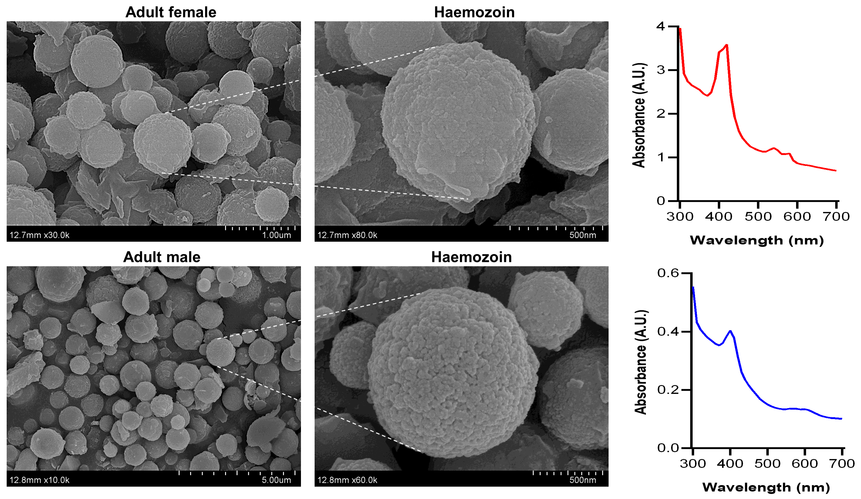
Haemozoin was extracted from adult H. contortus using a well-established method [25]. In brief, 100 adult worms were homogenized (30 min, 4°C) and ultrasonicated (5 min, 4°C). The homogenate was centrifuged at 12,000 g for 10 min. The collected supernatant was centrifuged at 12,000 g for 15 min. The pellet was washed and incubated with 1 mg/mL proteinase K (Yuanye, Shanghai, China) at 37°C overnight. After the sample was washed again and freezed drying, the samples were transferred into a silicon chip coated with gold (10 min), following by observing using a SU8010 FESEM (Techcomp, Shanghai, China) at an accelerating voltage of 5.0 kV.
Spectrophotometry analysis
The haemozoin was performed qualitative analysis as described previously [26]. Briefly, haemozoin extracts were dissolved in 0.1 M NaOH solution and the sample (200 µL) was added to the 96-well plates. The wavelength of haemozoin was determined at 300–700 nm or 400 nm using a multi-mode plate reader (BioTek Cytation 5, Winooski, Vermont, USA).
Protein and lipid extraction from adult worm
Protein and lipid samples were isolated using the well-established methods [27]. Briefly, female worms and male worms were respectively homogenized in PBS buffer (pH 7.4) with 1 M proteinase K inhibitor. The homogenate was centrifuged (10,000 g for 10 min) and the supernatant was collected. Protein concentration was assessed by a bicinchoninic acid assay (BCA) kit (Beyotime Biotechnology, Shanghai, China).
For lipid extraction, 1 mL of H. contortus homogenates (female or male) was fully mixed in an extract liquor with 4 mL of chloroform and 2 mL of methanol. The sample was vigorously shaken (5 min) and subsequently centrifuged (1000 g for 10 min). The upper aqueous phase was discarded and the lower organic phase was dried in the N2 stream, and the lipids extracted was gravimetrically determined by an analytical balance. The dried lipids were solubilized in 100% chloroform and stored at − 80°C until used.
Haem aggregation assay
To determine whether proteins and lipids promote the formation of haemozoin, the haem aggregation assay was carried out [27]. In brief, 100 mM of bovine derived haem (Beyotime, Shanghai, China) was dissolved in 0.5 M sodium acetate (pH 4.8) with 20 mg of protein and 80 µg of lipid, respectively. The reaction was incubated at 37°C for 24 h, prior to centrifuge at 12,000 g for 10 min. The final pellet was solubilized in 0.1 M NaOH buffer, and the wavelength of haemozoin was determined at 400 nm based on the above spectrophotometry analysis.
L4 culture in vitro
L4 were cultured in vitro based on a previous report with some modifications [21]. 100 L3 were cultured in 24-well plants with Luria Bertani and NCTC-109 medium, then supplemented with 20% fetal bovial serum (FBS) and 100 IU/mL of penicillin, 100 µg/mL of streptomycin and 25 µg/mL of amphotericin (Antibiotic-Antimycotic, Sigma-Aldrich, St. Louis, Missouri, USA). To promote xL3 into L4, the larvae were incubated in a 20% CO2 incubator at 39 ℃ for 7 days. Subsequently, the medium was added the goat 12.5% deflbrinated blood (DFB).
At 48 h of cocultivation, the haemozoin-like pigments were observed in L4 cultured with/without DFB and spectrophotometrically determined at 300–700 nm. Also, the wavelength of haemozoin was determined at 400 nm after 24 h, 48 h and 72 h of cocultivation. To determine the effect of red blood cell (RBC) concentration, we isolated the RBC from the goat's FBS. After adding the 107, 108 and 109 RBC, the wavelength of haemozoin was determined spectrophotometrically at 72 h.
Quinolone-drugs inhibition analysis
To assess the inhibition of quinolone-drugs, the different concentration (0 nM, 1 nM, 10 nM, 100 nM) mefloquine (Macklin, M845501-250mg, Shanghai, China) and quinine (Macklin, Q817159-100g, Shanghai, China) were added to the L4 (n = 100) cultured medium containing 12.5% DFB. The wavelength of haemozoin was spectrophotometrically determined at 72 h of cocultivation.
Statistical analysis
Statistical analysis was conducted using GraphPad Prism 8.0 software (GraphPad Software, San Diego, CA, USA). One-way ANOVA was applied to compare the statistical differences between groups. The * p < 0.033, ** p < 0.002, and *** p < 0.001 indicate the degree of significance.