Patient specimens
The patient information has been described previously [23]. In brief, tissues and adjacent nontumorous tissues were collected following a protocol from 75 CRC patients who underwent surgery at Ruijin Hospital from 2010 to 2011. We recorded clinical parameters, pathological data, overall survival (OS), and disease-free survival (DFS).
Cell Lines, Bevacizumab And Chemical
Human CRC cell lines RKO and HT29 cells were purchased from American Type Culture Collection (ATCC). HUVECs were purchased from Shanghai Institutes for Biological Sciences, Chinese Academy of Sciences. All cells were cultured in RPMI 1640 with 10% fetal bovine serum (FBS) and antibiotics at 37 °C with 5% CO2. Bevacizumab (a humanized monoclonal antibody that specifically binds to all VEGFA isoforms with high affinity) was purchased from MedChemExpress (Cat. No. HY-P9906). Anti-CCL2 (clone 2H5) antibody, an IgG monoclonal antibody neutralizing the bioactivity of human natural or recombinant CCL2, was purchased from BioLegend (Cat. No. 505913).
RNA Extraction And Quantitative RT-PCR
Total RNA was extracted by TRIzol (Invitrogen, USA) according to the manufacturer’s instructions. cDNA was synthesized using a reverse transcription kit (Invitrogen, CA). Quantitative PCR was performed using TaqMan® Gene Expression Assays (Thermo Fisher Scientific). The premiers were listed as follows: CXCL11: (forward) 5’-GACGCTGTCTTTGCATAGGC-3’, (reverse) 5’-GGATTTAGGCATCGTTGTCCTTT-3’; CXCL5, (forward) 5’-GAGAGCTGCGTTGCGTTTGTTTAC-3’, (reverse) 5′-CCGTTCTTCAGGGAGGCTACCACT-3’; CCL2, (forward) 5’-CAGCCAGATGCAATCAATGCC-3’, (reverse) 5’- TGGAATCCTGAACCCACTTCT − 3’; CCL13, (forward) 5’- TGCTGACCCAAAGGAGAAG-3’, (reverse) 5’- GCCAGAGGAGAATGGAAAAG-3’.
Western blot analysis
Western blotting was performed as previously described [23]. Briefly, cells were harvested at various time intervals and lysed in RIPA buffer in the presence of a Protease Inhibitor Cocktail (Pierce, USA) and a Protein Phosphatase Inhibitor Cocktail (New Cell & Molecular Biotech, China). One hundred micrograms of protein was separated by 10% SDS-PAGE gel and transferred to PVDF membranes (Tanon, China). The membranes were blocked with 5% bovine serum albumin (BSA) for 2 h and then incubated overnight with primary antibodies at 4 °C. The blots were probed with anti-pP38 (4511S, 1:1000, Boston, USA), anti-P38 (9212S, 1:1000, Boston, USA), anti-AKT (4691S, 1:1000, Boston, USA), anti-pAKT (4060S, 1:1000, Boston, USA), and anti-GAPDH (ab8245, 1:10000, Cambridge, UK); GAPDH was used as the internal control. Goat anti-mouse or goat anti-rabbit horseradish-peroxidase-conjugated IgG was used as the secondary antibody (Santa Cruz Biotechnology). The membranes were incubated with secondary antibody for 2 h at room temperature, and bands were visualized using an enhanced chemiluminescence detection system (Amersham Bioscience, Piscataway, NJ, USA) according to the manufacturer’s protocol.
Generation Of Gene Overexpressing And Knocked Down Cells
Lentiviruses for ETV5 and VEGFA overexpression and knockdown were purchased from Shanghai Bioegene Co., Ltd. (Shanghai, China). Lentiviral particles were transduced into CRC cells according to the manufacturer's instructions, followed by stable selection. Lentivirus transfection was performed as described previously [19]. Antibiotic puromycin was used to select stably transfected cells. The effects of overexpression and knockdown were evaluated by western blotting.
Cell Viability And Colony Formation Assays
Cell viability and colony formation assays were performed as described previously [19]. Approximately 3000 cells were plated in 96-well plates and cultured in a 37 °C/5% CO2 incubator. Cell viability was detected in a time series with CCK-8 (Dojindo Molecular Technologies Inc.). For the colony formation assay, 1,000 cells were plated per well in 6-well plates and cultured at 37 °C for 2 weeks. Colony formation was calculated by staining with 0.1% crystal violet in methanol for 30 min.
Endothelial Tube Formation Analysis
Human umbilical vein endothelial cells (HUVECs) were treated with supernatants of CRC cell lines in 96-well plates at a density of 1 × 10⁴ cells per well at 37 °C. Each plate was precoated with 50 µL Matrigel (BD Bioscience) at 37 °C for 30 min. After a 6-h incubation, tubules were observed by microscopy and analyzed using Image-Pro Plus software.
Chick Embryo Chorioallantoic Membrane (CAM) Assay
The CAM assay was performed as previously described [3]. Filter paper was placed on eggs, with a round window cut in the egg shell in advance, and then 30 µL of the cell culture supernatant was dropped onto the filter paper tray, which was sealed with a transparent tape for 3 days. On the 10th day, the eggs were imaged using a MacroPATH dissecting microscope (Milestone, Italy), and the number of blood vessels around the filter paper tray was counted.
In vivo PC xenograft tumor model
The CRC xenograft mouse model was performed as previously described, and all mice were euthanized one month after injection [23]. Tumors were weighed and fixed in formalin. All animal studies were approved by the Ruijin Hospital's Ethics Committee, Shanghai Jiao Tong University School of Medicine.
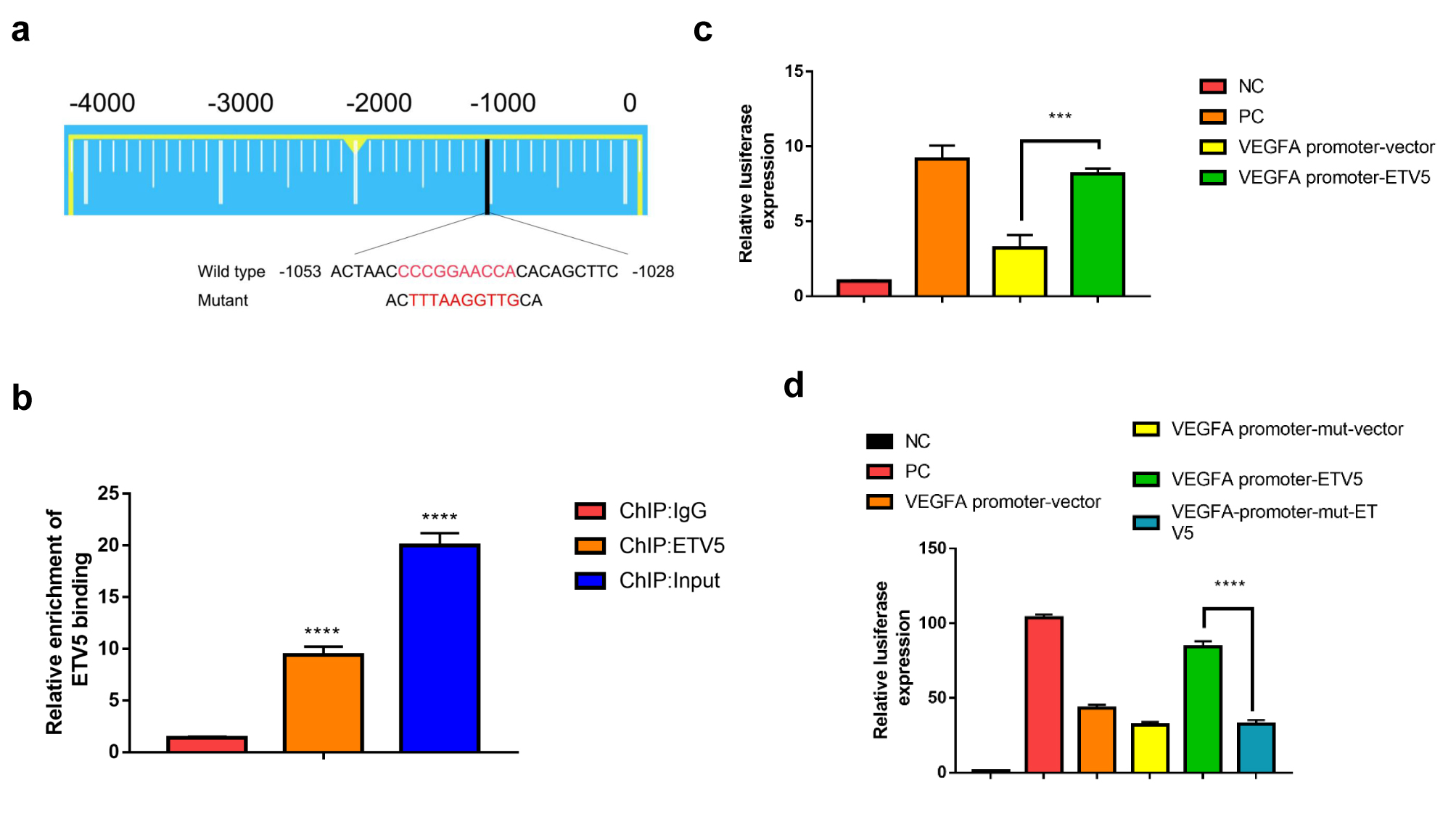
Chromatin Immunoprecipitation (ChIP)
ChIP assays were performed following the chromatin immunoprecipitation kit (Millipore) protocol, as previously described [19]. Briefly, an anti-ETV5 antibody (Santa Cruz Biotechnology, SC-22807) was used for immunoprecipitation. After purification of the precipitated DNA, the human VEGFA promoter was amplified by qRT-PCR. The premiers used to amplify the two site of VEGFA promoters were listed as: forward, 5’-TAGTGCTGGCGGGTAGGTTT-3’; reverse, 5’-CCAAGTTTGTGGAGCTGAGAA-3’.
ELISA
ELISA was performed according to previously described methods [24, 25]. In brief, CRC cell lines were seeded in six-well plates and incubated for several days. The VEGFA and CCL2 expression in supernatant was detected by using the VEGFA Human Biotrak ELISA system (Amersham Biosciences Corp., Piscataway, NJ) and Human MCP1 (CCL2) ELISA kit (ab179886, Abcam, Cambridge, UK).
Luciferase Report Assay
ETV5 promoter fragments were amplified from human genomic DNA and cloned into the pGL3-Basic vector. Luciferase activity was examined using the Dual-Luciferase Assay (Promega) following the manufacturer’s instructions.
Immunohistochemistry Assay
FFPE slides with CRC specimens and nude mice tumor tissue sections were stained as previously described [3, 19]. Antibodies used for IHC included antibodies against ETV5 (ab102010, Abcam), VEGFA (ab46154, Abcam), CD31 (3528, Cell Signaling Technology), Ki67 (1:200, Santa Cruz), and CCL2 (ab9669).
GSEA Analysis
The transcription data of raw count of 635 CRCs were downloaded from TCGA (The Cancer Genome Atlas) database (https://portal.gdc.cancer.gov/). Then, the transcripts per million (TPM) of every gene was calculated and normalized by Log2(TPM + 1). According to the median of ETV5 gene expression value, we divided all CRCs into High (n = 212), Moderate (n = 211) and Low (n = 212) groups. By Gene Set Enrichment Analysis (GSEA) software (https://www.gsea-msigdb.org/gsea/index.jsp), the enriched KEGG pathways were analyzed between High and Low groups.
Statistics
The genes significantly enriched in Chemokine Signaling Pathway were displayed by heatmap using “pheatmap” package in R software. The gene expression value of CXCL11, CXCL5, CCL2 and CCL13 were extracted from our previous RNA-Seq data of HT29/Vector and HT29/shETV5 cells, which had been deposited in GEO database (GSE112628). Data are expressed as the means ± SD. Analysis of variance (ANOVA) and Student's t-test were employed for comparisons among groups. The Mann-Whitney U-test was applied for the tumor volume comparison. Categorical data were evaluated with the chi-square test or Fisher's exact test. ROC curve was plotted to determine the cutoff values for ETV5, VEGFA and CCL2 expression. Log-rank test was performed to compare the survival curves of two or more groups. P-values less than 0.05 were considered significant. Statistical analyses were processed using