Biofilm conditions promote strong production of pulcherrimin, an iron chelator in B. subtilis.
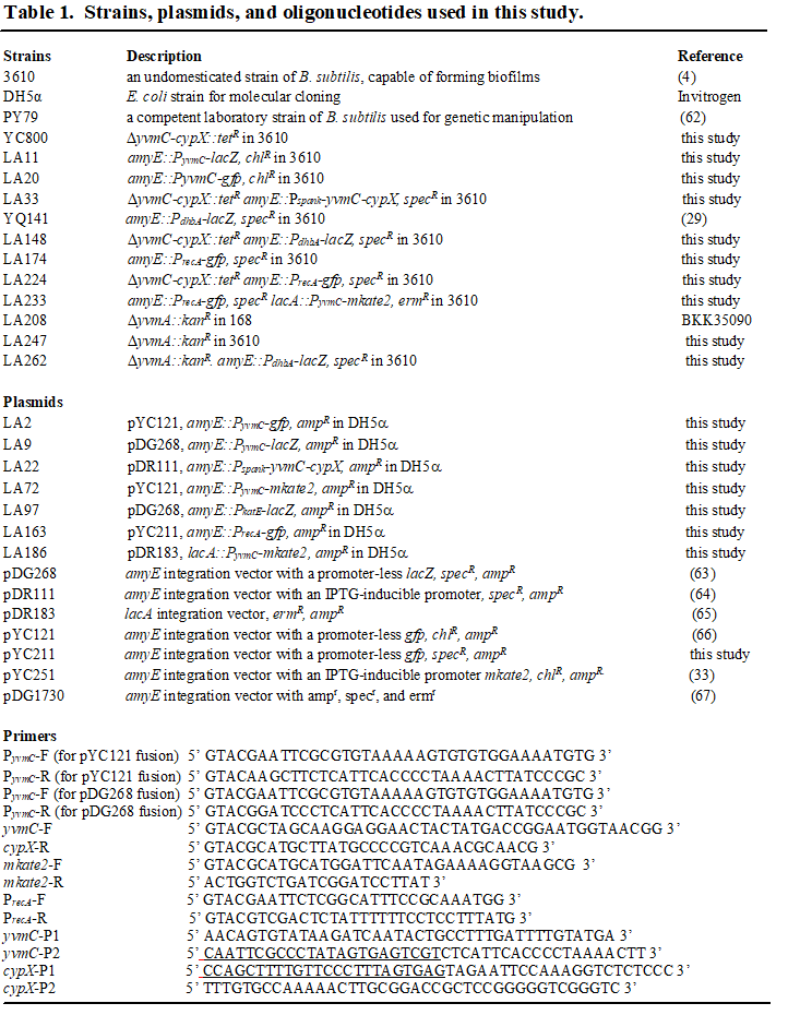
To study the red-colored, iron-binding pigment pulcherrimin, we first constructed a mutant strain in B. subtilis (ΔyvmC-cypX, YC800) that abolished the production of this iron chelator, as well as a complementation strain (LA33, yvmC-cypX under the control of an IPTG-inducible promoter was integrated at the amyE locus in YC800). We plated colonies of the mutant, the wild type strain NCIB3610 (abbreviated as wild type hereafter), and the complementation strain on the iron-supplemented biofilm-inducing media LBGM (LBGM + 0.2 mM FeCl3). After incubation for 48 hours, a reddish halo surrounding the colony biofilms was clearly observed in both the wild type and the complementation strain, but was completely absent in the mutant (Fig. 1A). This suggests that the main component of the reddish pigment is pulcherrimin. Overproduction or loss of pulcherrimin does not seem to impact growth of B. subtilis cells since no difference in growth was observed among the wild type, the pulcherrimin mutant, and the complementation strain (Supplemental Fig. 1).
Pulcherrimin production seems to increase over time during biofilm development (Fig. 1A). To learn about when and how the pulcherrimin biosynthetic operon (yvmC-cypX) is activated during biofilm development, we constructed two transcriptional reporters by fusing the promoter of the yvmC gene to the green fluorescent protein gene (gfp)(LA20), and to the β-galactosidase gene lacZ (LA11), and introduced the two reporter fusions into the wild type strain, respectively. Biofilm pellicles of the reporter strain bearing PyvmC-gfp were developed in LBGM, harvested every day over the course of 4 days, and imaged using fluorescence microscopy. The results show that there was a modest promoter activation in cells from the 24h pellicles, while the peak activation was observed at 48h. Cells from the 72h and 96h pellicles also displayed moderated fluorescence compared to the peak activation at 48h (Fig. 1B). In addition, population heterogeneity was observed for the fluorescent reporter activation, with a subpopulation of cells displaying strong fluorescence while others displayed modest fluorescence (48h, Fig. 1B). A similar expression profile, including peak activation, was observed when the PyvmC-lacZ reporter strain (LA11) was used and β-galactosidase assays conducted for pellicle samples similarly collected (Fig. 1C).
Synthesis of pulcherriminic acid remains constitutive upon changing iron levels.
Even though pulcherrimin is an iron-binding molecule, the pulcherrimin biosynthetic operon, yvmC-cypX, is not known to be regulated by Fur, a master regulator for iron homeostasis and biosynthesis of other iron-binding molecules such as bacillibactin in B. subtilis (12). Interestingly, however, when we grew wild type cells in LBGM, and LBGM supplemented with 0.05 mM or 0.2 mM FeCl3, we observed higher pigment yield when higher amounts of FeCl3 were added to the media (Fig. 2A). FeCl3 supplementation alone did not lead to a difference in the color of the media (data not shown). We spun down an equal number of cells from the above cultures and cell pellets showed strong color differences (note that pulcherrimin is largely insoluble and can be spun down by centrifugation)(Wild type, Fig. 2B)(41–43). Very little color difference was observed when the pulcherrimin mutant was used (ΔyvmC-cypX, Fig. 2B). This indicates that the color difference was due to different amounts of pulcherrimin present. These results suggest that the yield of pulcherrimin is significantly influenced by iron levels even though its biosynthetic genes are not regulated by the iron-responsive regulator Fur.
We then tested if iron concentrations in the media regulate the activity of the pulcherrimin biosynthetic genes. We cultured the PyvmC-lacZ reporter strain under biofilm conditions (48h development) as well as in shaking (24h growth), in LBGM and LBGM supplemented with different amounts of FeCl3. The results show that the activities of the pulcherrimin biosynthetic genes did not respond to varying concentrations of ferric iron in the media in both biofilm pellicles and shaking cultures (Fig. 2C-D). Thus, iron concentrations in the media influence pulcherrimin yield without regulating the expression of its biosynthetic genes (e.g. regulating secretion of pulcherriminic acids). Another possibility, which we favor, is that the colorless pulcherriminic acid molecules (iron-less) are produced and secreted in large quantities and are increasingly converted to pulcherrimin (iron bound) in the media in the presence of increasing amounts of ferric iron. The reason why we believe pulcherriminic acids are produced in large quantities is because the conversion to pulcherrimin was not saturated until the supplemented FeCl3 reached the millimolar level (mM)(data not shown).
Global transcription profiling suggests that pulcherrimin regulates genes involved in iron homeostasis and DNA damage response
To have a better understanding of transcriptional regulation, we characterized the global transcriptome of the pulcherrimin mutant compared to the wild type under biofilm conditions. Pellicle biofilms of the wild type and the mutant were grown in LBGM. Pellicles were collected after 72 hours. Global transcription profiling was performed by using RNA-Seq. Principal Component Analysis (PCA) suggested that in general, the wild type replicates clustered separately from the pulcherrimin mutant replicates (Supplemental Fig. 2). We applied a cut-off of log2 fold change (log2FC) of +/-1 for significantly up- and down-regulated genes, respectively. A volcano plot was generated where we could observe a total of 4,237 genes retrieved from the RNA-Seq analysis, 513 of which significantly downregulated (Table S1), and 179 upregulated (Table S2) in the pulcherrimin mutant (Fig. 3A). Both sets of genes were categorized according to known or predicted function, and the results were summarized in Fig. 3B. In the pulcherrimin mutant, we could observe a much higher number of upregulated genes related to general stress response (“Coping with Stress”, 28 genes), as well as DNA damage response (“DNA repair”, 10 genes). On the other hand, a significant number of genes related to iron acquisition/homeostasis (15 genes) were downregulated in the pulcherrimin mutant. More strikingly, hundreds of sporulation genes were downregulated in the pulcherrimin mutant, implying that the timing of sporulation during biofilm development was negatively impacted in the pulcherrimin mutant since the gene deletion does not impact final sporulation efficiency (data not shown). In other words, pulcherrimin could function as a development signal to modulate the timing of sporulation during B. subtilis biofilm development.
Pulcherrimin is known to be an iron chelator. Previous studies have shown that secreted pulcherrimin caused localized iron depletion in the media (17, 44). Results from our transcriptome profiling show that many iron homeostasis genes were downregulated in the pulcherrimin mutant, including the entire bacillibactin biosynthetic operon (dhbABCEF), bacillibactin transport genes (feuABC, fhuBCG) and the bacillibactin esterase gene besA (Fig. 3C). Bacillibactin is the main iron-scavenging molecule in B. subtilis, and it possesses extremely high affinity to iron ions (16, 45). BesA is a bacillibactin esterase, which catalyzes the hydrolysis of iron from bacillibactin and its release intracellularly (46). Lastly, the operons feuABC and fhuBCG are responsible for uptake of bacillibactin bound to iron from the extracellular environment (47). A plausible explanation for the above results, which is also consistent with previously published studies, is that in the wild type mature biofilm (day 3 pellicle), cells experience iron limitation stress due to chelation of ferric iron by pulcherrimin in the media, thus increasing the expression of those iron scavenging pathway genes (17). This also supports the idea that pulcherrimin itself is not likely a siderophore used by B. subtilis for iron acquisition.
Among the significantly upregulated genes in the pulcherrimin mutant, we observed a meaningful number of DDR genes. Many of these genes are controlled by the DDR regulator LexA (recA, yhaO, lexA, dinB, yneA and uvrB, Fig. 3D), and are part of the DDR pathway that is upregulated when cells experience DNA damage (37). We previously showed that in the wild type B. subtilis biofilm, DDR genes were modestly activated due to accumulation of reactive oxygen species (ROS) derived from metabolism (31). This result indicates that in the absence of pulcherrimin production, more significantly elevated DNA damage occurs in those cells in the mature biofilm.
Pulcherrimin protects cells from DNA damage.
Our RNA-Seq results indicated that multiple DDR genes were upregulated in the pulcherrimin mutant. To further investigate this, we constructed a DDR reporter by fusing the promoter of recA, a well-known DDR gene, with gfp. The reporter fusion was introduced, by integration at the amyE locus, into the wild type (LA174) and the pulcherrimin mutant (LA224), respectively. Pellicle biofilms were developed in LBGM, collected every day over the course of 4 days, and imaged using a compound fluorescence microscope. The wild type reporter strain displayed increasing fluorescence over time (Fig. 4A), which was expected due to increasing accumulation of toxic molecules derived from metabolism that could damage DNA as the biofilm ages (31). Interestingly, the pulcherrimin mutant displayed much higher fluorescence compared to the wild type at each of the four time points that the samples were collected (Fig. 4B). We quantified the fluorescence of hundreds of individual cells from both the wild type and the pulcherrimin mutant collected at the 48h using the MicrobeJ (48). Figure 4C is the violin plot generated from the quantifications, where it shows that the values of fluorescence representing the activities of the DDR reporter PrecA-gfp are significantly higher in the pulcherrimin mutant (red) than in the wild type (blue). These results indicate that in the mature biofilm, cells of the pulcherrimin mutant likely undergo elevated stress that leads to more DNA damage compared to the wild type.
To further assess the implication of the above results, the wild type and the pulcherrimin mutant reporter strains were challenged with two different DNA-damaging agents. One of them was mitomycin C, an antibiotic known to cause DNA double stranded breaks and widely used to study the DDR in B. subtilis, and the other was hydrogen peroxide (H2O2), a well-known ROS that causes damage of several cellular structures including DNA (49, 50). The assay consisted of growing cells under shaking conditions in LB broth for 2 hours followed by mitomycin C or H2O2 challenge for one hour at the final concentration of 0.25 µg/mL and 100 µM, respectively. Controls corresponded to untreated samples. After treatment, cells were collected, washed twice with PBS, and imaged under a fluorescence microscope. The PrecA-gfp reporter in the pulcherrimin mutant was found to display substantially higher fluorescence upon both DNA-damaging treatments compared to the wild type (Fig. 5A-D). These results suggest that lack of pulcherrimin rendered the mutant much more sensitive to external DNA-damaging agents than the wild type. We thus conclude that pulcherrimin plays a role in protecting cells by lowering DNA damage caused by both internal and external stresses.
To test whether levels of DNA damage are physically elevated in the pulcherrimin mutant, we applied assays of spontaneous rifampicin resistance frequency as a measurement for DNA mutation rate and levels of DNA damage (51). To do so, we grew both the wild type and the pulcherrimin mutant for 24h in LBGM supplemented with 0.2 mM FeCl3. We chose to culture the cells in an iron-overloaded medium so they would be more prone to oxidative stress through the Fenton reaction and consequently more DNA damages if a protective mechanism is lacking. Cells of each strain were then plated onto LB agar supplemented with rifampicin (5 µg/mL) and incubated overnight. Spontaneous rifampicin-resistant mutants appeared on the plates of the wild type strain as expected. Interestingly, on the plates of the pulcherrimin mutant, the number of the spontaneous rifampicin-resistant colonies appeared at a much higher rate than the wild type (Fig. 4D). This result supports our hypothesis that in the wild type, pulcherrimin protects cells by physically lowering DNA damages.
To further test if there are differences in cell survival under severe DNA damage stress between the wild type and the pulcherrimin mutant, we grew cells overnight in LBGM supplemented with 0.2 mM FeCl3 (to ensure high pulcherrimin yield in the wild type; Fig. 2A). Each overnight culture was then split into two flasks. In one of the flasks, H2O2 was added at a final concentration of 10 mM for 10 minutes with shaking. The second flask, as a control, was not exposed to H2O2. Untreated and H2O2-treated cultures were both plated on LB agar plates and incubated overnight at 37°C. CFUs were counted the next day and the percent survival calculated for both strains. Percent survival of the pulcherrimin mutant after exposure to H2O2 was approximately 20 times lower compared to the wild type (Fig. 5E). This result supports the idea that pulcherrimin protects cells and allows better survival of the cells under severe DNA damage stress.
Pulcherrimin production and DNA damage appear to be anticorrelated in cells in the biofilm.
We constructed a dual reporter strain (LA233) carrying both the DDR reporter PrecA-gfp as a proxy for DNA damage, and the PyvmC-mKate2 reporter for pulcherrimin production. We cultured this dual reporter strain in LBGM for pellicle development, harvested the pellicles after 48 hours (mature biofilm), mildly sonicated the pellicles to disrupt the bundles and chains, and imaged the cells under the compound fluorescence microscope applying the corresponding wavelengths for each reporter (Fig. 6A). We then quantified the fluorescence for each reporter in individual cells and plotted the results in a dot plot graph where the DDR reporter’s green fluorescence is displayed in the y axis, and the pulcherrimin reporter’s red fluorescence is on the x axis (Fig. 6B). Each dot in the panel corresponds to a single cell bearing both reporters. The results depicted show a significant amount of overlap among cells that displayed moderate fluorescence for both reporters, suggesting that a large proportion of cells in a mature biofilm express both reporters at intermediate levels. However, it is important to note that cells displaying high green fluorescence for the DDR reporter had very low red fluorescence for the pulcherrimin reporter (cells pointed by purple-colored arrows in panels in Fig. 6B), and vice-versa (cells indicated by white-colored arrows in the overlay image in Fig. 6B). Thus, we observed that high pulcherrimin production anticorrelates with levels of DDR and probably of physical DNA damage. This, again, supports our hypothesis that pulcherrimin can protect cells from DNA damage.
Pulcherrimin lowers ROS accumulation.
Our results suggest that pulcherrimin provides protection against DNA damage (Figs. 3–4). We wanted to further elucidate what could be causing this effect. ROS such as H2O2, superoxide radicals, and hydroxyl radicals, are byproducts of cell metabolism and are known to cause DNA damage such as double stranded breaks (26, 52, 53). We previously showed that ROS increasingly accumulates during biofilm development and triggers DDR in cells in the B. subtilis biofilm (31). We thus hypothesized that pulcherrimin may protect cells against ROS and thus ROS-induced DNA damage. We decided to directly measure ROS levels in the wild type and the pulcherrimin mutant by utilizing a kit for total ROS measurements (Enzo Biosciences), which includes a cell-permeable and non-fluorescent dye that, once in contact with ROS (hydrogen peroxide or hydroxyl radicals), generates green fluorescence. Cells can then be imaged using a compound microscope with a standard green filter (490/525nm).
Cells of the wild type and the pulcherrimin mutant were grown overnight in LBGM supplemented with 0.2 mM FeCl3 to enhance pulcherrimin yield. Cells were then incubated with the dye for 30 minutes in the dark, followed by three washes with PBS to remove excess dye, and then imaged. Figure 7A corresponds to representative fluorescence microscopy images acquired for each sample, where the green fluorescence levels in the pulcherrimin mutant were clearly higher than in the wild type. Fluorescence of individual cells was also quantified using Image J and plotted in a violin plot, which exhibited a significant difference between the two strains (Fig. 7B). These results suggest that pulcherrimin is able to lower ROS accumulation and thus prevent damaging levels of oxidative stress to happen in the cells.
Pulcherrimin production could reduce iron toxicity by lowering iron levels.
The results above provide both direct and indirect evidence that pulcherrimin reduces the oxidative stress in the cells and protects them from DNA damage. Finally, we can speculate that the reason why pulcherrimin can provide protection is probably due to its ability to sequester iron and lower iron bioavailability both outside in the environment and inside the cells. Robust biofilm formation by B. subtilis demands excessive amounts of iron in the media for reasons that are still not well understood (29, 54). Iron overload is known to cause toxicity because it triggers the Fenton reaction (55). Thus, we expect that iron sequestration by pulcherrimin leads to reduced Fenton reactions, less accumulation of hydroxyl radicals, and consequently, less DNA damage. Our RNA-Seq results show that lack of pulcherrimin led to downregulation of iron homeostasis genes, suggesting that pulcherrmin produced by the wild type not only chelates extracellular iron, but also indirectly lowers intracellular iron levels.
To test the above hypothesis that by sequestering extracellular iron, pulcherrimin production can lower intracellular levels of iron, a transcriptional reporter with the promoter for the bacillibactin operon, whose expression highly anti-correlates with intracellular iron levels, fused to lacZ was constructed (PdhbA-lacZ), and introduced into the wild type (YQ141) and the pulcherrimin mutant (LA148), respectively. Pellicle biofilms of the two reporter strains were developed and harvested daily over the course of 4 days. β-Galactosidase assays were performed on collected samples and results shown in Fig. 8A. It is evident that the pulcherrimin mutant displayed much lower activities of the PdhbA-lacZ reporter when compared to the wild type at every time point measured. This result supports the hypothesis that pulcherrimin production in the wild type significantly lowered intracellular iron levels and thus triggered upregulation of iron acquisition through bacillibactin and possibly other siderophores as well. It also supports the previously proposed hypothesis that pulcherrimin production causes iron depletion in the surroundings so that B. subtilis needs to scavenge more iron from the extracellular environment (17).
To further confirm that the observed difference in the PdhbA-lacZ reporter activity between the wild type and the pulcherrimin mutant is caused by the formation of pulcherrimin extracellularly, and consequently iron depletion, we introduced the same reporter into the pulcherrimin transporter mutant, ΔyvmA (LA262). This yvmA gene mutation significantly reduces the secretion, but not biosynthesis, of pulcherriminic acid to the extracellular environment, as less pulcherrimin is observed in the media when compared to the wild type (17, 22). Pellicle biofilms were similarly collected, and β-galactosidase assays performed to compare the activities of the PdhbA-lacZ reporter in the wild type, the pulcherrimin biosynthetic mutant, and the pulcherrimin secretion mutant. The results appeared as we expected. Both mutants (ΔyvmC-cypX and ΔyvmA) showed a much lower promoter activation when compared to the wild type, with the pulcherrimin biosynthetic mutant (ΔyvmC-cypX) having the lowest promoter activation of all three (Fig. 8B). This confirms that the reason why the PdhbA-lacZ reporter behaves differently between the wild type and the pulcherrimin mutant is indeed because of the formation of pulcherrimin extracellularly.
Together, the results described above suggest that pulcherrimin production leads to extracellular iron depletion in wild type biofilms. The iron depletion likely also significantly lowers intracellular iron levels because the promoter for the bacillibactin operon is strongly upregulated in the wild type compared to the pulcherrimin mutant, indicating an increased need for iron acquisition when pulcherrimin is present. To this end, we have presented several lines of evidence to support our working model on how pulcherrimin can function as an antioxidant to protect cells from oxidative stress and DNA damage during B. subtilis biofilm development (Fig. 8C).