Materials and chemicals
A human SPOP ELISA kit was purchased from Shanghai Jianglai Bio-Technology Co., Ltd (Shanghai, China). Copper chloride (CuCl2), L-Aspartic acid (L-Asp), sodium hydroxide (NaOH), hydrochloric acid (HCl), hydrogen peroxide (H2O2), sodium tetrachloropalladate (II) (Na2PdCl4), chloroplatinic acid (H2PtCl6·6H2O) was obtained from Aladdin (Shanghai, China). Lysozyme was obtained from Solarbio Life Science (Beijing, China). Glutathione (GSH), Horseradish Peroxidase (HRP), Catalase (CAT) were purchased from Sigma-Aldrich (St Louis, USA). IOSE80 cell line was obtained from Shengzhen Huatuo Bio-Technology Co., Ltd (Shengzhen, China). The other reagents used in this experiment were of analytical grade and all of the water used in the tests was obtained from a Millipore Mill-Q purification system(>18.2 MΩ cm, USA).
Apparatus and characterization
In this experiment, we use the conventional three-electrode system to perform the electrode detection tests. A glassy carbon electrode (GCE, 4 mm in diameter) was used as the working electrode, whereas a platinum wire was used as the auxiliary electrode and a saturated calomel electrode (SCE) was used as the reference electrode. The measurement of amperometric i-t curves and the cyclic voltammetry(CV) tests were performed using a CHI660E electrochemical workstation(Shanghai Chenhua Apparatus Corporation, China). The morphologies of the Cu@L-Asp hybrid nanoflowers and Cu@L-Asp/Pd-Pt nanocomposites were analyzed via field emission scanning electron microscopy (SEM, SU8010, Japan). Energy dispersive X-ray spectroscopy (EDS) were carried out using Oxford X-max50 microscope (Oxford England). X-ray photoelectron spectroscopy (XPS) measurements were performed with a Thermo scientific ESCALB 250 Xi spectrometer (Thermoelectricity Instruments, USA). Fourier transform infrared (FT-IR) was tested using a Nicolet 6700 FT-IR spectrometer (Thermo Nicolet, USA).
Synthesis of the Cu@L-Asp hybrid nanoflowers
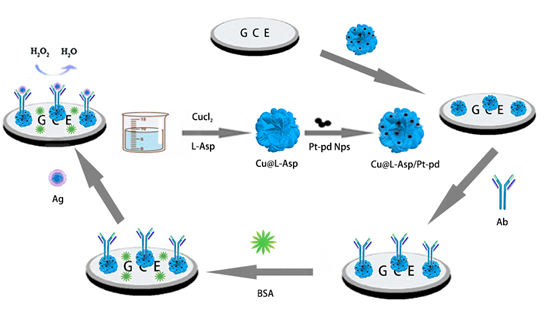
To synthesis three-dimensional jasmine-like Cu@L-Asp hybrid nanoflowers, 10mg CuCl2·2H2O and 15mg L-Asp were dispersed in phosphate buffered saline (PBS, 0.1M) with gently stirring overnight. Afterwards, the mixture was purified by centrifugation and washed with ultrapure water three times. Subsequently, the obtained solution was dispersed in ultrapure water and conserved in a refrigerator(4 °C) for further use. The preparation process of the three-dimensional jasmine-like Cu@L-Asp hybrid nanoflowers is demonstrated in Scheme 1.
Synthesis of Pd-PtNPs, PdNPs, PtNPs
To achieve signal amplification of the electrochemical immunosensor, Pd-PtNPs were used as a signal enhancer. Pd-PtNPs were synthesis according to the literature with slight modifications24. Briefly, 90 µl of H2PtCl6 (5%) and 119 µl of Na2PdCl4 (5%) was added to 10 mL of ultrapure water. Afterwards, the mixed solution was added with 10 mL of NaBH4 solution (0.28 mg/mL) and then gently stirred for 1h. Then, the complex solution was centrifuged at 12,000 rpm and washed with ultrapure water and ethanol three times, respectively. Finally, the mixture was dispersed in the 2 mL of ultrapure water for further use. Detailed synthesis methods of palladium and platinum nanoparticles will be provided in supplementary materials.
Preparation of Cu@L-Asp/Pd-Pt NPs, Cu@L-Asp/Pd NPs, Cu@L-Asp/Pt NPs
Since the prepared Cu @ L-Asp nanoflower has high specific surface area and rich amino group, it could be used as an excellent platform for in-situ assembly of Pt-Pd NPs. We added Cu@L-Asp and Pd-Pt NPs to ultrapure water in a ratio of 1:15 and stirred them continuously at room temperature for 12h. The resulting solution is then placed in a centrifuge at 3000 RPM for 5 minutes. Finally, the supernatant is sucked out with a pipette and washed with ultrapure water for three times. The synthetic methods of Cu@L-Asp/Pt NPs and Cu@L-Asp/Pd NPs is similar to that of Cu@L-Asp/Pd-Pt NPs, except that Pd-Pt NPs were displaced by Pd NPs and Pt NPs, respectively.
Cell thawing
First, the ultraviolet lamp was turned on to irradiate the sterile operating table for 30 minutes, and meanwhile the culture medium and PBS were placed in a 37℃ water bath box for the next experiment. Next, the sterile operating table was wiped with an alcohol cotton ball for disinfection, and then we prepared a cell culture medium containing 10% serum (10 mL serum +90 mL PBS) on the sterile operating table. After that, 2mL of the above culture medium was taken into a centrifuge tube for standby use. The frozen cells were then removed from the -80℃ refrigerator and quickly placed in a 37℃ water cup for melting and then centrifuged (800rpm, 4min). Finally, the well-prepared cells were transferred into a new cell culture bottle containing 3 mL culture medium and incubated overnight.
Cell subculture
The culture medium was poured out and rinsed with PBS for 3 times (3 mL PBS each time), then 1 mL trypsin was added, shaken from side to side for 15s, and the digestion was terminated with 2 mL culture medium. After that, the complex solution was centrifuged at 800 rpm for 4 min. Subsequently, the supernatant was discarded and transferred into the new culture bottle. Finally, they were put back to the incubator for the next round.
Cell lysis
The culture medium was poured out and then washed with PBS (pre-cooled) twice, and the adherent cells were scraped on the ice with cell scraper, then blown evenly with 1 mL PBS, and centrifuged at 1000rpm 4℃ for 5min. Subsequently, discard the supernatant and add 100ul RIPA and 1ul PMSF, then centrifuge at 12000rpm 4℃ for 20min. Finally, transfer the supernatant into a new EP tube for further use.
Fabrication of the electrochemical SPOP sensor
The fabrication process of the label-free immunosensor is shown in Scheme 1. First, we use the aluminum oxide powder with a diameter of 0.3μm to polished the surface of the electrode and each electrode lasted for 5 minutes. Then ultrasonic cleani ng was carried out on the polished electrode in the order of ultra-pure water, ethanol and ultra-pure water respectively, 5 minutes for each step. Finally, the electrode was polished again with aluminum oxide powder with a diameter of 50nm in accordance with the above steps. After the electrode was dried, 10μL of the Cu@L-Asp/Pd-Pt NPs nanocomposite solution was added to the surface of the pretreated clean electrode. After the electrodes have dried at room temperature, 6μL of anti-SPOP was dropped onto the electrodes and combined with Cu@L-Asp/Pd-Pt NPs by Pt-NH2 and Pd-NH2 bond and incubated for 4h at 37°C. In order to block the nonspecific sites, the electrodes were coated with a BSA solution(1%, w/v) at room temperature for 30min. Finally, the well-constructed electrodes were conserved in a refrigerator(4 °C) for further use.
Measurement procedure
Prior to the measurement, we dripped different concentrations of SPOP antigen onto the electrodes that had been constructed and incubated for 1h at 37 °C and after that, the ultrapure water was used to remove the unbound compounds. Then, the immunosensor were dried at room temperature before the following experiment. The i-t curve was carried out using -0.4V as the starting voltage, and 20 μL H2O2 was added to the PBS (0.1M,PH=7.4) after the background current was stable. The change of current was obtained according to the following formula: Δcurrent=current 1- current 0, where current 1 represents the current when different concentration of SPOP and 20 μL H2O2 was added, and current 0 is the background current.