Polylactic acid (PLA) is a biodegradable and biocompatible polymer commonly used in tissue engineering and bone regeneration 1. It is a promising material for these applications because it can be easily processed into various shapes and structures, such as scaffolds, films, and fibers 2–6. Additionally, PLA can be combined with other biomaterials, such as growth factors, to enhance its ability to support the growth and differentiation of cells. In tissue engineering and bone regeneration, PLA scaffolds are commonly used to provide a supportive structure for cells to attach and grow on 7. These scaffolds can be designed to mimic the extracellular matrix of the tissue or organ being regenerated, which helps to promote the growth and organization of the cells 8–10. Over time, the PLA scaffold will gradually degrade and replace the newly formed tissue or bone 11. One of the critical advantages of PLA is its biodegradability, which means that the body can break it down over time without causing any adverse effects. This makes it a potentially valuable material for medical implants that need to be gradually replaced as the body heals 11–13.
However, PLA is a relatively weak and brittle material 14, making it challenging to fabricate complex and highly porous scaffolds needed for some tissue engineering applications. Additionally, PLA may not be the most suitable material for applications that require high mechanical strength, such as load-bearing bone replacements. Another limitation of PLA is that its degradation rate can vary depending on the processing conditions and the presence of other materials, which makes it difficult to control the rate at which the scaffold degrades and is replaced by newly formed tissue or bone. In some cases, the scaffold may degrade too quickly, leading to a loss of mechanical support for the cells before the tissue or bone fully regenerates. In other cases, the scaffold may not degrade quickly enough, resulting in scar tissue formation and the eventual failure of the tissue-engineered construct. Mg, on the other hand, is a naturally occurring element that is an essential component of bone tissue. It has been shown to have several beneficial effects on bone cells, including promoting the growth and differentiation of osteoblasts (bone-forming cells) and inhibiting the activity of osteoclasts (cells that break down bone tissue) 15,16.
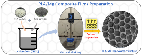
Polylactic acid (PLA) and magnesium (Mg) are biocompatible materials that have been studied for bone regeneration and other biomedical applications 17. In particular, their combination into a composite material has shown promise for improving PLA's mechanical properties and biocompatibility, making it more suitable for medical implants and other applications 18. Mg-doped PLA composites have been synthesized by blending Mg with PLA or by incorporating Mg into the polymer chain during the synthesis of PLA. The Mg content, particle size, and distribution of Mg within the composites can affect their properties 19. Mg-doped PLA composites have been shown to support the attachment, proliferation, and differentiation of various types of bone cells, such as osteoblasts and osteocytes, in in vitro studies 20. The Mg content of the composites can affect their biocompatibility, mechanical properties, and proliferation. Mg-doped PLA composites have also been demonstrated to stimulate the formation of new bone tissue in animal studies.
This research aims to develop a PLA/Mg composite printable for a direct ink 3D printing technique for biomedical applications. Direct ink printing is a 3D printing technique involving extruding material through a nozzle to build structures layer by layer 21,22. In this process, we produced some honeycomb structures with PLA/Mg composite that can have multiple applications. Finally, we successfully 3D printed the PLA/Mg composite ink using a direct ink printing technique into sample scaffolds.