Traditionally, open posterior lumbar interbody fusion (PLIF) and transforaminal lumbar interbody fusion (TLIF) have been established as the gold standard surgical treatments used in the management of degenerative lumbar spine conditions. However it comes with unavoidable complications such as damage to the posterior ligamentous complex and paraspinal muscles which are associated with the development of failed back syndrome.[6] In 2003, foley et al[7] developed the MIS-TLIF technique to overcome this problem using a 20-24 mm tubular retractor. However, with this technique it is difficult to observe the end plate during end plate preparation and it is relatively difficult to decompress the contralateral side. Lateral lumbar interbody fusion (LLIF) and Oblique lumbar interbody fusion (OLIF) have been developed to avoid injury to back muscles and improve visualization of the end plates [8], however these techniques are also associated with serious approach related complications such as potential injury to major vessels, ureter and lumbar plexus. [9-10]
Successful clinical results in a fusion surgery depends upon achieving a high fusion rate along with maintenance of disc height and sagittal balance. Failure to achieve bony fusion may result in implant loosening, breakage and back pain in the long term. Previously reported fusion rates for PLIF ranged from 56 to 100 %;[11,12] while fusion rate for open TLIF ranged from 86 to 100%.[13,14]. Despite the narrow operative field in MIS-TLIF; several scientific papers have demonstrated good fusion rates ranging from 92-100%.[15,16]
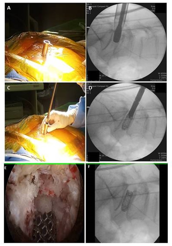
Successful fusion primarily depends upon the surface area of the fusion bed and the correct placement of the bone graft along with the interbody cage over fusion bed. The inaccurate placement of bone graft over unprepared end plates with intact cartilage often results in pseudarthrosis. Potter et al[13] have reported that in order to obtain a firm interbody fusion, exposure of more than 30% of the endplate is required. With the transforaminal approach 56% of endplate can be prepared. Whilst performing endplate preparation, it is important not to remove excessive amounts of subchondral bone. Overenthusiastic disc preparation can result into subsidence and recurrent foraminal stenosis; particularly in osteoporotic patients. As we used a 15 degree endoscope inside the intervertebral disc space which can be rotated 360 degrees, it allows access to 60-80% of the end plate.[17] We calculated the mean percentage of surface area of fusion mass to be 42.61% which is more than sufficient to achieve interbody fusion. We used 3D-printed porous titanium interbody cages which provide an osteo-conductive scaffold for new bone growth. The cages provide short term stability due to friction and long term stability due to osteoblast adhesion, proliferation and bone ingrowth. A single large cage improves the construct stability and bone ingrowth decreases subsidence due to load distribution. [18] Fusion rates can be further enhanced with the use of rh-BMP and various other osteoinductive materials.
In 2012, Osman et al[19] first reported the outcomes of 60 cases of endoscopic TLIF for the treatment of degenerative lumbar conditions with a 59.6% solid fusion rate with 36.2% of patients achieving a stable fixation. But the complication rate was upto 20%. Recently, Mongesterns[17] et al published the outcome in 30 patients operated with percutaneous TLIF with a mean follow-up period of 38 months. He reported a 100% fusion rate using rigid PEEK cages and an expandable titanium cage. Lee et al[20] performed percutaneous TLIF in 18 patients using titanium expandable spacers(B-Twin) under local anesthesia with conscious sedation. He achieved good clinical results with 88% fusion rates (16 out of 18 patients) with an average subsidence of 2.1 mm. Subsidence of the interbody cage with a loss of interbody disc height is expected in the post-operative period; however progressive subsidence can cause a reduction in foraminal height which results in recurrent foraminal stenosis and loss of sagittal balance also. In the present study we did not find any radiological signs of non-union, but we did experience a subsidence rate of 0.9 mm. There was a significant difference between pre-operative measurements and measurements at final follow-up(p < 0.01). Significant subsidence is defined as a decrease in disc height of more than 3mm. Although our subsidence rate was low; it does require close long term follow up.
Jacquet et al[21] considered the procedure controversial due to the technical difficulty and reported high complication rates (up to 36%). They also emphasized the need for technical improvements to the procedure. Most studies describe the utilization of the classical endoscopic transforaminal approach with expansile foraminoplasty for disc space preparation and cage insertion. Majority of these studies describe the development of the common postoperative complication of dysaesthesia due to irritation of DRG. This usually occurs whilst performing disc preparation and/or during the insertion of a large cage into the disc space. In our study we utilized a conventional Wiltse's approach which minimizes the need for retraction of the exiting root and DRG and thus we did not see this complication of post-operative dysaesthesia in our study population. However, we did have a complication rate of 3.6% secondary to dural tears in 2 patients which occurred over traversing root sleeve. These injuries occurred secondary to incomplete retraction of traversing root by the bevel tip. This complication can be minimized by carefully rotating the bevel tip and introducing it into the disc space, thereby safely retracting the traversing nerve root during disc space preparation and cage insertion.
Interbody fusion via the transforaminal approach has certain limitations such as inadequate foraminoplasty which could make cage insertion difficult, and also the iliac crest can act as an obstacle for the insertion of an interbody cage into the L5-S1 disc space. Such difficulties can often be overcome using the paraspinal approach. Despite the encouraging clinical results, endoscopic TLIF has certain limitations which needs further technical development; it is not suitable for grade 2 or more spondylolisthesis due to difficulty to achieve complete reduction and sagittal balance. In patients where the disc space is collapsed more than 50%, it is very difficult to introduce the endoscope into disc space. Osman et al[17] has advised prior pedicle screw instrumentation and distraction of the disc space prior to introduction of the endoscope to counter this problem. In our study we did not find a statistically significant difference in segmental balance from pre-operation to final follow-up. Although the technique of endoscopic TLIF is similar to open TLIF, its ability to restore segmental balance cannot be confirmed in the present study. Further technical developments in instrumentation may be required to achieve disc space distraction, and also in cage design perhaps with the use of expandable or hyperlordotic cages to help restore segmental balance. Lewandrowski et al[24] performed endoscopic TLIF with a standalone lordotic cage for a single level with good clinical results (77.8%). However, he reported cage subsidence in all the cases with revision surgery rate of around 8% (4 out of 48). Long term radiological studies with respect to subsidence rate, sagittal balance, effect at adjacent segment, fusion mass distribution and fusion rate will be helpful for future development of endoscopic instrumentation for fusion surgery. Certain studies have also proposed the feasibility of using biportal endoscopic surgery for interbody fusion with encouraging clinical results.[22,23].
This is a retrospective study with a small sample size providing preliminary results. We ideally need to compare an Endoscopic TLIF group with a matched control group like an open or MIS TLIF group in order to more accurately compare the effectiveness of Endoscopic TLIF compared with existing lumbar fusion techniques. A prospective study with large sample sizes will give more comprehensive results.