Incorporation of molecular scissors to capture intricate dynamic conformations on Parkin
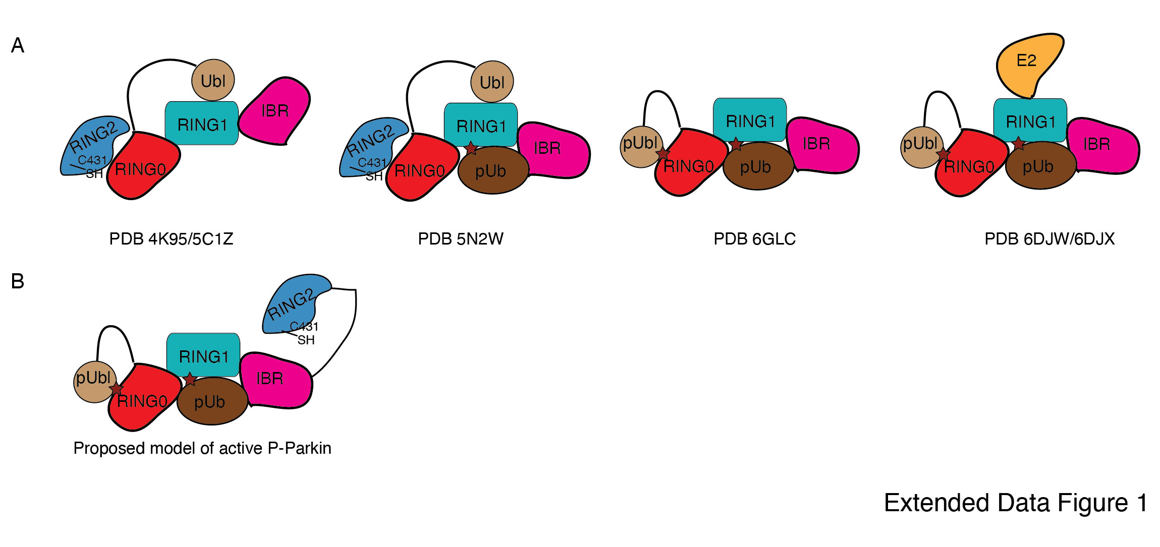
Previous studies using various biophysical methods showed that upon phosphorylation of the Ubl domain of Parkin, phospho-Ubl (pUbl) does not interact with the core of Parkin, lacking the Ubl domain 25–27. However, the crystal structure of phospho-Parkin missing the RING2 (1-382) showed pUbl domain bound to the basic patch (K161, R163, K211) on the RING0 domain 24, 29. RING2 shared a large surface with RING0, and the superimposition of phospho-Parkin (1-382, PDB 6GLC) and WT-Parkin (PDB 5C1Z) structures showed steric clashes between RING2, ACT, and pUbl (Fig. 1A). Therefore, we hypothesized whether the RING2 domain competes with the pUbl domain and thus blocks the interaction of pUbl with RING0. The latter hypothesis would also explain why previous attempts to study pUbl interactions show weak or no interactions between pUbl and Parkin in trans.
To capture crystal structures of protein-protein complexes, researchers use fusion constructs to allow the expression of two proteins in a single polypeptide chain. The fusion method increases the effective net concentration of two proteins in solution compared to mixing two proteins separately, thus stabilizing the interactions between two proteins. Earlier binding assays on Parkin failed to capture interactions in trans, and we speculated that this might be due to the lower net concentration of the domain in trans compared to the high net concentration of the fused domain. We hypothesized that untethering (cleavage of peptide bond) upon protease treatment would solve the above problem and enable us to capture the binding in trans using biophysical methods. To understand the above intricate mechanism, we introduced molecular scissors (human rhinovirus type 3C (HRV 3C) protease and tobacco etch virus (TEV) protease on Parkin constructs (Fig. 1B) to analyze the Ubl and RING2 domain rearrangements under native or phosphorylated states. We introduced HRV 3C (between 140th -141st residue) or TEV (382nd -383rd) sites in the loop regions of Parkin (Fig. 1B) to avoid any artifacts due to perturbations in native interactions on protein.
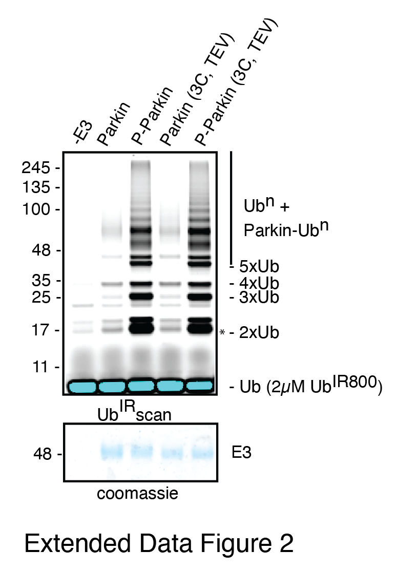
First, we tested the ubiquitination activity of Parkin (3C, TEV)to ensure that the inclusion of protease sites did not affect the protein folding or function, which is confirmed by the similar activity of Parkin (3C, TEV) as of the native Parkin construct (Extended Data Fig. 2). Furthermore, we noticed that Ubl-linker (1-140) co-elute with R0RBR (141-465) in native Parkin (3C, TEV) after treatment with 3C protease, suggesting a stronger interaction between Ubl and the Parkin core (Fig. 1C). However, in phosphorylated Parkin (3C, TEV) treated with 3C, pUbl-linker (1-140) did not form a complex with R0RBR (141-465), suggesting a poor/no interaction between phospho-Ubl with the core of Parkin (Fig. 1C). Furthermore, in native Parkin (3C, TEV) treated with TEV, RING2 (383-465) co-eluted with Parkin (1-382), suggesting a stronger interaction between RING2 and the Parkin core in the native Parkin (Fig. 1D). However, in phospho-Parkin (3C, TEV) treated with TEV, RING2 (383-465) eluted separately from the Parkin (1-382), suggesting that phosphorylation of the Ubl domain results in the displacement of the RING2 domain (Fig. 1D). All the above data confirmed that inclusion of molecular scissors on Parkin constructs do not affect Parkin folding. Previous observations that phosphorylation of Ubl weakens Ubl and Parkin interaction, and displacement of RING2 in phospho-Parkin, were validated using our assay. Also, respective proteases only cleaved (untethered) the peptide bond without affecting the native interactions between Parkin domains.
Phospho-Ubl domain and RING2 domain have a competitive mode of binding on RING0 domain
Previous models of Parkin activation suggested permanent displacement of RING2 after Ubl phosphorylation (Extended Data Fig. 1B). We wanted to test whether RING2 and pUbl affect the binding of each other on Parkin, which would suggest a competitive binding mode between pUbl and RING2 on the RING0 domain, and a dynamic displaced or bound states of pUbl and RING2. To test the competitive mode of binding between pUbl and RING2 on RING0, and thus affecting the binding of each other, we performed the SEC assay after sequential treatment with HRV 3C and TEV on Parkin (3C, TEV). Interestingly, pUbl-linker (1-140) co-eluted with Parkin core (141-382) upon 3C treatment on fractions that were collected after TEV treatment on phospho-Parkin (3C, TEV) which led to displacement of RING2 (383-465) (Fig. 2A). Similarly, RING2 (383-465) co-eluted with Parkin core (141-382) upon TEV treatment on fractions that were collected after 3C treatment on phospho-Parkin (3C, TEV) which led to displacement of pUbl-linker (1-140) (Fig. 2B). This data confirmed that pUbl and RING2 competitively bind on RING0. The binding of one negatively affected the binding of the other, unlike previous observations, which only showed phosphorylation of Ubl leading to RING2 displacement.
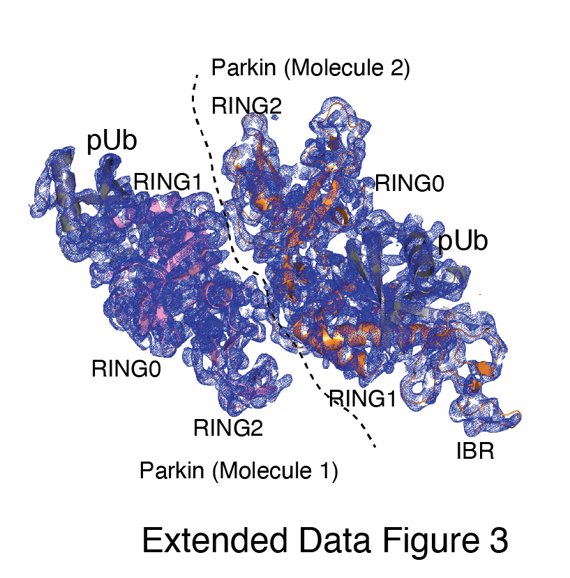
Our data in Fig. 2B suggested dynamic displacement of RING2 as untethering of RING2 after pUbl wash-off resulted in stabilization of interactions between RING2 and Parkin core. To further confirm, we crystallized the phospho-Parkin (3C, TEV)-pUb complex after treatment with 3C protease. Treatment with 3C led to displacement of the pUbl-linker (1-140) from the Parkin core (141-465). The overall structure of pUbl-linker (1-140) depleted Parkin (141-465)-pUb complex was determined at 3.3 Å (Extended Data Table 1), and showed similar conformation as seen in previously solved structures of Parkin in autoinhibited state (Fig. 2C). The crystal structure showed RING2 bound to RING0, which confirmed that RING2 was only transiently displaced from the RING0 domain in phospho-Parkin and returned to its original position after removal of pUbl-linker (Fig. 2C), further confirming our SEC data (Fig. 2B). The crystal structure also revealed that the REP element is bound to the RING1, similar to the autoinhibited state of Parkin (Fig. 2C). Phospho-ubiquitin was bound to the basic patch between RING0 and RING1, and resulted in similar conformational changes in IBR and helix (connecting RING1-IBR domains) (Fig. 2C). In the asymmetric unit, two molecules of Parkin bound to pUb were seen; however, in one of the Parkin molecules, no density was observed in the IBR region (Extended Data Fig. 3). Overall, this data suggested that pUbl and RING2 exist in a dynamic state in phospho-Parkin (pUbl binding<->RING2 open<->pUbl displaced<->RING2 closed), and compete for binding on RING0.
K211N mutation on Parkin perturbs RING2 displacement, not pUbl displacement
As phosphorylation of Ubl resulted in displacement of pUbl from Parkin core (Fig. 1C), we wondered whether interactions between pUbl and the basic patch (comprising K161, R163, and K211) on RING0 played a key role in pUbl displacement from RING1. Interestingly, similar to phospho-Parkin (3C, TEV) (Fig. 2C), pUbl-linker (1-140) remained flexible in phospho-Parkin K211N (3C, TEV) and eluted separately from Parkin core (141-465) on SEC (Fig. 3A). This data suggests that binding of pUbl with the basic patch on RING0 domain may not be the driving force for pUbl displacement. Further, to confirm that displacement of the RING2 domain is mediated by pUbl binding in the basic patch (K161, R163, and K211) on the RING0 domain, we tested the RING2 displacement using phospho-Parkin K211N (3C, TEV). N211 resulted in stabilization of the RING2 (383-465) domain on phospho-Parkin K211N (1-382) upon TEV treatment, and the two fragments co-eluted on SEC (Fig. 3A). Although pUbl was displaced in phospho-Parkin K211N, Parkin activity was drastically reduced (Fig. 3B), suggesting RING2 displacement, not Ubl displacement, is a major cause of Parkin activation. We also noticed a basal level of Parkin activity in the lanes without any activator (pUb), which was reduced in the Parkin K211N mutant (Fig. 3B). To understand the conformational changes upon mutation in the basic patch on RING0, we also crystallized phospho-Parkin R163D/K211N/Q347C (3C)-pUb complex after treatment with 3C protease, which washed off pUbl-linker (1-140) from Parkin core (141-465). This complex resulted in better crystals diffracting up to 2.35 Å. The overall structure of the pUbl-linker (1-140) depleted Parkin R163D/K211N/Q347C (141-465)-pUb complex (hereafter R0RBR R163D/K211N-pUb complex) was similar to the autoinhibited structure wherein RING2 was bound on RING0 and REP element was bound on RING1 (Fig. 3C).
Untethering of the linker connecting IBR and RING2 allows pUbl binding in trans
We next investigated whether the competitive binding between pUbl and RING2 to the RING0 could explain previous reports 19-30 observing the lack of interaction between pUbl and Parkin (lacking Ubl domain) in trans. To test this, we used phospho-Parkin K211N, which would not allow the binding of pUbl in the RING0 pocket of the same molecule, and tested its interaction with ∆Ubl-Parkin. However, no complex formation between phospho-Parkin K211N and ∆Ubl-Parkin was seen on SEC (Fig. 4A). We next validated this finding using isothermal titration calorimetry (ITC), which did not show any detectable interaction between phospho-Parkin K211N and ∆Ubl-Parkin (Fig. 4A), consistent with previously published reports.
As our data suggested that the fused domain outcompetes the untethered domain (Fig 2), we wondered whether this may explain the lack of detectable binding in trans. To test this, we used ∆Ubl-Parkin (TEV) treated with TEV as acceptor Parkin, which overcomes the problem of higher net concentration of the fused competing RING2 domain. Acceptor ∆Ubl-Parkin (TEV) was treated with TEV, and TEV was removed using an affinity column followed by SEC. SEC showed co-elution of ∆Ubl-Parkin (77-382) and RING2 (383-465), confirming that TEV cleaved (untethered) the peptide bond (connecting IBR and REP-RING2) without affecting the native interactions between ∆Ubl-Parkin (77-382) and RING2 (383-465) (Fig 4B). Incubation of phospho-Parkin K211N with untethered ∆Ubl-Parkin (TEV) led to the displacement of RING2 (383-465) from ∆Ubl-Parkin (77-382), and a stable trans complex between phospho-Parkin K211N and ∆Ubl-Parkin (77-382) by SEC analysis (Fig. 4B). The ITC showed a strong affinity (Kd = 1.1 ± 0.3 µM) between phospho-Parkin K211N and untethered ∆Ubl-Parkin (TEV) (Fig. 4B), which further supported the SEC data.
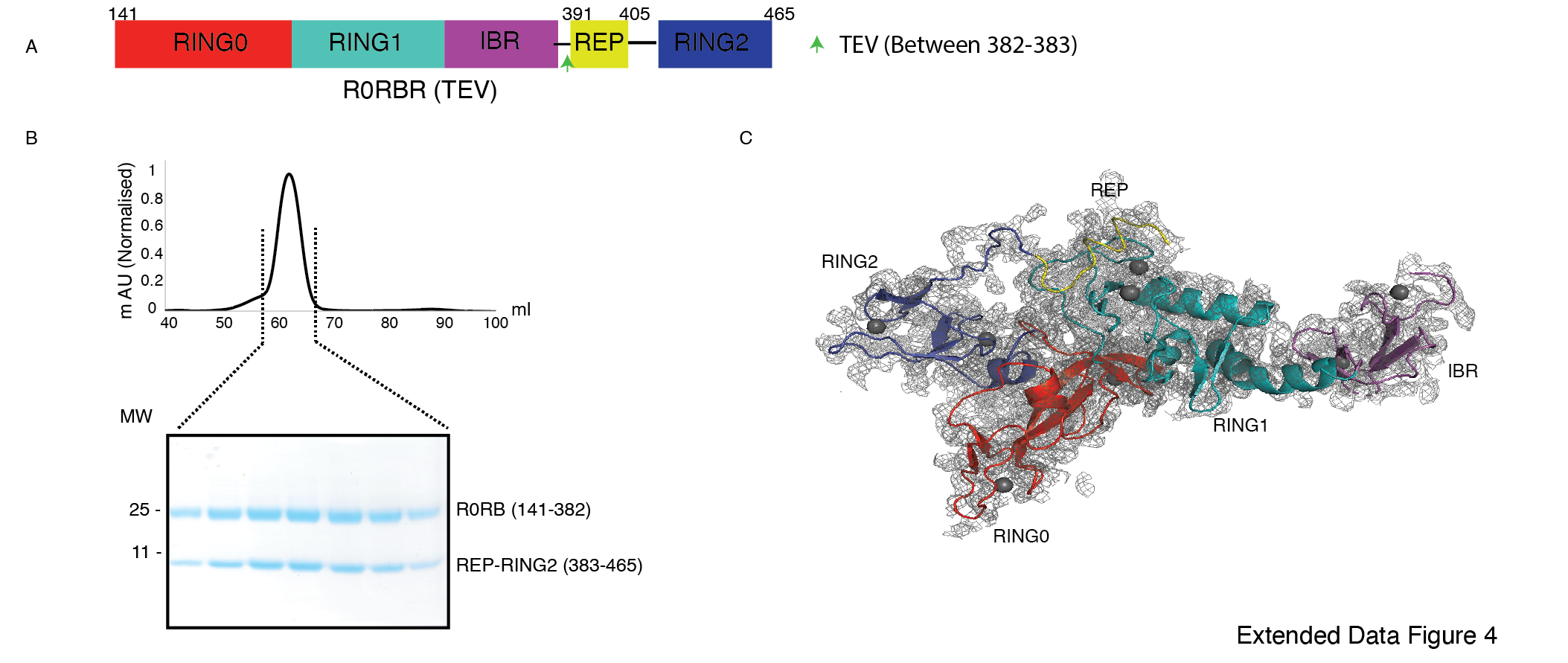
Further, to confirm that untethering does not affect the native interactions between RING2 and RING0 domains, we purified and determined the structure of untethered R0RBR (TEV) Parkin (Extended Data Fig. 4A). Co-elution of R0RB (141-382) and RING2 (383-465) fragments on SEC (Extended Data Fig. 4B) and crystal structure analysis showing intact native interactions between RING2 and RING0 (Extended Data Fig. 4C) excluded the possibility of artifact.
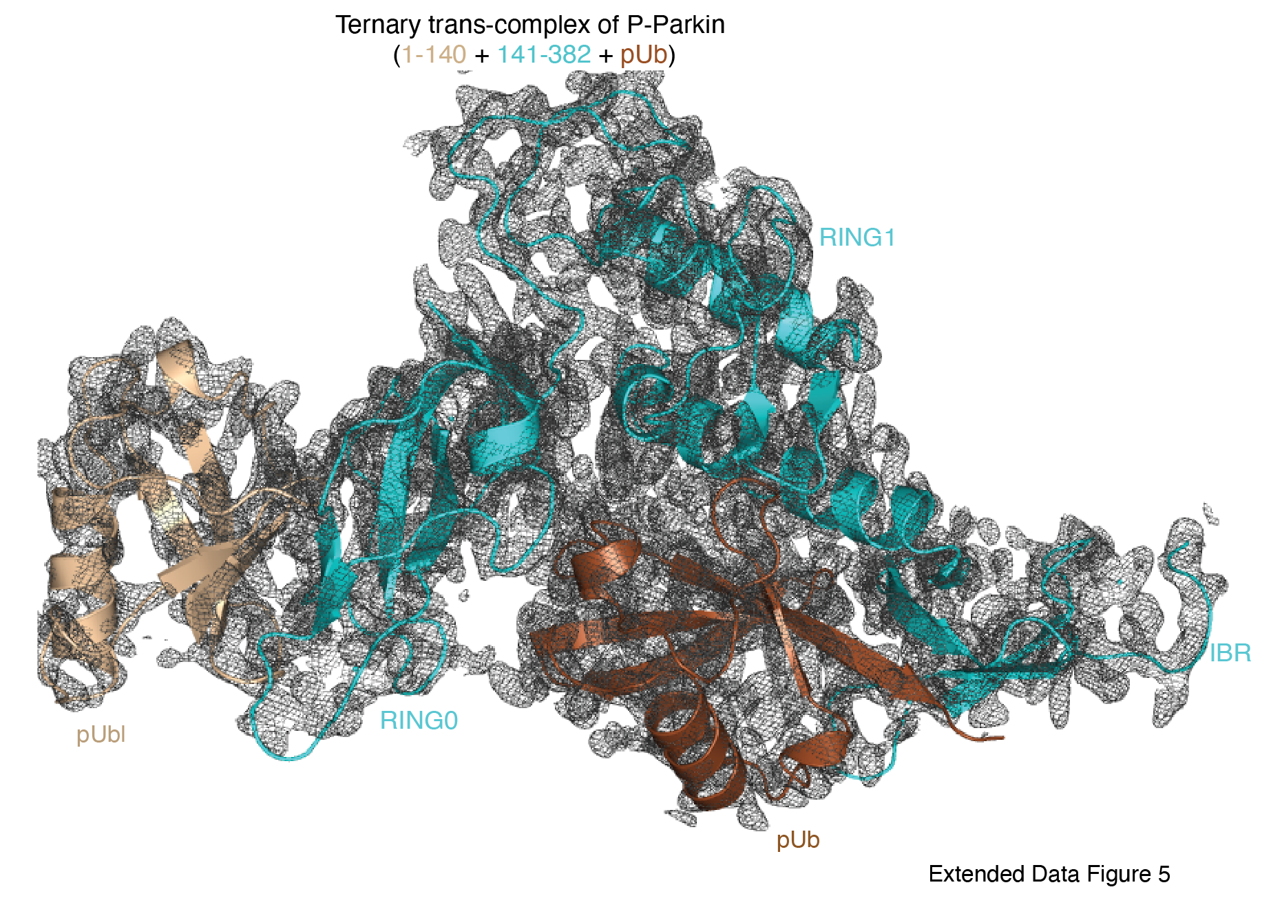
To understand the molecular details of the complex observed in Figure 4B, we used Parkin K211N (3C) as a donor of pUbl-linker (1-140) and R0RBR Q347C (TEV) Parkin as an acceptor of pUbl-linker (1-140). Phospho-Parkin K211N (3C) formed a stable complex with untethered R0RBR Q347C (TEV), and RING2 (383-465) was removed from R0RBR Q347C (TEV) (Fig. 4C). The fractions containing the complex of phospho-Parkin K211N and R0RB (141-382) upon treatment with 3C protease followed by incubation with pUb-3Br showed co-elution of components of the ternary trans-complex (R0RB (141-382), pUbl-linker (1-140), and pUb) on SEC (Fig. 4C). The crystal structure of the ternary trans-complex of phospho-Parkin (pUbl-linker (1-140) + R0RB (141-382) + pUb) was solved at 1.92 Å (Extended Data Table 1) which further confirmed trans-complex formation between Parkin molecules (Fig. 4D, Extended Data Fig. 5). In the crystal structure, the pUbl domain from the donor molecule (phospho-Parkin K211N (3C)) was bound to the basic patch of RING0 on the acceptor molecule (untethered R0RBR (TEV)) (Fig. 4D) in trans. The conformation observed in the trans complex was similar to phospho-Parkin (1-382) structure with fused pUbl domain and untethered/truncated RING2 in a cis molecule 24,29. Interestingly, the linker connecting pUbl and RING0 remained disordered in all the structures 24,29. Therefore, it would be difficult to say whether, in the previous cis structures, the pUbl bound to RING0 was from the same molecule or different molecules. Moreover, the fusion of pUbl with RING0 and untethering/truncation of RING2, as in the earlier structures 24,29, could favor pUbl binding with RING0 in cis. Our data established that keeping pUbl and RING2 untethered from their binding partner RING0, thus reducing the artifact due to the higher net concentration of the fused domain with RING0, is ideal for measuring trans interactions using biophysical methods.
Phospho-Parkin activates native Parkin in trans
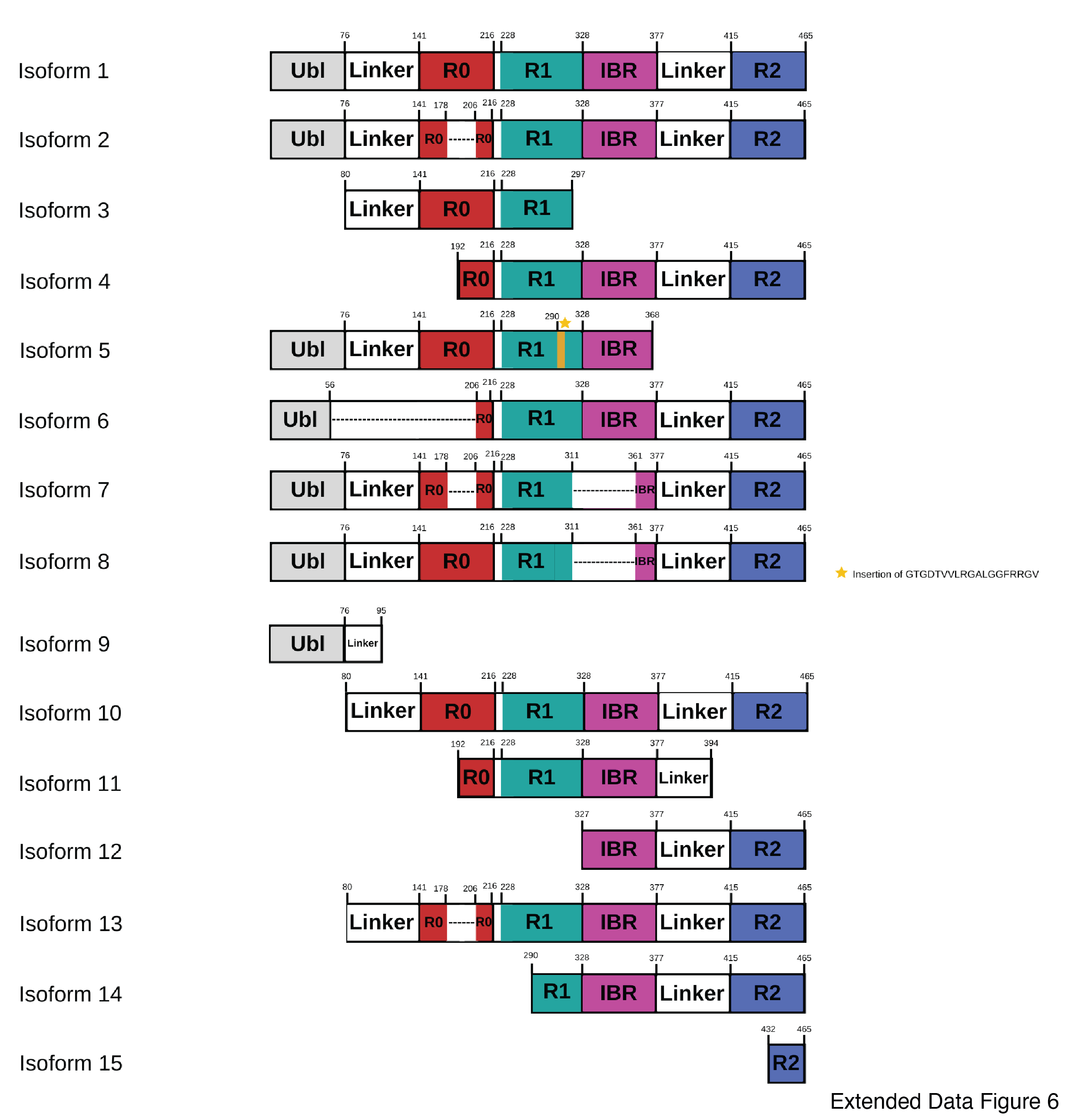
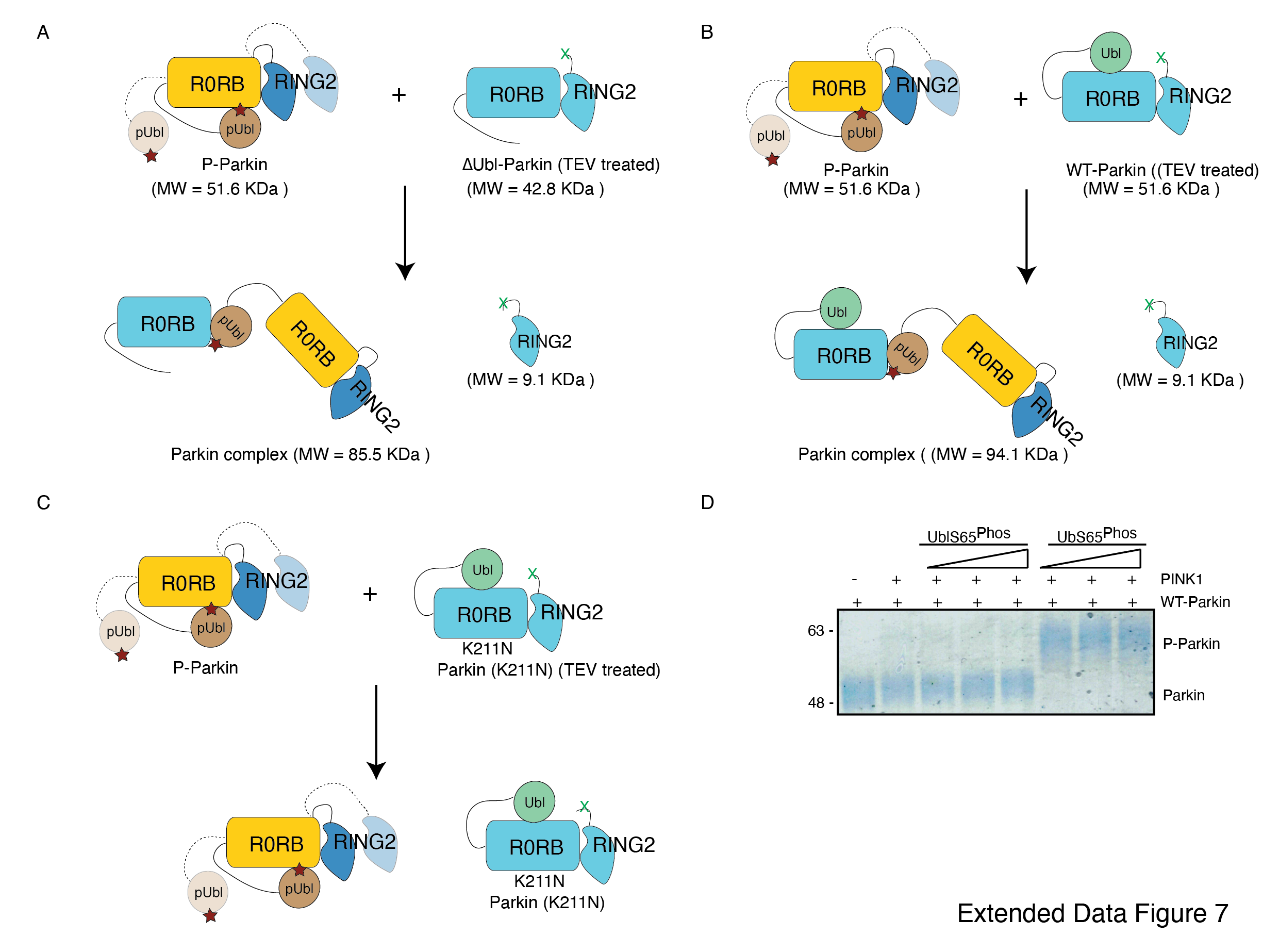
As the pUbl domain remained dynamic in both native phospho-Parkin and phospho-Parkin K211N (Fig. 1C, Fig. 3A), we wondered whether a trans-complex was formed between native phospho-Parkin. The latter could also be helpful in the context of activation of various Parkin isoforms lacking either the Ubl domain or RING2 domain (Extended Data Fig. 6). To test trans complex formation between native Parkin molecules, we used native phospho-Parkin (1-465) as a pUbl donor and untethered (processed with TEV protease) ∆Ubl-Parkin (TEV) as a pUbl acceptor on RING0. Interestingly, phospho-Parkin formed a stable complex with ∆Ubl-Parkin (77-382) and RING2 (383-465) was displaced from untethered ∆Ubl-Parkin (TEV) (Fig. 5A, Extended Data Fig. 7A), similar to the interaction between phospho-Parkin K211N and untethered ∆Ubl-Parkin (TEV) (Fig. 4B). The SEC data was confirmed by ITC measurements showing similar affinities between phospho-Parkin or phospho-Parkin K211N and untethered ∆Ubl-Parkin (TEV) (Fig. 5B, Fig. 4B).
We further tested the binding of phospho-Parkin with untethered WT-Parkin (TEV). Similar to untethered ∆Ubl-Parkin (TEV), untethered WT-Parkin (TEV) formed a complex with phospho-Parkin, and resulted in the removal of RING2 (383-465) from WT-Parkin (1-382) (Fig. 5C, Extended Data Fig. 7B). However, unlike untethered WT-Parkin (TEV), untethered Parkin K211N (TEV) failed to form the complex with phospho-Parkin (Fig. 5C, Extended Data Fig. 7C). This latter findingconfirmed that interactions between pUbl and the basic patch on the RING0 domain form a trans-complex. To further validate this, we also confirmed complex formation using SEC-MALS (size-exclusion chromatography coupled with multi-angle light scattering). MALS analysis further confirmed complex (Phospho-Parkin and WT-Parkin (1-382), Observed M. W. = 94 ± 3 Kda) formation between phospho-Parkin (Observed M. W. = 53 ± 2 Kda) and untethered WT-Parkin (TEV) (Observed M. W. = 52 ± 3 Kda) (Fig 5D, Extended Data Fig. 7B).
As our binding experiments suggested interaction between phosphorylated Parkin and native Parkin, we next checked whether phosphorylated Parkin can activate native Parkin. To test phospho-Parkin mediated Parkin activation in trans, we used a catalytic-inactive version of phospho-Parkin T270R/C431A with mutations in both the E2 binding site (T270R) and catalytic site (C431A). Interestingly, we observed that WT-Parkin ubiquitination/autoubiquitination activity was increased with increasing concentrations of phospho-Parkin T270R/C431A (Fig. 5E). Although, we were not expecting activation of WT-Parkin by phospho-Parkin as Ubl of WT-Parkin would block the E2 binding site on RING1 in WT-Parkin, activation of WT-Parkin with phospho-Parkin T270R/C431A suggested that a significant inhibition on Parkin is mediated by RING0 blocking RING2, which was released upon pUbl binding.
Further, we wondered whether pUbl would enhance Parkin phosphorylation similar to pUb 32. To test this, we checked Parkin phosphorylation by PINK1 in the presence of pUbl or pUb. However, unlike pUb, pUbl did not enhance Parkin phosphorylation by PINK1 (Extended Data Fig. 7D), confirming that pUbl and pUb binding lead to unique conformational changes in Parkin. Overall, this data demonstrates pUbl-mediated dimerization of Parkin molecules leading to Parkin activation in trans.
Assessment of Parkin activation in cells
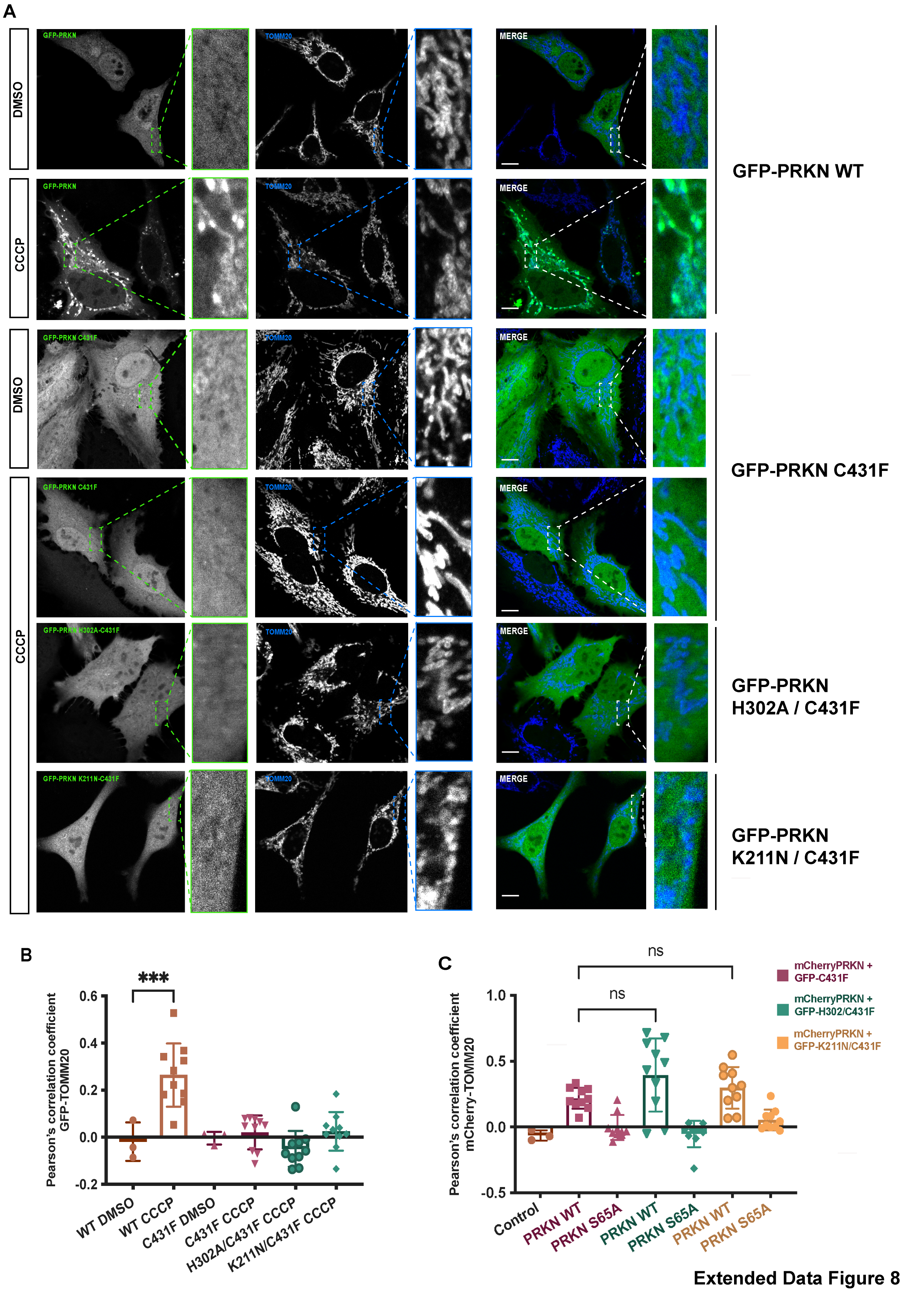
It has previously been reported that pUb may interact with the RING0 domain of Parkin and that loss of this interaction underlies loss of Parkin recruitment to the mitochondria in cells expressing Parkin K211N 39. However, we recently showed that pUb does not bind in the RING0 pocket (comprising K161, R163, and K211), and pUb binds specifically in the RING1 pocket (comprising K151, R305, and H302) 33, unlike phospho-Ubl binding in the RING0 pocket and displacing RING2 in trans (Fig. 5). Biophysical assays also revealed that unlike the tight binding of pUb in the RING1, pUbl binding in the RING0 pocket was very transient. Furthermore, K211N mutation in the RING0 pocket resulted in loss of Parkin activity by both loss of pUbl-mediated interactions (Fig 5) and by N211-driven conformational changes leading to loss of Parkin activity independent of pUb binding 33. This loss of Parkin activity would lead to a reduced amount of pUb, resulting in loss of Parkin recruitment to mitochondria. Therefore, we decided to test an activity-independent Parkin recruitment to impaired mitochondria using a Parkin translocation assay in HeLa cells 10,14-16,31. Consistent with previous studies, 10,14-16,31 full-length wild-type but not catalytic-inactive GFP-Parkin C431F was recruited to mitochondria following carbonyl cyanide m-chlorophenyl hydrazone (CCCP) treatment (Extended Data Fig. 8A-B). Similarly, we did not observe the recruitment of GFP-Parkin C431F/H302A or GFP-Parkin C431F/K211N mutants to impaired mitochondria when expressed alone (Extended Data Fig. 8A-B).
We observed that co-expression of mCherry-tagged-Parkin WT with GFP-Parkin C431F enabled GFP-Parkin C431F recruitment to the mitochondria, similar to a previous study 31 (Fig. 6A, D). Under these assay conditions, we strikingly observed that mutation of the pUb binding pocket in the RING1 completely abolished recruitment of the double mutant GFP-Parkin C431F/H302 to the mitochondria when co-expressed with mCherry-tagged-Parkin WT (Fig. 6B, D). This excluded a significant role for the RING0 pocket in pUb binding in the context of full-length parkin expressed in cells following mitochondrial damage (Fig. 6B, D). In line with this, mutation of the RING0 binding pocket produced a moderate defect in recruitment of the double mutant GFP-Parkin C431F/K211N to the mitochondria when co-expressed with mCherry-tagged-Parkin WT (Fig. 6C, D), suggesting that the transient interaction between pUbl and RING0 of Parkin in trans acts in concert with pUb binding to RING1 pocket for optimal Parkin recruitment to sites of mitochondrial damage (Fig. 6C, D). Under all transfection conditions, we did not observe a significant difference in mCherry-tagged Parkin WT (Extended Fig. 8C). Furthermore, co-expression of GFP-Parkin C431F or GFP-Parkin C431F/K211Nor GFP-Parkin C431F/H302A with the non-phosphorylatable mCherry-tagged-Parkin S65A failed to rescue recruitment to the mitochondria (Fig. 6A, B, C, D). These findings were in line with our biophysical data and highlight the importance of phospho-Ubl domain-mediated interactions in Parkin recruitment to the mitochondria.
ACT improves enzyme kinetics of Parkin
A previous study identified a small region (101-109) in the linker between Ubl and RING0 as an activator element (ACT) required for Parkin activity 24. To further explore the role of the ACT, we tested whether the omission of ACT affects the binding of Parkin with the charged state of E2 (E2~Ub). We observed a tight complex formation between phospho-Parkin, pUb, and E2~Ub on SEC assay (Fig. 7A). Interestingly, deletion of the ACT did not affect the complex formation with E2~Ub, as phospho-Parkin ∆ACT co-eluted with pUb and E2~Ub (Fig. 7A). As the displacement of RING2 is a crucial process during Parkin activation, we tested whether the removal of the ACT affects the displacement of the RING2 domain using our TEV-based SEC assay. We observed that phospho-Parkin ∆ACT (TEV) after treatment with TEV resulted in a shift where RING2 (383-465) was displaced from the Parkin core (1-382, ∆ACT), resulting in the elution of two fragments of Parkin separately on SEC (Fig. 7B). As the deletion of ACT did not show any functional defect in Parkin, we hypothesized that the presence of ACT at the interface of RING0 and RING2 might affect the dynamic nature of RING2, thereby regulating the enzyme kinetics. To test this hypothesis, we compared the phospho-Parkin ∆ACT ubiquitination activity over different time points. We observed that the deletion of ACT slowed the kinetics of Parkin activity, doubling the time for phospho-Parkin ∆ACT to reach a similar level of activity as phospho-Parkin (Fig. 7C).
ACT is more efficient in cis
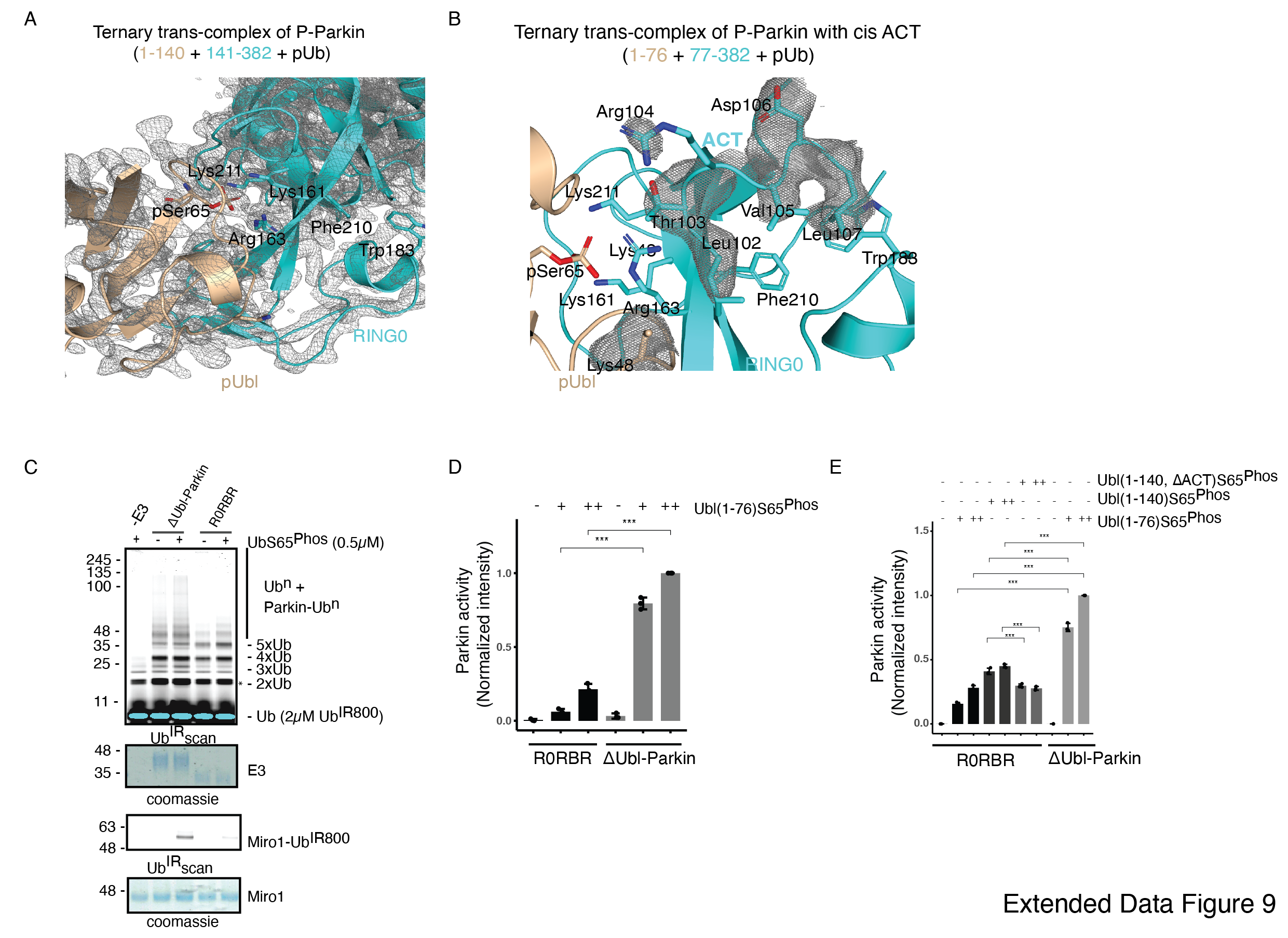
The ternary trans-complex of phospho-Parkin (1-140 + 141-382 + pUb) structure in this study was solved at a similar resolution and in the same space group as the previously solved structure of phospho-Parkin (1-382) in complex with pUb 24. In the previous structure of phospho-Parkin (1-382)-pUb complex (PDBID:6GLC), the ACT region was clearly shown to occupy the hydrophobic pocket on RING0 (Fig. 8A). However, we did not see any density of the ACT region in the ternary trans-complex structure of phospho-Parkin (1-140 + 141-382 + pUb) (Fig. 8A, Extended Data Fig. 9A). Interestingly, we observed that in the ternary trans-complex structure of phospho-Parkin, K48 of the pUbl domain occupied the same pocket that R104 of the ACT region occupied in the structure of phospho-Parkin-pUb complex (Fig. 8A, Extended Data Fig. 9A). Also, the side-chain of K48 of the pUbl domain was disordered in the previous structure of phospho-Parkin (1-382)-pUb complex (Fig. 8A).
We wondered whether the lack of density in the ACT region was due to the preference of ACT to remain associated with the cis molecule rather than to be complemented by the trans molecule. To test this hypothesis, we determined the crystal structure of the ternary trans-complex of phospho-Parkin with cis ACT using phospho-Ubl (1-76) and ∆Ubl-Parkin Q347C (TEV). pUbl formed a stable complex with untethered ∆Ubl-Parkin Q347C (TEV) and resulted in the displacement of RING2 (383-465) (Fig. 8B). Fractions containing trans-complex of phospho-Parkin (1-76 + 77-382) with cis ACT were mixed with pUb-3Br to get the crystals of the ternary complex. The ternary trans-complex of phospho-Parkin (1-76 + 77-382 + pUb) with cis ACT was crystallized, and structure was determined at 2.6 Å (Extended Data Table 1). Interestingly, in the structure of the ternary trans-complex of phospho-Parkin with cis ACT, we could observe the electron density of the ACT region (Fig. 8C, Extended Data Fig. 9B). Furthermore, K48, which occupied the ACT region in the ternary trans-complex structure of phospho-Parkin with trans ACT, was disordered in the ternary trans-complex structure of phospho-Parkin with cis ACT, similar to what was seen previously in the phospho-Parkin structure (Fig. 8A, C, Extended Data Fig. 9B).
To validate crystal structures, we compared the ubiquitination activity of R0RBR (141-465) and ∆Ubl-Parkin (77-465) in the presence or absence of pUb. The presence of linker (77-140) containing ACT in ∆Ubl-Parkin (77-465) made it more active compared to R0RBR (141-465) (Extended Data Fig. 9C). We then compared the activation of R0RBR and ∆Ubl-Parkin using pUbl (1-76) in trans. We observed that pUbl (1-76) efficiently activated ∆Ubl-Parkin (77-465); however, R0RBR (141-465) activation by pUbl (1-76) was very poor (Fig. 8D, Extended Data Fig. 9D). Further, we tested whether pUbl-linker (1-140) with or without ACT would affect the activation of ∆Ubl-Parkin (77-465) in trans. Interestingly, ubiquitination assays performed using increasing concentrations of pUbl (1-76) or pUbl-linker (1-140), or pUbl-linker-∆ACT (1-140, ∆101-109) showed that ∆Ubl-Parkin activation was not affected by the linker (77-140) or ACT region in trans (Fig. 8E). However, compared to pUbl (1-76), pUbl-linker (1-140) showed better activation of R0RBR (141-465) (Fig. 8F, Extended Data Fig. 9E). Also, in contrast to pUbl-linker (1-140), pUbl-linker-∆ACT (1-140, ∆101-109) showed poor activation of R0RBR (141-465) which was similar to pUbl (1-76) (Fig. 8F, Extended Data Fig. 9E). However, the activity of R0RBR (141-465) complemented with pUbl-linker (1-140) was less than the activity of ∆Ubl-Parkin (77-465) complemented with pUbl (1-76) (Fig. 8F, Extended Data Fig. 9E). Overall, our data suggested that ACT can be complemented in trans; however, ACT is more efficient in cis.
Crystal structure of pUbl-linker (1-140) depleted R0RBR (R163D/K211N)-pUb complex reveals a new ubiquitin-binding site on Parkin
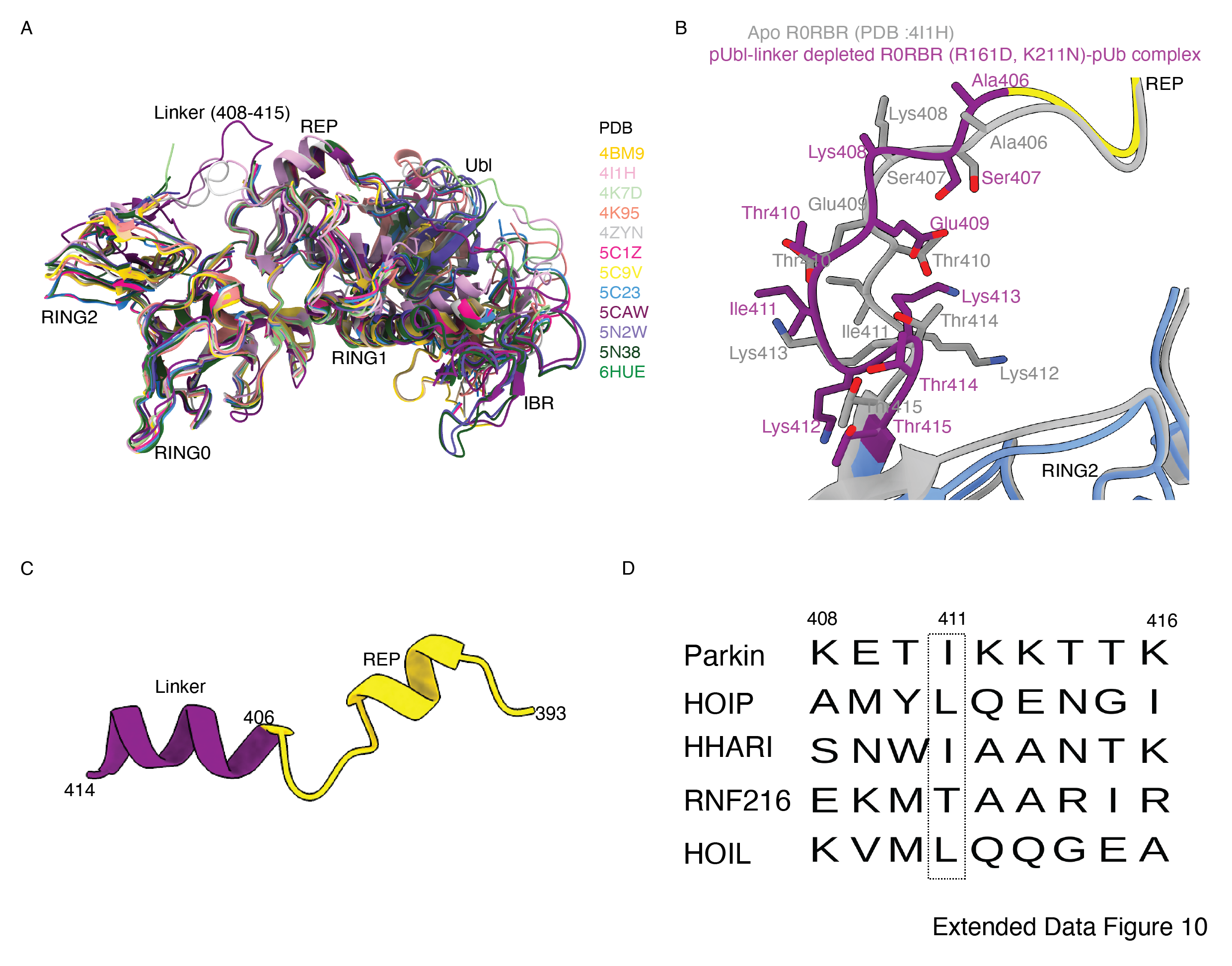
In the last few years, several structures of Parkin or Parkin complexes were solved in various conditions and from different species. However, the linker (408-415) between REP element and RING2 was mostly disordered, except in structures (PDB 4I1H, 5CAW, 4ZYN) where the above region was modeled in different conformations (Extended Data Fig. 10A), highlighting its flexible nature. A pathogenic mutation T415N was also found in the linker (408-415), which abolished Parkin activity. However, the role of this small linker region on Parkin remains elusive. Therefore, we decided to inspect all the structures solved in the present study. In the crystal structure of R0RBR (R163D/K211N)-pUb complex, out of two molecules of Parkin in the asymmetric unit, one molecule of Parkin showed nice electron density of the linker (408-415) region of Parkin (Fig. 9A, B). We further noticed conformational changes in the linker (408-415) region in the structure of the R0RBR (R163D/K211N)-pUb complex when compared to the previously solved apo R0RBR structure (PDB 4I1H) (Extended Data Fig. 10B). While T410, I411, and K412 were facing outwards in the apo R0RBR structure, in the structure of R0RBR (R163D/K211N)-pUb complex these residues were present in the core (Fig. 9B, Extended Data Fig. 10B). Interestingly, we noticed interactions between the linker (408-415) of Parkin and pUb from the neighboring molecule of the asymmetric unit (Fig. 9C). The core of interactions between the Parkin linker and ubiquitin was mediated by I411, which was involved in hydrophobic interactions with the hydrophobic pocket of ubiquitin (Fig. 9C). Other interactions between Parkin and ubiquitin included ionic interactions mediated by K412, and H422 (Fig. 9C). Water-mediated interactions between linker (408-415) and ubiquitin included T410 with the carbonyl group of R72 of ubiquitin, and T415 with the carbonyl of G35 of ubiquitin (Fig. 9C). Furthermore, E409 formed a salt-bridge with K413 (Fig. 9C), which could be required for maintaining the structure of the linker region for ubiquitin binding. Also, residues in the linker region interacting with ubiquitin were highly conserved in Parkin across different species (Fig. 9D), suggesting their functional importance. Our data in Fig. 2 suggested that RING2 was flexible (open and closed states) mediated by pUbl binding in the basic patch. As R0RBR (R163D/K211N)-pUb complex structure was captured in the closed state of RING2, we wondered whether the linker connecting REP and RING2 may adopt an alternate conformation dependent upon RING2 position (open or closed). The crystallization of the open state of phospho-Parkin remains challenging due to the flexible/multiple possible conformations of the REP-RING2 region. Therefore, we used AlphaFold 2 34 to predict the model of the linker connecting REP and RING2of Parkin. Interestingly, the AlphaFold model predicted helical structure in the linker region of Parkin (Extended Data Fig. 10C) in the RING2 open state of Parkin, indicating the flexible nature of this region under different states (RING2 closed <-> RING2 open) of Parkin. The latter also suggested that the conformation of the linker observed in the crystal structure could be one of the intermediates.
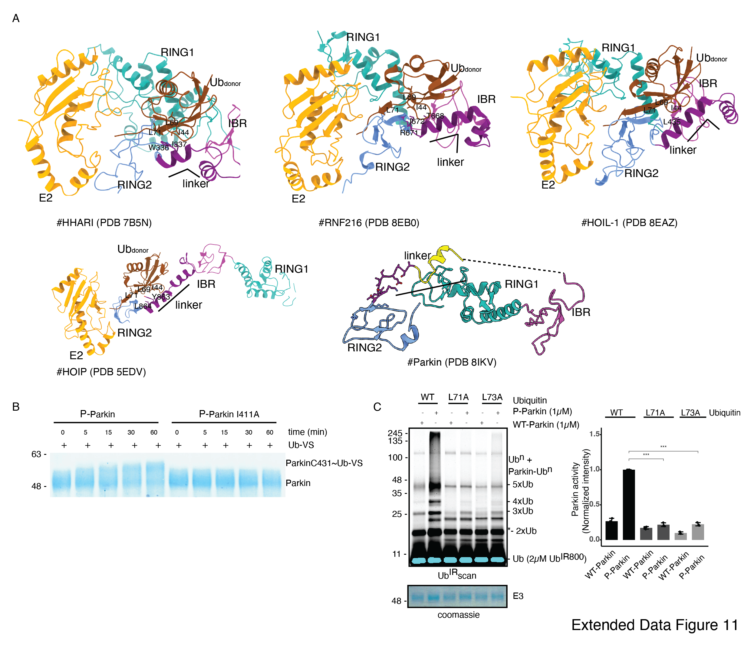
To validate the observations from structural analysis, we mutated these residues and compared their ubiquitination activity. In contrast to WT-Parkin, E409A and H422A drastically reduced Parkin activity, whilst I411A, T415N, and K416A resulted in the complete abolishment of Parkin activity (Fig. 9E). Further inspection revealed that although the linker region of Parkin is not conserved across different members of RBR family E3-ligases (Extended Data Fig. 10D), hydrophobic nature at the corresponding position of I411 on Parkin is conserved among various RBRs except RNF216 (Extended Data Fig. 10D). Also, the crystal structures of HOIP, HOIL, HHARI, and RNF216 solved with E2~Ub 35–37 showed interactions between linker region and donor ubiquitin (Ubdon) (Extended Data Fig. 11A). To test whether the linker between REP and RING2 of Parkin binds with donor ubiquitin (Ubdon), we performed binding assays using E2~Ubdon. Interestingly, unlike phospho-Parkin, which formed a stable complex with E2~Ubdon on SEC and co-eluted with E2~Ubdon and phospho-ubiquitin (Fig. 9F), phospho-Parkin I411A did not show interaction with E2~Ubdon (Fig. 9F). Furthermore, the SEC data was confirmed by ubiquitin-vinyl sulfone (Ub-VS) assay 16,24,25 where unlike phospho-Parkin, phospho-Parkin I411A did not react with Ub-VS (Extended Data Fig. 11B). We also tested Parkin activity using ubiquitin mutants (L71A or L73A) which would perturb the interactions of ubiquitin and Parkin linker as suggested by structure in Fig. 9C. Compared to native ubiquitin, ubiquitin mutants show loss of Parkin activity (Extended Data Fig. 11C) which nicely corroborated with our data. Overall, our data showed that the linker region between REP and RING2 interacts with donor ubiquitin and plays a crucial role in Parkin function.