Collecting of Mushrooms
Mushroom samples were collected locally in the Mansehra district of Khyber Pakhtunkhwa, Pakistan. They were identified as Daedalea sp by the Department of Plant Sciences at Quaid-I-Azam University in Islamabad, Pakistan. With a surgical blade, the fresh biomass was cut into tiny fragments, rinsed thoroughly with distilled water to remove any dust particles, and then dried in the shade. Shade dried biomass was ground into powder and stored at room temperature for later use using a Willey mill. In an Erlenmeyer's conical flask, 20 gm of the powder was suspended in 200 mL of distilled water and heated at 80°C for 3 hours on a magnetic stirrer. The extract prepared was dually filtered using nylon cloth and later refiltered on Whatman filter paper to get pure extract. The prepared aqueous extracts were stored at a low temperature of 5 ºC for future experimentation (Zhao, Jin, & Jin, 2018).
Mycosynthesis of Silver Nanoparticles (Ag-NPs)
To make silver nanoparticles (AgNPs), 100 mL of aqueous mushroom extract was gradually added to 100 mL of 1 mM AgNO3 (ACS reagent, 99.0%, Sigma-Aldrich) solution, followed by a reduction incubation at room temperature (20°C 2). De-ionized water containing only silver nitrate solution was used as the negative control in separate flasks, and pristine extract was used as the positive control. The change in color from light brown to darkish brown within 24 hours was the first indication of AgNP synthesis. After centrifugation at 15000 rpm for half an hour, the supernatant was removed, and the nanoparticles were washed three times with centrifugation at 10000 rpm for 10 minutes. The thick suspension was cast into a petri dish after thorough washing and dried overnight in a 70 oC oven. The dried particles were then grind into a fine powder with a mortar and pestle before being stored at room temperature for physical and chemical characterization as well as biomedical assays (Abdel-Aziz & Rizwan, 2019).
Characterization of Mycosynthesized Ag-NPs
Diverse characterization techniques were employed to investigate the physicochemical properties of Daedalea sp synthesized AgNPs, including UV spectroscopy, Fourier transform infrared spectroscopy (FTIR), X-ray diffraction (XRD), scanning electron microscopy (SEM), transmission electron microscopy (TEM), energy dispersive x-ray (EDX) analysis. The biosynthetic reaction between the extract and silver nitrate solution was observed in the range of 200 to 700 nm UV-Visible. XRD technique was used to determine the crystalline nature of biosynthesized NPs. The XRD pattern was obtained using PANalytical X’Pert X-ray diffractometer. Scherer’s equation was used to calculate the crystallite size as follows.
D = k λ/ β Cosθ
D represents crystal size, k denotes shape factor (0.94), λ depicts X-rays wavelength of 1.5421Å while β and θ refers to FWHM in radians and Bragg’s angle, respectively. To determine functional groups associated with NPs, FTIR spectroscopy was carried out in the spectral range of 400 cm − 1 and 4000 cm − 1.
Morphology was examined by SEM (JSM-7600F, Japan) and TEM (JEM-2100F, Japan) while elemental analysis was conducted using Energy Dispersive X-Ray Spectroscopy (EDX). Furthermore, Thermal stability was investigated by thermo gravimetric analysis using Q500 thermo gravimetric analyzer. 10 mg of the tested sample was decomposed under flowing nitrogen gas from 40°C to 500°C at 10°C/min heating rate.
Biological applications
Antibacterial activity of AgNPs, Non-coated and AgNPs coated Antibiotics against Tested Strains
Collection of bacterial species
A total of four multidrug resistant strains were isolated from UTI (urinary tract infection) patients, including Klebsiella pneumonae, Echerichia coli, Pseudomonas aeruginosa, and Staphylococcus aureus. These isolates were collected from the Hayat Abad Medical Complex in Peshawar, Pakistan, and were previously identified at the hospital using biochemical tests and a molecular technique (16s rRNA).
Preparation of Antibiotic Discs Coated with AgNPs
A powdered AgNPs stock solution was made by combining 10mg of AgNPs residue in 1 mL of distilled water. After thorough mixing, approximately 5 L of the suspension were taken from the stock solution and poured onto an antibiotic disc within the petri plates, which were then placed in a 70oC oven for 10 minutes to dry. Each antibiotic was coated using the same method.
Agar Well Diffusion Assay
The 8 mm diameter wells were punched into the Nutrient Agar (NA) media, which was then bacterial lawn prepared. The wells were filled with a 100 L suspension of AgNPs. The petri dishes were then placed in a 37oC incubator for 24 hours. The potency of AgNPs against the tested MDR bacterial strains was determined immediately after incubation by measuring zones of inhibition in millimeters.
Disc Diffusion Assay for Ag-NPs Coated and Non-Coated Antibiotics
The standard Kirby-Bauer disc method was used to assess the potency of both coated and non-coated antibiotics against tested MDR bacterial strains. Nutrient agar was prepared, and NA media and Petri dishes were autoclaved according to standard SOPs. After sterilisation, the media was cooled to 50oC before being poured into sterilized Petri dishes in the biosafety cabinet. After the media had solidified, bacterial cultures were prepared on Nutrient Agar (NA) plates, and antibiotic discs with and without AgNPs coating were applied. The petri plates were then incubated for 24 hours at 30°C. The potency of coated and non-coated antibiotics against test MDR bacterial strains was determined after incubation by measuring zones of inhibition in millimetres (Wei, McGrath, Hayden, & Kutcher, 2015).
Anti-Fungal Assay
AgNPs were tested for fungicidal activity against spore-forming fungi such as Aspergillus flavus (ATCC 9643), Aspergillus fumigatus (FCBP 66), Fusarium solani (FCBP 434), Aspergillus niger (ATCC 1015), and Mucor species (FCBP 300). For each fungal strain, spore suspension from stock cultures was prepared in Tween 20 solution (0.02% v/v). A 100 L aliquot of the suspension was poured onto multiple petri plates containing sterile SDA media and swabbed well. Following that, under highly sterilized conditions, tested samples (10 L) were introduced into each well within the solidified media. Clotrimazole and DMSO were used as positive and negative controls, respectively, followed by a 48-hour incubation to examine the Zone of Inhibition (ZOI). The ZOI was calculated using a Vernier caliper.
Antileishmanial Assay
A well-established protocol was used to assess the leishmanicidal activity of mycosynthesized AgNPs against both promastigote and amastigote forms of Leishmania tropica. Incubated cultures of the parasite L. tropica KWH23 strain were tested for leishmanicidal potential (in MI99 medium supplemented with 10% FBS). In brief, each well in a 96 well plate was filled with 10 L of tested solution, and then 150 L of 150 L aliquots of culture suspension (seeding density: 1 106 cells/mL) were added. The mixture was incubated at 30 degrees Celsius for 71 hours. Positive and negative controls were amphotericin-B and DMSO (1%) in PBS, respectively (Abou El-Nour et al., 2010). The incubation was followed by the addition of 10 µL MTT solution (4 mg/mL in dH20) to each well and re incubation of the culture plate for 3 hrs at 30 oC. Absorbance was then recorded at 540 nm via a microplate reader. % inhibition was calculated as follows:
$$\text{%} \text{I}\text{n}\text{h}\text{i}\text{b}\text{i}\text{t}\text{i}\text{o}\text{n} =\left[1-\left\{\frac{\text{A}\text{b}\text{s}\text{o}\text{r}\text{b}\text{a}\text{n}\text{c}\text{e} \text{o}\text{f} \text{s}\text{a}\text{m}\text{p}\text{l}\text{e}}{\text{A}\text{b}\text{s}\text{o}\text{r}\text{b}\text{a}\text{n}\text{c}\text{e} \text{o}\text{f} \text{c}\text{o}\text{n}\text{t}\text{r}\text{o}\text{l}}\right\}\right]\times 100$$
Furthermore, the sample was analyzed at different concentrations and the assay was repeated thrice. The values of IC50 were determined by using Table curve software 2D v5. 01.
Protein kinase inhibition assay
With slight modifications to a protocol elucidated in, this bioassay was performed to verify the protein kinase inhibitory ability as a preliminary assay to screen the anticancerous potentials of biogenic AgNPs. Streptomyces 85E was used as a test strain in this study. A volume of 100 L was then added to the plates containing sterile ISP4 medium from the refreshed culture of Streptomyces 85E. Each 5mm well was filled with NPs (5 L) and labelled appropriately. The positive control was surfactin, and the negative control was DMSO. Following that, the plates were incubated for two days at 28 degrees Celsius. The presence of a clear and bald zone around wells indicates that phosphorylation, mycelia, and spore formation have been inhibited (Abou El-Nour et al., 2010). The zones were measured to the nearest millimeter with a vernier caliper. Clear zones demonstrate Ag-NPs' cytotoxic potential and the test strain's death (Srikar, Giri, Pal, Mishra, & Upadhyay, 2016).
Antidiabetic assay
The antidiabetic potential of mycosynthesized AgNPs were evaluated by using ɑ-glucosidase and ɑ-amylase inhibition assay.
𝛼-amylase inhibition assay
To assess the -amylase inhibition potential of AgNPs, a well-established protocol was followed with minor modifications. This assay was carried out in a 96-well microplate. Phosphate buffer (15 L), -amylase (20 L), sample (10 L), and starch (30 L) were added to each test well. The plate was then incubated at 50 oC for 30 minutes. Finally, each well received 20 L of 1M HCl and 80 L of iodine solution. The negative control was DMSO, the positive control was acarbose, and the blank contained buffer solution and starch instead of IONPs. A microplate photometer was used to measure absorbance at 520 nm. The inhibition was calculated as a percentage using the formula.
$$\text{%} \text{E}\text{n}\text{z}\text{y}\text{m}\text{e} \text{i}\text{n}\text{h}\text{i}\text{b}\text{i}\text{t}\text{i}\text{o}\text{n}=\left(\frac{\text{A}\text{b}\text{s} \text{S}\text{a}\text{m}\text{p}\text{l}\text{e}-\text{A}\text{b}\text{s} \text{n}\text{e}\text{g}\text{a}\text{t}\text{i}\text{v}\text{e} \text{c}\text{o}\text{n}\text{t}\text{r}\text{o}\text{l}}{\text{A}\text{b}\text{s} \text{b}\text{l}\text{a}\text{n}\text{k}-\text{A}\text{b}\text{s} \text{n}\text{e}\text{g}\text{a}\text{t}\text{i}\text{v}\text{e} \text{c}\text{o}\text{n}\text{t}\text{r}\text{o}\text{l}}\right)\times 100$$
𝛼-glucosidase inhibition assay
The anti-diabetic capacity of mycosynthesized AgNPs was further assessed using a -glucosidase inhibition bioassay based on a previously published protocol with minor modifications. To dissolve -glucosidase, 50 mL of phosphate buffer (pH 6.8) supplemented with 100 mg of BSA (bovine serum albumin) was used in the experiment (Saccharomyces cerevisiae, Sigma-Aldrich). A reaction mixture of 10 L of tested sample, 490 L of phosphate buffer (pH 6.8), and 5mM of p-nitrophenyl-D-glucopyranoside (250 L) was incubated at 37 oC for 5 minutes. Following that, samples were incubated at 37°C for 15 minutes with 250 L-glucosidase (0.15 unit/mL). Absorptions were measured with a UV-Vis spectrophotometer at 400 nm after the reaction was stopped by adding 2 mL Na2CO3 (200 mM) solution (Rafique, Sadaf, Rafique, & Tahir, 2017). The assay measures the amount of p-nitrophenol released from p-nitrophenyl-D-glucopyranoside. The experiment used acarbose as a positive control and was repeated three times.
$$\text{%} \text{E}\text{n}\text{z}\text{y}\text{m}\text{e} \text{i}\text{n}\text{h}\text{i}\text{b}\text{i}\text{t}\text{i}\text{o}\text{n}=\left(\frac{Abs Sample-Abs negative control}{Abs blank-Abs negative control}\right)\times 100$$
Antioxidant assays
Total antioxidant capacity determination (TAC)
The assay was used to determine the sample's total antioxidant capacity. A micropipette was used to add 100 L of sample to the Eppendorf tubes during the experiment. The TAC reagent (0.6 M sulphuric acid, 25 mM sodium phosphate, and 4 mM ammonium molybdate in 50 mL dH20) was then transferred to eppendorff tubes containing the tested samples. The reaction mixture was incubated in a water bath for two hours at 80 degrees Celsius; after cooling, the absorbance of the samples was measured in a microplate reader at 630 nm. TAC was calculated as g ascorbic acid equivalent/mg of sample after three repetitions of the experiment.
Total reducing power determination (TRP)
This activity was performed in triplicate to determine the total reducing power of the sample. The test sample (100 L) was added to the eppendorff tubes along with 400 L of 0.2-molar phosphate buffer (pH 6.6) and potassium ferric cyanide (1% w/v), which was then incubated in a water bath at 55 oC for 30 minutes. Following that, 400 L of 10% w/v trichloroacetic acid was added to each Eppendorf tube, followed by 20 minutes of centrifugation at 4000 rpm. The supernatant (140 L) of each mixture was added to the corresponding wells of a 96-well plate already containing 60 L of ferric cyanide solution (0.1% w/v). The absorbance of the samples was then measured at 630 nm with a microplate reader. The same procedure, as mentioned earlier, was followed both for positive and negative controls.
Free radical scavenging assay (FRSA)
The previously reported protocol was adopted with minor modifications. Using the DPPH reagent at concentrations ranging from 12.5 L to 400 L, the potential free radical scavenging ability of Ag-NPs was investigated for their antioxidant potential. Each well of a 96-well plate was filled with tested samples (10 L). The DPPH reagent (190 L) was then added to each well containing the sample. It was then incubated for 60 minutes in the dark at 37 o C. Ascorbic acid was considered a positive control, while DMSO was considered a negative control. Furthermore, absorbance rates at 515nm were measured using a microplate photometer. The mycosynthesized Ag-NPs' FRS potential was expressed as a percentage.
$$\left(\text{%}\right) \text{F}\text{R}\text{S}\text{A} =\left(1-\frac{Abs}{Abc} \right)\times 100$$
Abc and Abs indicates the absorbance of negative control and sample respectively.
Trolox Antioxidant Assay (ABTS)
The antioxidant potential of the biosynthesized assay was assessed using a modified ABTS assay. Potassium per sulphate (2.45 mM) and ABTS salt (7 mM) were mixed in equal parts and incubated at room temperature overnight. Samples were loaded into the mixture immediately after incubation and kept in the dark for 15 minutes at room temperature. At 734nm, the absorbance of the sample in the reaction mixture was measured using a BioTek ELX800. Trolox reagent was considered + ve for this assay, while DMSO was considered -ve. The tests were run in triplicate, and the results (antioxidant potential) were expressed as TEAC.
Biocompatibility studies
The hemolytic activity assay for RBCs-NPs interaction was conducted to evaluate the toxic nature of biosynthesized silver NPs using freshly obtained human blood. 1 cc of blood was collected from volunteers (students) who had no other health issues. To prevent coagulation, blood was collected in an EDTA tube (Khalil et al. 2017). Blood was centrifuged at 14000rpm for 5minutes to obtain fresh RBCs and separate out plasma. RBCs were obtained in a pellet after 2–3 times of centrifugation. Later, 9.8 ml PBS (phosphate buffered saline) was added to a 200µl of freshly obtained pellet erythroctes. Then mixing was done by proper shaking of solution in PBS.NPs sample of four different concentrations 20µl, 40µl, 60µl and 80µl respectively was added to suspended erythrocytes in 1.5ml Eppendorf tube. The mixture was then incubated at 35C˚ for 1 hr. Then after, Centrifugation of incubated sample was carried out at 10000rpm for 10minutes. A supernatant was created, and 100 l of it was poured into 96-well plates (Kamal, Saba, & Farooq, 2023). The absorbance of the obtained supernatant was measured at 541nm using a spectrophotometer. Triton X-10 was used as a positive control and DMSO as a negative control.
Calculation of percentage hemolytic activity of RBCs by NPs was evaluated through following percentage formula,
% Hemolysis = (Verma & Maheshwari, 2019)×100
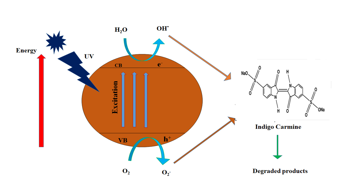
Photo catalytic study
20 ppm of indigo carmine dye was prepared in 50 ml of double-distilled water. The original concentration was taken in 5ml, and the remaining solution contained 30mg of Ag-NPs catalyst, which was added to 45ml of dye solution. After keeping the solution in the dark for 20 minutes to achieve the adsorption desorption equilibrium, it was exposed to UV light and samples were taken every 20 minutes. After centrifuging the sample at 10000 rpm for 10 minutes, the photodegradation was studied in a UV-Visible Spectrophotometer (Siddiqi, Husen, & Rao, 2018). The following formula was used to calculate the percentage of dye degradation.
(%) = \(\frac{Co-Ct}{Co}\) × 100 (1)
(%) = \(\frac{Ao-At}{Ao}\) × 100 (2)
Where Co and Ao is the initial concentration of dye and Ct and At after time interval.