CircRNAs are initially reported by Hsu and Coca-Prados (26). Nevertheless, these CircRNAs were thought to be transcription errors. Now, researchers have discovered thousands of circRNAs which may serve as diagnostic or predictive biomarkers, such as hsa_circ_0005075 in hepatocellular carcinoma (27), as well as hsa_circ_002059, hsa_circ_0000190, and circPVT1 in gastric carcinoma (28–31), and all of this were benefited from bioinformatics analysis.
CircRNAs can regulate many cellular processes, including sponging miRNAs (32, 33), the assembly and transport of cellular proteins (34), alternative splicing and gene expression (35), possessing protein-coding activity, modulating generation of rRNAs/tRNAs (35), and antiviral immunity (36). Specifically, ciRS-7 is the first identified CircRNA sponging miR-7 with direct binding (32, 33). Foxo3 circular RNA (circ-Foxo3) binds to ID-1 (inhibitor of differentiation-1), the transcription factor E2F1, HIF-1α (hypoxia-inducible factor-1α ) and FAK (focal adhesion kinase ), and inhibits their translocation (34). The knockdown of circRNA EIF3J or circRNA PAIP2 significantly reduces the expression level of their parental genes (37). circRasGEF1B, which is conserved between human and mouse circRNA, positively regulates lipopolysaccharide (LPS) response (36).
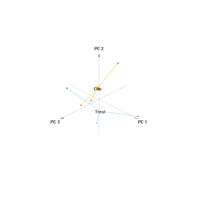
However, no study has focused on circRNA regulation in EC with progesterone treatment. Therefore, in this study, we applied RNA-seq to investigate the differentially expressed circRNA in EC with progesterone treatment, and the results revealed that a total of 87 circRNAs were differentially expressed in MPA-treated-ISK cells compared control cells (|fold change| ≥2.0, p < 0.05). Furthermore, the expression levels of the top 20 significant expressed circRNAs were validated by qRT-PCR. Particularly, hsa_circ_0046843 was significantly down-regulated in the ISK cell line treated with progesterone, while hsa_circ_0001860 was significantly up-regulated. In addition, we performed GO and KEGG pathways analysis. Most of the BP terms in our study have been reported to be closely associated with cancer, including response to cholesterol, epithelial to mesenchymal transition, and base-excision repair. Firstly, cholesterol has been reported to play a role in the synthesis of estrogen and progesterone. 3β-Hydroxysteroid-Δ24 reductase (DHCR24), the final enzyme in the cholesterol biosynthetic pathway, has been demonstrated to aggravate cancer invasion and progesterone resistance in EC (18). Additionally, dietary cholesterol consumption, including saturated fatty acid, unsaturated fatty acid, and cholesterol intake, influences EC risk by regulating the production, metabolism, and excretion of endogenous hormones (6). Secondly, Epithelial-to-Mesenchymal Transition (EMT) is an important step towards the invasion and metastasis of cancer (19). Hsu et al. demonstrated that epithelial cell adhesion molecule (EpCAM)-regulated transcription exerted influences on nanomechanical properties of EC cells, which promotes EMT (20). Last, a hospital-based case-control study examined the association between various polymorphisms in base excision repair (BER) DNA pathway genes (OGG1, MUTYH, XRCC1, APEX1, and PARP1) among Japanese postmenopausal women with and without endometrial cancer and found some worthful interaction (21). In MF terms, WNT-activated receptor activity and WNT-protein binding were significantly enriched.
Circular RNAs act as ceRNAs to regulate the expression of their targeted miRNAs. Additionally, miRNAs promote the degradation of target messenger RNAs (mRNAs) or inhibit their translations by recognizing specific binding sites on the 3′-untranslated region (UTR) of mRNAs in either a completely or partially complementary fashion (38). Therefore, these three elements constitute an associated pathway to modulate physiological function. The previous study has reported many miRNAs dysregulated in EC, which may be used as prognostic markers, treatment assessment markers, or treatment targets (39). miRNAs play essential roles in the ontogenetic processes of EC, including cell proliferation, migration, and metastasis (40). For example, mir-505 suppresses EC cell proliferation, invasion, and metastasis by targeting TGFA (41). miR-30c enhances the proliferation of EC cells, and the low expression level of miR-194 contributes to poor prognosis. The expression levels of miR-200c and miR-205 are significantly increased in EC compared with normal tissue (42–44). The silence of miR-124, a novel tumor suppressor miRNA, reverses EMT, and the invasive properties, by attenuating the expression of IQGAP1 (IQ Motif Containing GTPase Activating Protein 1) oncogene (45). Thus, to further understand the impact of the circRNA-related ceRNA crosstalk on MPA-treated EC cells, we used miRNA-circRNA interaction data to construct a circRNA-miRNA network. Hsa_circ_0046843, hsa_circ_0001860, hsa_circ_0020028, hsa_circ_0105045 and hsa_circ_0001993 were the top 5 significant circRNAs in EC cells treated with MPA. Each circRNA paired with five miRNAs, and all these circRNAs could regulate the expression of hsa-miR-4753-3p. Among the miRNAs, miR-296-3p, miR-144-5p, and miR-1236-3p have been reported to play a role in tumor progression. miR-1236-3p is associated with ovarian cancer metastasis (24). miR-296-3p plays a critical role in cell growth and multi-drug resistance in glioblastoma by targeting ether-à-go-go (EAG1) (23). As is known to all, drug resistance and recurrence are the limitations of the clinical application of progesterone treatment. The objective response rate of patients with advanced or recurrent EC is approximately 15 to 20% (46). Thus, miR-296-3p can be further used for progesterone-induced drug resistance. miR-144-5p acts as a tumor suppressor in bladder cancer cells by regulating CCNE1 and CCNE2 (25). miR-296-3p pairs with hsa_circ_0105045, while miR-144-5p and miR-1236-3p pair with hsa_circ_0001860, suggesting the two circRNAs may be associated with cancer progression or drug resistance.
Understanding the mechanism of circRNA-miRNA may provide critical insights into potential strategies for overcoming the shortcomings of progesterone treatment in EC. However, this study also has some limitations. First, this study examined the circRNAs in small EC cell lines treated with progesterone. In order to increase the accuracy of the results, tissues and plasma levels should also be studied respectively in EC patients treated with progesterone. Second, the mechanisms of circRNA-miRNA interaction are intricate. However, we only constructed a circRNA-miRNA network using five circRNAs. Further study should be concentrated on this interaction. If breakthrough this bottleneck, we can get great achievements in the progesterone treatment of EC.
