The major conclusions derived from the current studies are that altered gastric flora and elevated bile acids might aggravate the injury to the gastric mucosa and even exacerbate Correa's cascade process. We base this conclusion on three lines of evidence. First, we demonstrated that opportunistic pathogenic bacteria colonized the stomach mucosa of patients with cirrhosis. Next, we provided metabolic evidence to support the conclusion that gastric mucosal cells had an impaired energy metabolism. Finally, the HIF-1 pathway was found to upregulate in the gastric mucosal cell by transcriptome analysis.
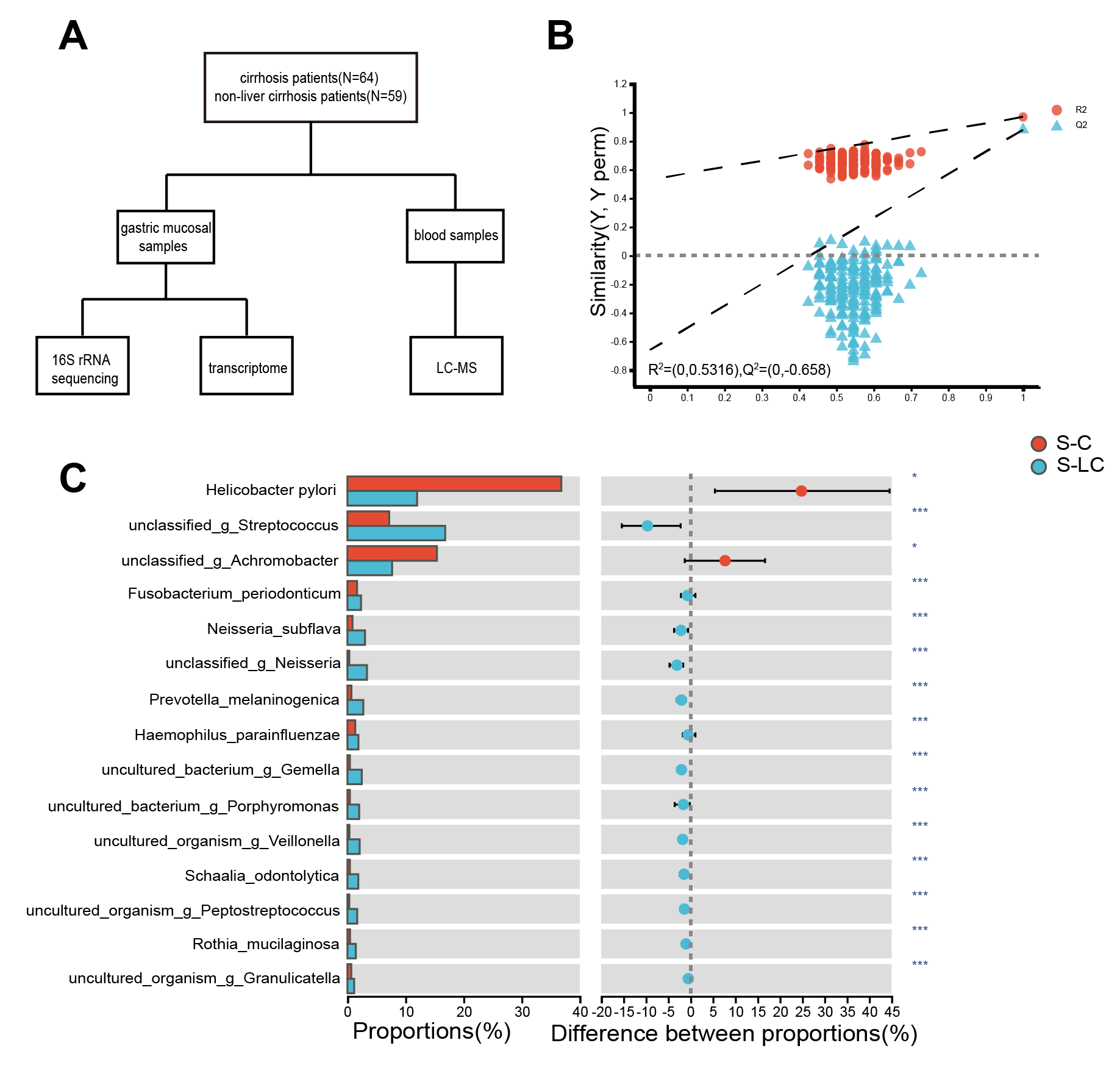
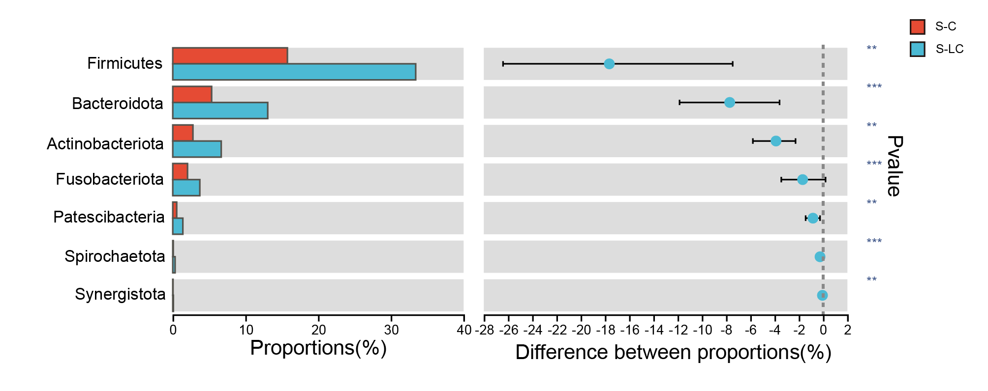
The microbiota alteration in the skin, intestinal mucosa, ascites fluid, serum, and the oral cavity have been studied before, except for the gastric mucosa. Our data demonstrate that higher bacterial diversity and increased relative abundance of multiple bacterial genera characterize S-LC microbial dysbiosis. In our study, only Helicobacter pylori (Hp) was detected in the Helicobacter; therefore, we consider the Helicobacter to be Hp in the following. There was a greater relative abundance of Streptococcus sp. and Prevotella_melaninogenica, Neisseria spp., and Fusobacterium_periodonticum, with lower Hp. These significantly increased bacteria are pathogenic oral bacteria[18–20] that have the potential to elicit an inflammatory response in epithelial cells.
Interestingly, emerging findings suggest the magnitude of roles of specific oral and gastric microbiota correlated with inflammation in the development of early-stage gastric adenocarcinoma[13]. Furthermore, as the diseases develop into more severe stages, such as atrophic gastritis, intestinal metaplasia, and gastric adenocarcinoma, the dominance of Hp begins to be displaced by other bacteria, including Streptococcus, Prevotella, and other bacteria[13, 21]. Taken together, we hypothesize that bacteria in the stomach of patients with cirrhosis may originate from the oral cavity, which may induce gastric mucosal abnormalities similar to Correa's cascade process.
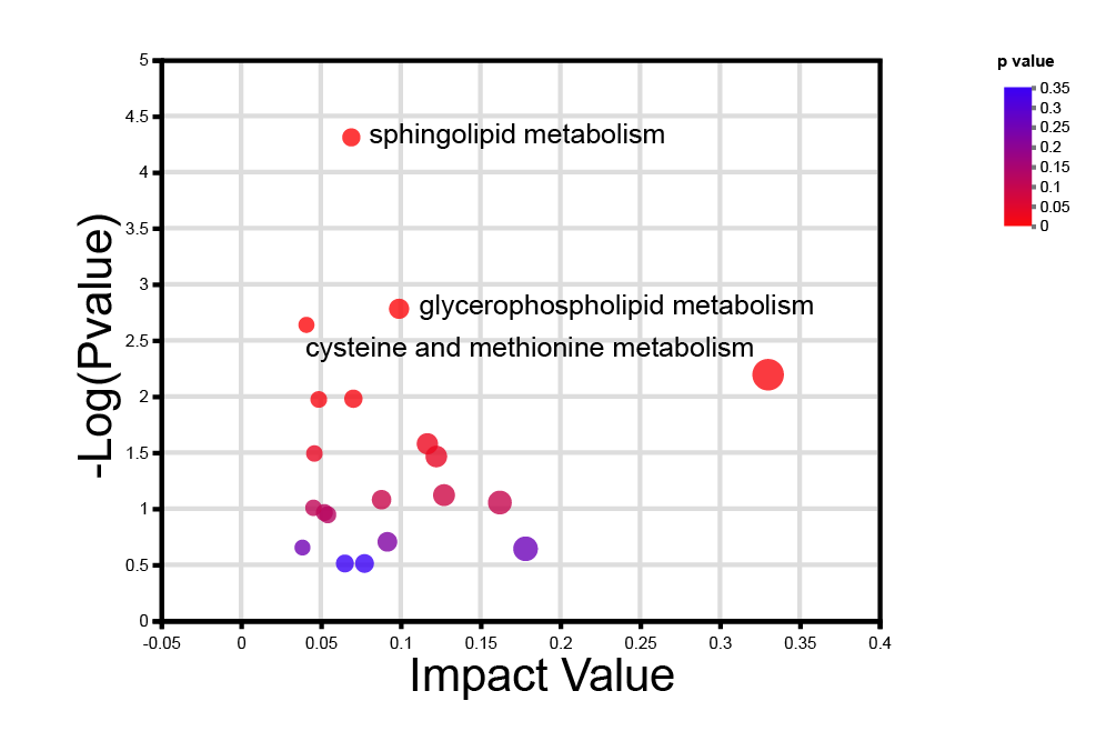
Previous studies have demonstrated that BAs modulate intestinal immunity, inflammation, and tumorigenesis[22]. We found that serum BAs were predominantly conjugated, and primary BAs were elevated. BAs can alter membrane lipid composition, and increased BAs concentrations can solubilize membranes and dissociate integral membrane proteins[23]. Of note, we also found that long-chain ACs were increased and positively correlated with most gastric mucosal flora, except Hp. In the process of β-oxidation, acylcarnitine transports acyl groups (organic acids and fatty acids) from the cytoplasm to the mitochondria so that they can be broken down to produce energy for cell activities[24]. According to the Human Metabolome Database, the primary function of most long-chain acylcarnitine is to ensure long-chain fatty acid transport into the mitochondria. Blood accumulation of long-chain ACs is a marker for incomplete fatty acid oxidation. Moreau R. et al. found that acute-on-chronic liver failure was characterized by extra-mitochondrial glucose metabolism through glycolysis and depressed mitochondrial ATP-producing fatty acid β-oxidation, which may contribute to the development of organ failures[10]. The evidence above indicates impaired energy utilization in the microcirculation of cirrhotic patients who did not have other organ complications at enrollment.
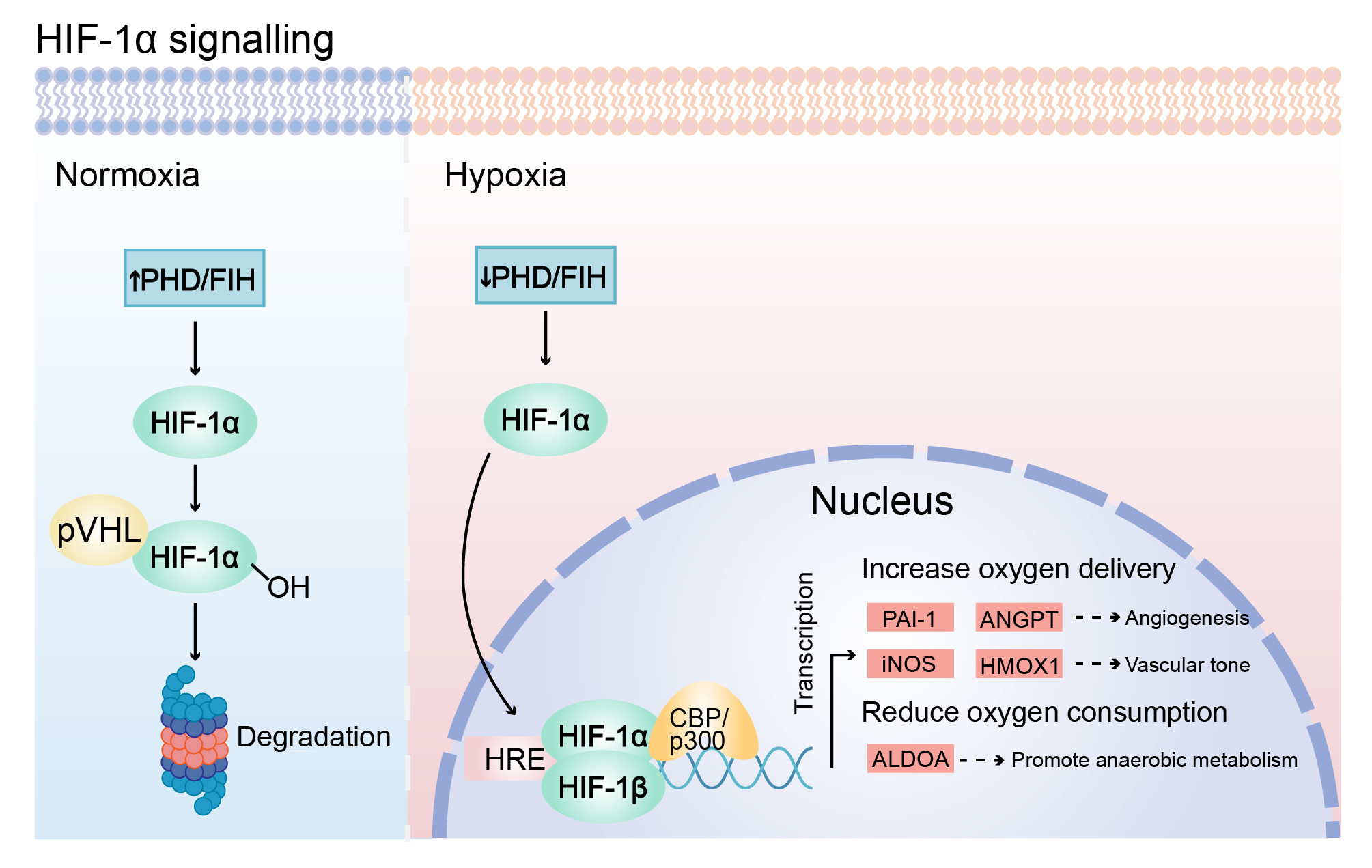
We performed a small sample transcriptome analysis to validate further the alterations in gastric mucosa—upregulation of genes associated with gastric mucosa malignancy of cirrhotic patients identified in differential gene analysis. We also found that the gene encoding DEFA5 upregulates, contributing to direct antimicrobial, mucosal host defense, and immunomodulatory properties[25]. Furthermore, the over-expression of defensins in multiple types of cancer, such as colon cancer, lung cancer, and renal cell carcinomas, suggests a potential involvement of defensins in cancer development[25–27]. Another study indicates that DEFA5 produced from metaplastic Paneth cells may accelerate the initiation of Barrett's esophagus, which is thought to be a precancerous lesion of esophageal adenocarcinoma[28]. An ex vivo animal study shows the down-regulation of the DEFA5 gene in gastric cancer cells and that DEFA5 inhibits the growth of gastric cancer cells[29]. The underlying mechanisms of DEFA5 in the initiation and progression of gastric cancer await further studies. Based on the pathway enrichment analysis of DEGs from gastric mucosa transcriptome, we found that the HIF-1 signaling pathway was significantly upregulated in the LC group. In a hypoxic environment, HIF-1α accumulates in cells and binds to HIF-1β to form HIF-1, and HIF-1 binds to hypoxia-responsive element to participate in multiple signaling pathways, leading to the response of cells to hypoxia. To adapt to hypoxia, cells activate hypoxia-inducible factors signaling and increase the expression of HIFs target genes, including those involved in cell survival, proliferation, angiogenesis, invasion, metastasis, and cancer stemness[30]. In the present study, angiogenesis-related genes (i.e., plasminogen activator inhibitor-1, angiopoietin) and vascular tone-related genes (i.e., inducible nitric oxide synthase, heme oxygenase 1) were upregulated in the LC group, which indicated that gastric mucosa increases oxygen delivery. Meanwhile, the upregulation of aldolase gene expression suggests an increase in anaerobic metabolism. Upregulation of these genes is an adaptive change in gastric mucosal cells in response to hypoxia.
There are some limitations of the study that are important to the trial. First, this is a single-center study with a limited sample size. Second, the etiology of LC is complex and diverse, which may yield different flora microenvironments and serum metabolites. Meanwhile, this difference can reflect the generality of the study's conclusions. Other factors, such as drinking and smoking, were not adjusted, which may impact gastric flora and damage to the gastric mucosa, but relevant antibiotic exposures have been excluded.
In conclusion, this study is the first work on integrated metabolomic, transcriptomic, and microbial analyses to identify critical metabolites and provide insight into the molecular and metabolic mechanisms underlying the alteration in gastric mucosa. Importantly, we showed that members of gastric pathogenic taxa were accumulated, which might originate from the oral cavity. The over-represented bacteria and serum bile acids co-exacerbated the damage of gastric mucosa and even accelerated the Correa's cascade process via regulating the HIF-1 pathway. Our study highlights the enrichment of the HIF-1 pathway in gastric mucosal cells in the context of LC, which may potentially serve as therapeutic targets for preventing gastric lesions.