Synthesis and characterization of Fe-CDs
In the present work, Fe-doped carbon dots (Fe-CDs) were derived from a Food and Drug Administration (FDA)-approved green iron supplement, i.e., ferrous gluconate, via an environmentally friendly one-step hydrothermal reaction. Our previous reports revealed that this kind of gluconic acid complex-based metal salt was suitable for the preparation of metal ions-doped CDs (MCDs) [23, 24]. Recently, MCDs had shown growing bioapplication prospects in the fields of diagnosis and therapy, due to the synergistic features of the nano-size effect, biocompatibility of CDs and specific functionality of metal ions [25]. Iron metabolism in the tumor-related cells was closely related to the occurrence and development of cancer, which inspired plenty of works on Fe-based anti-tumor nanomaterials, bringing cancer therapy into an iron age [26–28]. In this regard, it was speculated that the as-synthesized Fe-CDs might have potential as a novel iron agent for tumor treatment. First, the morphology and structure of Fe-CDs were investigated, as shown in Fig. 1a, the transmission electron microscopy (TEM) image showed that Fe-CDs appeared as uniform nanoparticle with an average diameter of 2.2 nm, such a small nanosize would facilitate the rapid entry of Fe-CDs into cells. The high-resolution TEM image revealed lattice fringes with 0.21 nm spacing, which was corresponding to the (100) crystal plane of graphite, indicating the graphitized carbon core within Fe-CDs [29] The photoluminescence (PL) spectrum of Fe-CDs exhibited an typical excitation wavelength-dependent blue fluorescence property (Fig. 1b) [30], and the maximum emission wavelength was 440 nm when excited under 358 nm light (Fig. 1c). There were two peaks observed in the UV-Vis absorption spectra of Fe-CDs that located at 252 and 316 nm, which could be attributed to the π→π* transition of C = C bond and n→π* transition of C = O bond, respectively [31]. Fourier transform infrared spectroscopy (FTIR) was adopted to study the functional groups of Fe-CDs, the stretching vibrations of O-H at 3386 cm− 1, C = O at 1610 cm− 1 and C-O at 1089 cm− 1 (Fig. 1d), demonstrating the abundant oxygen-containing groups on the surface of Fe-CDs that endow it with good water dispersibility.
Then, the existing states of Fe within Fe-CDs was further determined. X-ray diffraction analysis of Fe-CDs displayed a broad peak at around 20°, corresponding to amorphous carbon (Fig. 1e) [32]. There were no characteristic peaks of iron oxide or elemental iron, which might point to the fact that Fe dopants in Fe-CDs were inoic form. This was possibly because the hydrothermal conditions were relatively mild and the insoluble oxide from the as-prepared products were also subjected to post-treatment by filtration. The existence of Fe in Fe-CDs was confirmed by energy dispersive spectrometer (EDS), which determined the element weight percentage was 72.1%, 18.2%, and 9.7% for C, O, and Fe, respectively (Additional file 1: Figure S1). Besides, the Fe ion content was also accurately analyzed as 6.46 wt% by inductive coupled plasma emission spectrometer (ICP) (Additional file 1: Figure S2), similar to the EDS result. In order to further understand the elemental composition of Fe-CDs and doping forms of Fe, X-ray photoelectron spectroscopy was conducted, which confirmed the existance of C, O, and Fe in Fe-CDs with the atomic percentage (At.%) of 64.9%, 28.9%, and 6.2%, respectively (Fig. 1f). The deconvoluted Fe 2p spectra of Fe-CDs possessed the signals of Fe 2p3/2 and Fe 2p1/2 electronic configurations at the binding energy of 711.6 and 724.9 eV, respectively, along with the adjacent satellite peaks (Fig. 1g). These were associated with the presence of ionic Fe3+ according to previous reports [33], which further confirmed the XRD analysis. The C 1s spectra was divided into four types of carbon, i.e., sp2 C (C = C), sp3 C (C-C), C-O, and C = O at 284.4, 285.1, 286.3, and 288.4 eV, respectively (Fig. 1h), which was consistent with the FTIR results. The deconvoluted peaks in the O 1s spectra at 532.2 and 531.5 eV could be ascribed to C = O and C-O/Fe-O bonds, respectively (Fig. 1i). In summary, a type of Fe ions-doped fluorescent CDs was successfully synthesized, in which Fe ions could be bound to oxygen-containing functional groups on the surface of CDs through electrostatic interactions or ionic bonds. It would be of great interest to investigate the role of this novel nano-iron agent in the regulation of TME and the application in anti-tumor.
In vivo evaluation of anti-tumor effects of Fe-CDs
An orthotopic tumor model was constructed by subcutaneous injection of 4T1 cells (mouse breast cancer cells) in the back of Balb/C mice, which was a classic tumor model suitable for preliminary evaluation of the Fe-CDs anti-tumor effect. First of all, the in vivo metabolic kinetics of Fe-CDs was studied by intravenous injection of Fe-CDs chemically coupled with Cyanine 5.5 (Cy5.5), which could achieve near-infrared intravital imaging with low interference of tissue autofluorescence. As shown in Fig. 2a, the fluorescence signal was mainly concentrated in the liver area of mouse during the first 3 h, consistent with the previous reports that CDs being metabolized by liver and kidney [34]. Then, Fe-CDs gradually accumulated to the tumor site from 6 h after injection until 72 h (white dashed area), which could be due to the enhanced permeability and retention effect (EPR) effect of nano-sized Fe-CDs, or the increased permeability of tumor blood vessels leading to the local uptake of Fe-CDs [35].
The circulating administration of Fe-CDs (2 mg/20 g body weight per day) through tail vein was started when the subcutaneous tumor reached about 100 mm3, and the control group was an equal volume of PBS buffer solution injected intravenously (Fig. 2b). As we expected, the diameter of the resected tumor had grown to 12.56 ± 1.97 mm in the control group, while the tumor size of Fe-CDs group decreased to 6.74 ± 1.48 mm. Surprisingly, the tumors had completely disappeared in two groups of tumor-bearing mice (Fig. 2c), demonstrating the superior ability of Fe-CDs to resist tumors. The mean tumor volume gradually decreased and eventually shrinked to about half of the initial value when Fe-CDs were administered every 3 days (Fig. 2d). However, the tumor volume in the PBS-injected group increased by more than five times. Also, the average tumor weight of the PBS group was about 3.7 times that of the Fe-CDs treatment group, indicating that Fe-CDs significantly inhibited the in-vivo proliferation of solid tumors (Fig. 2e).
Moreover, systemic circulatory dosing of Fe-CDs had no significant influences on the mice weight, which demonstrated the extremely low toxicity of Fe-CDs (Fig. 2f). The hematoxylin-eosin staining (H&E) of tumor tissue illustrated that there were obvious necrotic areas and substantially remission of immune environment within the tumor area in Fe-CDs group compared with the PBS group, confirming that Fe-CDs could selectively induce tumor necrosis and ablation (Fig. 2g). It was worth mentioning that such an anti-tumor effect could be achieved by intravenous administration of Fe-CDs, without integrating other chemotherapy or phototherapy moieties, and targeting molecules. In order to further verify the biocompatibility of Fe-CDs to normal tissues, the vital organs (heart, liver, spleen, lung, kidney) of mice were dissected out for H&E staining, and the sections displayed no obvious changes of cell states and inflammatory infiltration in both the control group and Fe-CDs group, which also proved the excellent biocompatibility of Fe-CDs (Fig. 3). Therefore, a type of biocompatible Fe-CDs with exceptional anti-tumor activity was simply synthesized, which showed great potential as a target-free, low-toxicity, and effective anti-tumor nanodrug. It was necessary to explore and clarify the mechanism for the efficient tumor treatment effect of Fe-CDs, so as to provide guidance and reference for the preparation of Fe-based anti-tumor nanomaterials.
Cytotoxicity and ability of Fe-CDs to induce tumor cell apoptosis
Before conducting in vitro anti-tumor related experiments, the cellular localization and cytotoxicity needed to be assessed. Prussian blue staining was used to locate Fe-CDs in human triple-negative breast cancer cells (MDA-MB-231) by labeling ferric irons (Fig. 4a). After 6 h of co-culture with Fe-CDs, there were visible blue markers around partial cells, confirming initial cellular uptake of Fe-CDs. In addition, some cells showed morphological changes, nuclear pyknosis, and destruction of cell membrane after 12 h. With the extension of incubation time till 48 h, the content of Fe-positive labeled cells and necrotic cells gradually increased, which suggested that Fe-CDs could be enriched in MDA-MB-231 cells and induce cell death. Moreover, TEM images of tumor cell sections that co-cultured with Fe-CDs also clearly showed the presence of Fe-CDs in the state of aggregated particles within the cytoplasm in 2 h (Additional file 1: Figure S3), indicating that they could enter the tumor cells quickly.
Then, the cytotoxicity experiment of Fe-CDs was performed by MTT assay, and the raw material ferrous gluconate was used as a control to define the specific role of preparing Fe-CDs. It could be seen that Fe-CDs significantly led to MDA-MB-231 cell death with a time- and concentration-dependent manner (Fig. 4b), when the culture condition was 400 µg/mL of Fe-CDs for 3 d and 100 µg/mL for 5 d, the cell viability reached (53.8 ± 0.031)% and (55.9 ± 0.019)%, respectively, in other words, nearly half of the cancer cells died under Fe-CDs treatment. In contrast, the raw material compound, i.e., ferrous gluconate (FeG), had almost no cytotoxicity to tumor cells, similar to the control group. It was speculated that due to the improved water solubility and cell entry mode of iron drugs after nanometerization, which made it easier to be taken up by cells, and the exposure of iron on the surface of carbon dots was also conducive to its effect. It was worth mentioning that Fe-CDs could selectively kill tumor cells without showing obvious toxicity to normal cells, such as human dental pulp stem cells (hDPSCs) and human umbilical vein cell fusion cells (EA.hy926), with cell viability above 80% at all concentrations and time gradients (Additional file 1: Figure S4-5), indicating the the potential of Fe-CDs as biocompatible antitumor drugs.
In order to confirm how Fe-CDs caused tumor cell death, the effects of different inhibitors on cell activity were investigated. The cell ferroptosis inhibitor (Fer1) and necrosis inhibitor (Nec) showed negligible effects on the decrease of cell viability caused by Fe-CDs, while cell apoptosis inhibitor (Zvad) significantly reversed the Fe-CDs induced cell death (Additional file 1: Figure S6). Generally, ferroptosis was mainly caused by lipid peroxidation catalyzed by divalent Fe ions [36]. Although the as-prepared Fe-CDs was a type of Fe-doped nanomaterials, the Fe within Fe-CDs existed mainly in Fe3+ form, so the tumor cell death caused by Fe-CDs was not through ferroptosis way. In addition, the doping amount of Fe in Fe-CDs was relatively low, it was believed that the nano-structures and surface groups of CDs also played an important synergistic role in the improved anti-tumor function and biocompatibility of Fe-CDs. Further, Annexin-V and PI double staining was introduced to detect apoptosis by flow cytometer, which was shown in Fig. 4c. When the concentration of Fe-CDs was low (< 25 µg/mL), early apoptosis of MDA-MB-231 cells mainly occurred (Q3) and the apoptosis rate elevated with the increase of Fe-CDs concentration. Then late apoptosis started to rise (Q2) and became dominant as the concentration of Fe-CDs further increased to 100 µg/mL, representing that the progression of apoptosis was exacerbated by high Fe-CDs concentration. It was further verified by Western Blot that Fe-CDs induce tumor cell apoptosis rather than other death pathways. The addition of Fe-CDs activated the expression of apoptosis-related protein including PARP, Caspase 3, Caspase 9, and BAX with a time-dependent characteristic (Fig. 4d, Additional file 1: S7). Moreover, immunohistochemical staining of apoptosis-related markers was performed in tumor tissue sections, namely B-cell CLL/lymphoma 2 (Bcl-2) and Caspase 3, to further determine the ability of Fe-CDs in inducing tumor apoptosis. The up-regulation of apoptosis level in Fe-CDs group could be clearly observed as compared to the negative control (NC) group (Fig. 4e). The above results demonstrated that Fe-CDs possessed the function of inducing tumor cell apoptosis, which contributed to its excellent anti-tumor effect in vivo.
Ability of Fe-CDs to activate the immunity of antitumoral macrophages
Immunotherapy had become the most promising anti-cancer therapies in the clinic because it utilized the mobilization of autoimmune systems to achieve tumor eradication with extremely low side effects and recurrence rates. In tumor mircoenvrionment (TME), Fe was preferentially taken up by the surrounding tumor-associated macrophages (TAMs), forming iron-carrying macrophages [37, 38]. Previous reports had found that an FDA-approved iron agent, i.e., ferumoxytol (nano-iron oxide), could inhibit tumor cell proliferation by modulating macrophages functions [28, 39]. Regarding this, the effects of Fe-CDs on TAMs immunity were further explored. Similarly, the localization of Fe-CDs in mouse mononuclear macrophage leukemia cells (RAW 264.7) was also achieved by Prussian blue staining, showing that Fe-CDs began to enter cells within 1 day, and the degree of entry further increased at 3 days (Fig. 5a), which confirmed that Fe-CDs could be enriched in macrophages. Unexpectedly, both Fe-CDs and FeG showed no significant cytotoxicity to macrophages in a certain concentration range (Fig. 5b), that is, Fe-CDs could selectively kill tumor cells without side effects on other kinds of cells, and this performance was extremely important for the practical application of anti-tumor nanomedicines.
Then, quantitative real-time polymerase chain reaction (qPCR) and enzyme linked immunosorbent assay (ELISA) was implemented to analyze the expression level of Interleukin-10 (IL-10) in macrophages under the presence of Fe-CDs. The cytokine IL-10 was found to be abundantly secreted by activated macrophages in TME, making the phenotype of TAMs tend to be anti-inflammatory M2, which could inhibit the immune response and specific tumor killing [40, 41]. Here, it could be seen that Fe-CDs had no significant effect on IL-10 generation in macrophages under normal conditions (Fig. 5c). However, when macrophages cultured in tumor cells culture medium were used to mimic TAMs, the mRNA transcription and protein expression of IL-10 were obviously down-regulated by the addition of Fe-CDs. In other words, the differentiation of pro-inflammatory M1 type TAMs was promoted through inhibiting IL-10 secretion by Fe-CDs, which could awaken the immune function of antitumoral macrophages to selectively kill tumor cells. Noteworthy, the primitive drug FeG showed a relatively weaker activity than Fe-CDs due to the poor solubility and uptake rates. The results of immunofluorescence staining further proved the conclusion that the tumor cell culture significantly stimulate macrophages to secrete IL-10, which could be effectively restrained by Fe-CDs (Fig. 5d). Moreover, another two typical soluble mediator secreted by M2 macrophages, Arginase-1 (Arg-1) and phosphorylation of P38 (p-P38), could serve as the specific markers for M2 polarization [41, 42]. It could be clearly seen that the presence of Fe-CDs significantly reduced the expression of Arg-1 and p-P38 in macrophages cultured in tumor cell medium, whereas FeG had almost no effect on Arg-1 and p-P38 levels (Fig. 5e, Additional file 1: S8-9). In addition, compared with untreated TAMs (Additional file 1: Figure S10-11), Fe-CDs also markedly inhibited the phosphorylation levels of signal transducer and activator of transcription 3 (STAT3), c-Jun N-terminal kinase (p-JNK), extracellular regulated protein kinases (ERK), and P65 with a time-dependent tendency (Fig. 5f, Additional file 1: S12), which both belonged to the Mitogen-activated protein kinases (MAPK) signaling pathway. According to the previous reports, the activation of MAPK was essential for the antitumoral macrophages [43]. Moreover, the in vivo histochemical staining results of IL-10 and Arg-1 in tumor tissues further demonstrated that Fe-CDs stimulated antitumoral macrophages by inhibiting the expression of IL-10/Arg-1 (Fig. 5g). Based on the above results, it was concluded that the as-prepared Fe-CDs could modulate tumor immunity by the macrophage IL-10/Arg-1 routes, and promoted the transformation of macrophages to the tumor-specific killer M1 phenotype through the MAPK pathway (Fig. 5h), especially the regulating ability was stronger than that of FeG due to the improved internalization efficiency after nanoization.
Ability of Fe-CDs to inhibit epithelial-mesenchymal transition (EMT) process
The metastasis of tumor cells, which led to the recurrence and spread of malignant tumors, had been considered as the most important factor in the incurability of cancer [44]. In recent years, it was found that the programmed activation of epithelial-mesenchymal transition (EMT) could make tumor cells lose the tight junction and adhesion between cells, and then switched to the morphology of mesenchymal cells with strong migratory ability [45]. Surprisingly, the as-synthesized Fe-CDs possessed the unique ability to inhibit tumor cell migration and EMT. First, the scratch assay images of MDA-MB-231 cells cultivated with Fe-CDs exhibited a significant reduction of proliferation rate compared to the control group that incubated in phosphate buffered saline (PBS) (Additional file 1: Figure S13a). The migration rate of the control group in scratch assay was 57%, while the migration rate of cells treated with Fe-CDs was reduced to almost 6% (Additional file 1: Figure S14a). Then, the morphology of tumor cells observed under microscope changed from cobblestone-like to spindle-like shape, indicating the loss of cell polarity and increased migratory capacity (Additional file 1: Figure S13b). However, the existence of Fe-CDs (50 µg/mL) could partially reverse the cytoskeletal changes to a certain extent, representing its potential in suppressing EMT of tumor cells. In addition, cobalt chloride (CoCl2) was introduced to further explore the function of Fe-CDs in suppressing EMT, as an appropriate amount of CoCl2 had been reported to induce mesenchymal cell transformation [46]. Similar to the above experiments, it could be seen that although CoCl2 accelerated the cell migration process, the addition of Fe-CDs could still effectively prevent tumor cells from infiltrating into the scratch (Fig. 6a). The migration rate of cells treated with CoCl2 was 50%, while the migration rate of tumor cells co-treated with CoCl2 and Fe-CDs was reduced to 8% (Additional file 1: Figure S14b). Also, the exacerbated process of cell morphological transformation was alleviated by Fe-CDs, that is, in the presence of Fe-CDs, the tumor cells could be converted from aggressive spindle shape to conservative round shape (Fig. 6b). These results were further confirmed by the transwell migration experiments shown on the left side of Fig. 7b, which clearly depicted that the number of chemotactic tumor cells in both the control and CoCl2 groups was significantly higher when Fe-CDs was absent.
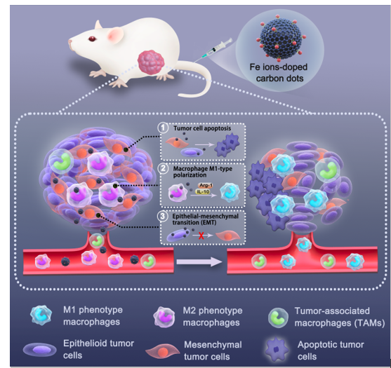
Furthermore, several EMT-related markers were performed by WB analysis, including Fibronectin (FN1), N-cadherin, Vemintin, α-Smooth muscle actin (α-SMA), Snail, which had been reported to induce EMT, and also Zonula occludens-1 (ZO-1), Occludin (OCLN), which could inhibit EMT (Additional file 1: Figure S15) [47]. It could be seen that Fe-CDs treatment significantly reduced the expression of EMT positive-associated proteins and showed an increase in the expression level of EMT inhibitory proteins with a concentration-dependent manner (Additional file 1: Figure S16). The obvious up-regulation of p-P38 and p-ERK with the addition of Fe-CDs also suggested that EMT process might be inhibited through related pathways. Similarly, when CoCl2 was introduced, the above proteins were also subjected to WB test, which confirmed that they could accelerate the EMT process of tumor cells over time (Additional file 1: Figure S17), whereas Fe-CDs effectively counteracted the progress of EMT (Fig. 6c, Additional file 1: S18-19). Furthermore, the function of Fe-CDs to inhibit EMT was also verified in vivo by staining with the antibodies of E-cadherin, OCLN, N-cadherin, Vimentin and Snail, similar to the WB assay in cells (Fig. 6d). The above results demonstrated that Fe-CDs might have the potential to suppress cancer migration by blocking the EMT process of tumor cells. Since the as-synthesized Fe-CDs had been proved to be equipped with three-step cancer therapy functions, i.e., inducing tumor cell apoptosis, modulating macrophage immunity, and inhibiting EMT, leading to the superior anti-tumor effect of Fe-CDs in vivo without integrating other moieties and external stimuli, it was believed that Fe-CDs could serve as a promising alternative to tumor immunotherapy agents and anti-tumor nanoplatform.