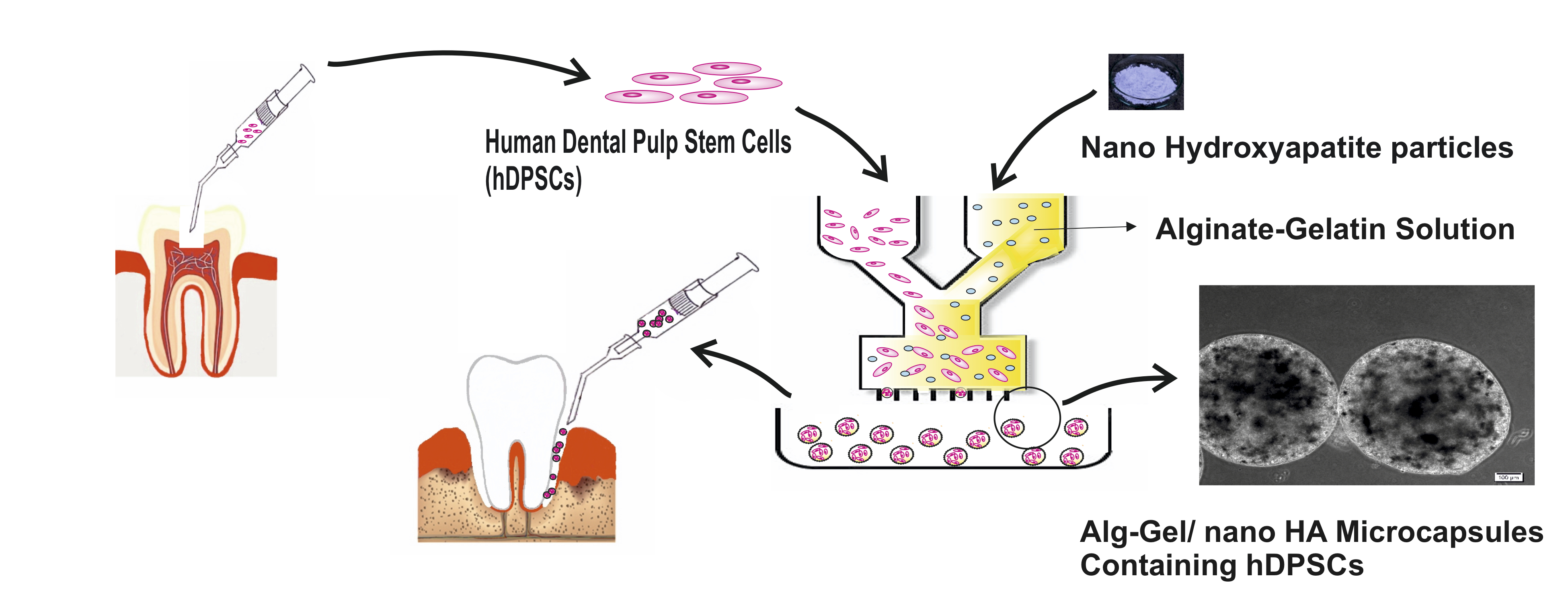
Microcapsules are widely used in tissue engineering including regeneration of different tissues and organs such as liver, cartilage, skin, neural tissue, and bone(9, 48–52). These microstructures with different characteristicsincluding immunoisolation, micrometer size, and providing 3D microenvironment considered a promising approach in regenerative medicine(9, 53). These modular microcarriers could directly be injected and transplanted to the defect side(54). This property is important for the reconstruction of hard tissues. The injectable microcarriers could adjust to bone defects with irregular shape and geometry, occupying the available spaces, precluding the invagination of the adjacent tissues and promoting tissue repairing(55, 56). As shown in graphical abstract, the current study aimed to use the available source of stem cells in the oral cavity and microcapsulating of theses cells for modular tissue engineering approach of bone defects in the oral and maxillofacial region.
The main findings of this study is the high osteogenic differentiation capacities of hDPSCs in Alg/Gel/nHA microcapsules. Moreover, both ALP and Alizarin red staining of micro carriers showed a greater extent of mineral deposition in nano- hydroxyl apatite modified microcapsules.
In the current study we investigated the osteogenic potential of hDPSCs as an available source of mesenchymal stem cells in Alg/Gel/nHA microcapsules for the first time.
Alginate is a natural biopolymer that has been widely used for drug delivery, dental impression materials, and tissue engineering (4, 57).
Gelatin is derived from collagen, the major organic component of the extracellular matrix of bone tissue, with adequate properties for bone tissue regeneration and revascularization (58, 59).
In the structure of Alg/Gel microcapsules, a rapid reaction between carboxyl groups of alginate and barium cations makes it possible to form an ion-crosslinking hydrogel. Microcapsules were cross-linked by the ionotropic gelation in the presence of BaCl2 that has shown high stability and low degradability in comparison to CaCl2 (60). Indeed, adding nHA to the system increases the crosslinking of the polymeric network, leading to an enhanced homogeneity and strength of the microcapsules (55).
Nano-hydroxyapatite (nHA) as the major inorganic mineral part of bone could increase the homogeneity and strength of microcapsules. Moreover, nHA increased the crosslinking of the polymeric network (55). These features besides, the facilitation of bone formation in the presence of nHA turn it to the important component of BTE (27).
The interaction between calcium ions of nHA and G-blocks of alginate makes a strong matrix with the rougher surface which improves cell adhesion, proliferation, and differentiation compared to smooth alginate surface. In the current study, the proliferation of hDPSCs in the Alg/Gel/nHA microcapsules was higher than Alg/Gel and control groups. Moreover, the cell viability in the micro-carrier structures was influenced by the size and mass too, which can diffuse inner layers (47). The viability and proliferation of hDPSCs in Alg/Gel and Alg/Gel/nHA microcapsules increased during the study. This viability is a result of an appropriate bead size, which could provide an immobilized matrix with a permeable membrane that facilitates waste, nutrients, and oxygen transmission for cells. This could avoid the crowd of toxic wast and lead to appropriate activity and proliferation of cells. These results were in accordance with other previous studies that used microcapsulation methods for cell transferring (7, 27).
The aim of BTE is not only to provide 3D structutres and cell proliferation without cell cytotoxicity or foreign body reaction but also the regeneration of new bone by provoking osteogenic differentiation of stem cells (4, 61).
In the oral and maxillofacial regions for the regeneration of bone defects, the non-invasive injectable methods for stem cell delivery are more useful (62). The osteo/odontogenic differentiation of hDPSCs on various scaffolds was evaluated previously (41, 43, 63, 64). However, none of these scaffolds evaluated non-invasive injectable carriers for these cells. In the current study, we transferred these accessible stem cells in the oral region to spherical injectable microcapsules, which could be useful for bone regeneration of this area.
The osteogenic differentiation of hDPSCs in Alg/Gel and Alg/Gel/nHA group was assessed by the relative expression of BMP–2, Osteocalcin, Osteonectin, and RUNX–2. Various extracellular ligands such as BMPs, WNTs, and FGFs controlled osteogenic differentiation of different multipotent mesenchymal stem cells (65). These ligands direct the three main stages of osteogenic differentiation, which associate with the expression of some genes like BMP–2, RunX–2, Osteocalcin, and Osteonectin (46).
The expression of BMP–2 as the most important growth factor in bone formation is known as an early indicator of calcified tissue generation and osteoblastic differentiation (61, 66). The expression of this factor was significantly higher in Alg/Gel/nHA group after 21 and 28 days.
Osteocalcin and osteonectin are noncollagenous proteins in the extracellular matrix of bone. Osteocalcin has an important role in the maturation of mineralized tissues and the regulation of osteogenic differentiation of mesenchymal stem cells (67). The microcapsulation of hDPSCs has positive effects on the expression levels of these genes. However, these effects were greater in Alg/Gel/nHA group.
RUNX–2 is known as a critical transcription factor associated with bone formation and plays an important role in the differentiation of pre-osteoblastic cells to mature osteoblasts. Also, this factor upregulates the VEGF factor which is important in the angiogenesis of bone tissue (29, 68). The upregulation of this gene as a late indicator of osteogenesis was higher than other genes after 21 and 28 days.
Moreover, DSPP, a classic odontogenic differentiation marker, gene expression was assessed in order to investigate any odontogenic differentiation of hDPSCs in microcapsules. As demonstrated in results, the up-regulation of this gene was not increased as well as osteogenic genes in microcapsules, which indicates the osteogenic potential of Alg/Gel/nHA microcapsules rather than odontogenic potential.
In general, hDPSCs in Alg/Gel/nHA microcapsules showed higher expression of these genes after 21 and 28 days. These results were in compliant with other previous studies, which evaluated the different alginate- nano hydroxyapatite-based scaffolds on osteogenic differentiation of stem cells (55, 61, 69).
To evaluate the mineralized deposition of hDPSCs in Alg/Gel/nHA microcapsules Alkaline phosphatase and Alizarin red staining were performed in the current study. The formation of mineralized nodules as shown in our results indicate the late stage of osteogenic differentiation. In summary, our results indicated the notable enhancement in mineralization of hDPSCs in Alg/Gel/nHA microcapsules after 21 days.
The combination of calcium with alizarin red makes orange nodules, which showed mineralization sites (41). These nodules were clearly observed in Alg/Gel/nHA group.
ALP enzyme activity as a major factor in the beginning phase of bone matrix mineralization was quantified by the hydrolysis of p-nitrophenyl phosphate to p-nitrophenol (70).This enzyme is a universal marker for the indication of mineralization. ALP deposition occurs during osteoblast maturation and bone matrix mineralization (71). Therefore, the ALP activity could demonstrate the biological activity of osteoblasts (41). Moreover, The formation of the bone-like mineralized tissue due to calcium deposition is essential to bonding of newly regenerated bone with the former bone tissue (55, 72).
The results observed for physical and osteoconductive properties of nHA particles in the hDPSCs-laden alginate hydrogels were in agreement with the reported results of other previous studies (6, 26, 41, 44), indicating the importance of nHA bioactivity in the composite hydrogel.
In general, our results showed that the proliferation of hDPSCs increased and the expression of all osteogenesis genes upregulated in nano-hydroxyapatite containing microcapsules. Moreover the calcium deposition was increased in the presence of nHA. These results are in compliance with other studies, which used these microcapsules on different stem cells (45, 48, 73). However, this study evaluated these modular microcapsules in osteogenic differentiation of hDPSCs for the first time, based on authors’ information.