Structural and morphological characterization
The structural and morphological characterizations of PI, PI/TiO2 and PI/TiO2/Ag flexible nano/microfibers were comprehensively demonstrated through a suite of analytical techniques including Fourier transform infrared spectroscopy (FTIR), Thermogravimetric analysis (TGA), X-ray diffraction (XRD), Energy dispersive X-ray spectroscopy (EDX), Scanning electron microscopy (SEM), Transmission electron microscopy (TEM) and X-ray photoelectron spectrum (XPS).
The FT-IR spectroscopy validates the distinctive structure and chemical bonds of PAA, PI, and PI/TiO2 flexible fibers. As exhibited in Fig. 1a, the characteristic absorption peaks at 1598 and 1502 cm− 1 can be attributed to the C = O stretching vibration and N-H vibration in PAA 20. The characteristic absorption peaks at 1771, 1724, 1550, 1353 and 735 cm− 1 are associated with the asymmetric stretching, the symmetric stretching, the aromatic C = N in the triazine unit, the stretching vibration of C-N-C in the five membered imide rings and the bending vibrations of the C = O bond in the imide unit of PI, respectively 21. The broad peaks observed in the range of 400 to 800 cm− 1 align with the stretching vibration of Ti-O-Ti, demonstrating the in-situ surface-grown of TiO2 on PI 22. These findings provide evidence that the primary chemical structure and bonding skeleton of PI remain unaffected by the TiO2 deposition.
The thermal stability and quantitative analysis were investigated through TGA. As shown in Fig. 1b, the imidization reaction of PAA initiates at approximately 200°C. As the temperature reaches 300°C, the macromolecular chains within the PAA nanofibers undergo rearrangement, promoting the formation of a more stable conformation and ultimately resulting in the formation of PI nanofibers. When comparing the thermal weight loss curves of PAA and PI nanofibers at 300 ℃, it can be seen that PI nanofibers no longer suffer from thermal weight loss and hence there is no imidization reaction. From that we can infer, after 2 hours of thermal imidization process of PAA nanofibers at 300 ℃, the internal molecular chain of PI has formed a stable conformation, showing good thermal stability. At about 580°C, an obvious inflection appears because the molecular chains of PI nanofibers begin to break. At this point, the thermal weight loss rate reaches maximum due to the carbonization of PI nanofibers. At about 800 ℃, they are almost fully carbonized into carbon fibers 19. Noteworthy, the weight loss of PI/TiO2 is less than that of PI, which can be ascribed to the addition of TiO2. The above results show that our prepared PI and PI/TiO2 nanofibers have good thermal stability.
The generated samples were subjected to XRD analysis to investigate their crystal structures and phase transition. Figure 1c depicts the XRD patterns of PI, PI/TiO2 and PI/TiO2/Ag flexible fibers. The prominent peak at 2θ value of 27.9°is related to the interlayer π-π stacking of PI, specifically corresponding to the (001) facet of PI 23. The primary diffraction peaks of PI/TiO2 and PI/TiO2/Ag are comparable. The typical diffraction peaks at 27.4°, 36.1°, 39.2°, 41.2°, 44.1°, 54.3°, 56.6°, 62.7°, 64.0°, 69.0° and 69.8° can be attributed to the crystal planes of crystalline rutile TiO2 (JCPDS NO. 21-1276) 24. But oddly, the XRD patterns of PI/TiO2/Ag exhibit an absence of the diffraction peaks corresponding to Ag. That may be explained by the low amount of Ag present or by the fact that the diffraction peaks of Ag QDs are obscured by those of TiO2. Nevertheless, the EDX spectrum of PI/TiO2/Ag-0.07 can prove the existence of Ag. As shown in Fig. 1d, Ag, along with Ti, O, C, and N elements are detected. The aforementioned results demonstrate the formation of PI/TiO2/Ag.
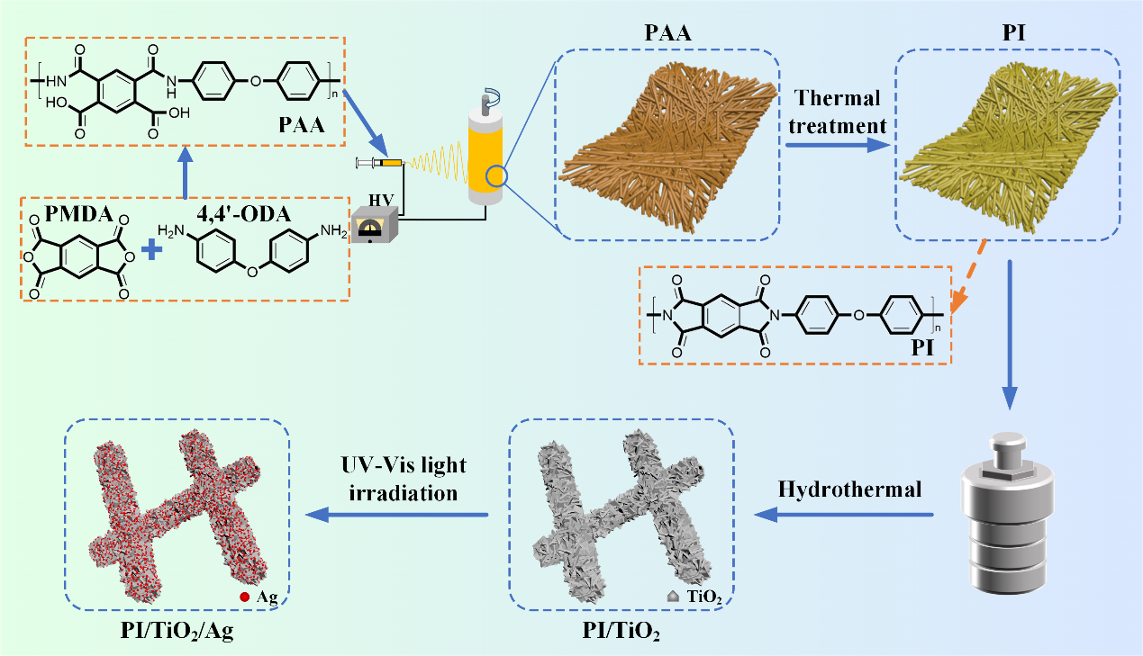
Afterwards, the morphologies of the produced PAA, PI, PI/TiO2 and PI/TiO2/Ag were examined using SEM. As illustrated in Fig. 2a and 2b, the PAA and PI nanofibers all exhibit smooth and uniform surfaces. But the diameter of PI nanofibers (about 200 nm) shrinks, compared with the PAA nanofibers (about 250 nm). This owes to the volatilization of DMF solvent and thermal imidization reaction during calcination. And as seen from Fig. 2c, TiO2 nanorods (NRs) with a petal-like appearance are uniformly grown on the surface of PI nanofibers. However, Ag QDs are not readily visible in Fig. 2d-2f.
Further, the microstructures of PI/TiO2 and PI/TiO2/Ag were investigated using TEM and High-resolution TEM (HRTEM) images. Figure 3(a1-d1) affirm that, in PI/TiO2, TiO2 nanorods (size: about 250 nm) are uniformly and compactly grown on the surface of PI nanofibers. Figure 3(a2-d2) also show that the number of Ag DQs on the surface of PI/TiO2 gradually increases with the increasing concentration of AgNO3. Figure 3(a3-d3) demonstrate that Ag DQs are bonded to the PI/TiO2 with an average size of 15 ± 5 nm, and the PI/TiO2/Ag samples include both the TiO2 NRs and Ag DQs crystal lattices. The lattice spacing of 0.32 nm observed in the figure matches to the (110) crystal plane of TiO2 (JCPDS NO. 21-1276) 25. The lattice distance of 0.24 nm in the figure corresponds to the (111) crystal plane of Ag (JCPDS NO. 87–0597) 26. The elemental mapping of PI/TiO2/Ag-0.07 microfibers is depicted in Fig. 3e, which indicates that the C, O, N, Ti and Ag elements are well-distributed throughout the PI/TiO2 nanofibers. Additionally, the distribution diameter of C and N elements in PI is smaller compared to that of Ti elements in TiO2, and the distribution diameter of Ti elements in TiO2 is smaller than that of Ag elements, indicating the formation of hierarchical core-shell structure.
Furthermore, the valence states and elemental composition of those prepared samples were verified through XPS analysis. As observed in Fig. 4a, the peaks corresponding to the elements C, N and O in PI/TiO2 and PI/TiO2/Ag are in agreement with which in PI. The peaks with binding energies of 458.5 eV and 464.2 eV observed in Fig. 4b are assigned to Ti 2p3/2 and Ti 2p1/2, and the 5.7 eV splitting between them shows the existence of natural form of Ti (Ti4+) 27. The Ag 3d XPS spectra of PI/TiO2/Ag-0.07 is shown in Fig. 4c. The identified peaks at 368.7 eV and 374.7 eV belong to Ag 3d5/2 and Ag 3d3/2 of metallic Ag (0), respectively. Notably, the observed energy difference of 6.0 eV between the Ag 3d3/2 and Ag 3d5/2 binding energies is a characteristic feature of the metallic Ag 3d state 28. Figure 4d displays the C 1s XPS spectra of PI, PI/TiO2, and PI/TiO2/Ag-0.07, which are characterized by three peaks located at 288.2 eV, 285.8 eV, and 284.6 eV, respectively. The binding energies of the peaks are calibrated relative to the C 1s peak at 284.6 eV, which is assigned to carbon contamination or the sp2 C-C/C = C bonds. In comparison, the peaks observed at 288.3 eV and 285.8 eV are attributed to the O-C = O bonds in the triazine rings and the C-N bonds in PI, respectively 29. As can be seen from Fig. 4e that N 1s peaks at 400.4 eV are associated with the N-(C)3 bond of PI 30. In Fig. 4f, the O 1s spectrum of PI displays two peaks at 532.0 and 533.2 eV, which can be attributed to C = O and C-O or adsorbed water, respectively. In comparison, PI/TiO2 and PI/TiO2/Ag have one additional peak at 529.8 eV, which corresponds to Ti-O bond 31, 32. Noteworthy, in Fig. 4d-4f, the peaks of PI/TiO2 exhibit a redshift relative to PI, and the peaks of PI/TiO2/Ag exhibit a redshift relative to PI/TiO2, indicating the successful incorporation of PI and TiO2, as well as PI, TiO2, and Ag. In addition, the binding energies of Ti 2p and O 1s in PI/TiO2/Ag are redshifted towards lower energy compared to PI/TiO2, indicating the electrons transfer from Ag to TiO2.
Photodegradation of TC
To ascertain the specific surface area and pore structures of the synthesized photocatalysts, Nitrogen adsorption-desorption isotherms are completed. According to Fig. 5a, the isotherms of PAA, PI, PI/TiO2 and PI/TiO2/Ag flexible fibers all exhibit type IV isotherms with type H3 hysteresis loops, implying that a slit-like mesoporous structure has been formed. The specific surface areas of PAA, PI, PI/TiO2 and PI/TiO2/Ag flexible fibers are estimated to be 17, 17, 21, and 29 m2/g, respectively. And clearly, PI/TiO2/Ag flexible microfibers have the highest specific surface area. As demonstrated in Fig. 5b, a significant proportion of pores with diameters ranging from 0 to 15 nm is observed, which aligns with the observations from SEM and TEM images. The presence of these abundant active sites in the hierarchical mesoporous nanostructure is believed to contribute to the high photocatalytic activity, thereby facilitating photocatalytic reactions 33.
As seen in Fig. 5c, the photocatalytic performance of PI, PI/TiO2 and PI/TiO2/Ag flexible fibers with different Ag QDs deposition amounts has been investigated by the photocatalytic degradation of TC under simulated sunlight irradiation. Under the simulated sunlight irradiation, the PI and PI/TiO2 exhibit very poor catalytic performance in degrading TC (16.2% and 25.7%, after 120 min), due to their inefficient photogenerated carrier separation and carrier transfer. Then, we start to load Ag QDs on the PI/TiO2 microfibers. As the content of AgNO3 increases from 0.05 M to 0.07 M, the photocatalytic degradation efficiency of TC has been increasing from 77.7–93.1%. However, as the concentration of AgNO3 further increases to 0.09 M, the photocatalytic degradation efficiency does not continue to increase but rather decreases to 63.3%. The enhanced photocatalytic efficiency is attributed to multiple factors 26. Firstly, the formation of a Schottky barrier between Ag and TiO2 facilitates the efficient separation of photogenerated electrons and holes. Additionally, the remarkable surface plasmonic resonance (SPR) and visible light absorption properties of Ag contribute to the enhanced photocatalytic activity and solar energy utilization efficiency. Moreover, the immobilization capability of PI and the incorporation of Ag QDs result in an enlarged specific surface area, further enhancing the photocatalytic performance. However, when more Ag QDs (0.09 M AgNO3) was located, the excessive amount of Ag QDs tended to occupy parts of reactive sites, which hinders the formation of the photocatalytic intermediates, handicaps the surface’s adsorption capability of TC, and consequently diminishes the photocatalytic performance 34, 35. Simultaneously, PI/TiO2/Ag is employed for degrading Rhodamine B (Rh B), Methylene Blue (MB) and Methyl Orange (MO). As shown in Fig. 5d, PI/TiO2/Ag exhibits excellent photocatalytic degradation activity towards these three pollutants (100%, 95.5%, and 100%), validating its high efficiency as a versatile photocatalyst suitable for the removal of diverse pollutants.
Photoreduction mechanism of TC
The light absorption properties and band gaps (Eg) of PI, PI/TiO2 and PI/TiO2/Ag-0.07 flexible fibers were determined using Ultraviolet-visible diffuse reflectance spectra (UV-vis DRS) and corresponding Tauc plots. In Fig. 6a, the spectral absorption of PI and PI/TiO2 exhibits a significant presence in the UV region, while it decreases rapidly in the visible band. When Ag QDs are deposited, as shown in the UV-vis DRS of PI/TiO2/Ag-0.07, the light absorption in the visible band is hugely enhanced, which is attributed to the SPR absorption of Ag QDs 36. The inserted plots in Fig. 6a depict the Tauc curves corresponding to PI, PI/TiO2 and PI/TiO2/Ag-0.07. From the graph, it can be observed that their respective Eg values are 2.59 eV, 2.96 eV and 2.14 eV, respectively.
The effectiveness of photocatalytic activity and the separation of photogenerated charge carriers in the obtained samples are evaluated by measuring Photoluminescence (PL) spectra with an excitation wavelength of 350 nm and Electrochemical Impedance Spectroscopy (EIS). In Fig. 6b, PI exhibits a prominent peak within the wavelength range of 400–500 nm. Similar peaks in PI/TiO2 and PI/TiO2/Ag-0.07 are centered at around 410 nm. This is attributed to the fact that the Ag 3d energy level functions as an electron acceptor, effectively capturing the charge carriers during the recombination of photogenerated electrons and holes 37, 38. Noticeably, the PL intensity of PI/TiO2/Ag-0.07 is much lower than PI/TiO2, suggesting a reduced rate of electron-hole recombination rate and improved photocatalytic activity. In other words, the synergy of Ag QDs and TiO2 NRs interlayer facilitates the transport of photogenerated charge carriers. Here we also like to point out, charge transfer not only helps the photocatalytic efficiency of PI/TiO2/Ag, but also improves its SERS performance 39. Figure 6c further shows Nyquist plots for various flexible nano/microfibers. The radius of the arc reflects the reaction rate at the surface of the flexible composite fibers, which can be interpreted as the charge transfer resistance (Rct). It’s discovered that PI/TiO2/Ag-0.07 has much smaller Rct values than PI/TiO2 and PI, revealing that the PI/TiO2/Ag-0.07 flexible microfibers possess the highest efficiency in separating photogenerated electron-hole pairs and facilitating rapid interface charge transfer 40.
Furthermore, the photocurrent response (i-t) experiments are performed under simulated sunlight irradiation with on/off cycles of 20 s. As illustrated in Fig. 6d, the as-prepared samples possess fairly stable photocurrent responses. Especially, the PI/TiO2/Ag-0.07 flexible microfibers exhibit much higher photocurrent and transient photocurrent density than PI/TiO2 and PI, indicating it has the most effective separation of photogenerated electrons. This enhanced performance can be attributed to the effective interfacial charge transfer facilitated by the tight bonding and synergy of TiO2 NRs and Ag QDs. Furthermore, this unique combination contributes to the excellent photocatalytic and SERS activity of PI/TiO2/Ag-0.07.
The semiconductive type and flat band (Fermi energy level, EF) potentials are determined via electrochemical Mott-Schottky (MS) testing. Figure 6e illustrates the potentials of PI, PI/TiO2 and PI/TiO2/Ag-0.07, which are 0.29 V, -1.31 V and − 0.60 V (vs. Ag/AgCl), respectively. These values can be converted into the Normal Hydrogen Electrode (NHE) potential by applying the blow formula:
E NHE = EAg/AgCl + 0.198 V (1)
Thus, the calculated EF potentials of PI, PI/TiO2 and PI/TiO2/Ag-0.07 are 0.49 V, -1.11 V and − 0.40 V vs. NHE. Further, the VB-XPS spectra (Fig. 6f) are used to determine the relative potentials of the valence band (VB) vs. Fermi energy level, and the estimated values for PI, PI/TiO2, and PI/TiO2/Ag-0.07 are 1.53 eV, 2.75 eV, and 1.98 eV, respectively. The VB of PI, PI/TiO2 and PI/TiO2/Ag-0.07 are calculated as 2.02 V, 1.64 V and 1.58 V, and the conduction bands (CB) are − 0.57 V, -1.32 V and − 0.56 V. Based on the previously mentioned band structure values, a possible photocatalytic degradation mechanism of TC by PI/TiO2/Ag is illustrated in Fig. 6g. Under the simulated sunlight irradiation, Ag QDs generate intense electric field near their plasmon frequency, which causes the electrons of TiO2 being excited to transition from the VB to the CB 41. Subsequently, the electrons transfer from the CB of TiO2 to Ag could convert O2 into·O2− (superoxide anion radical) (-0.33 V vs NHE). Nonetheless, the holes generated from the valence band of TiO2 are not able to convert H2O into ·OH (hydroxyl free radical) due to the negative position of the valence band of TiO2 compared to the potential of ·OH/H2O (2.34 V vs. NHE) 42, 43. 2H+, e− and ·O2− can further react to produce a small amount of ·OH. Importantly, the energy levels of Ag and the CB of TiO2 are different. As a result, electrons migrate from the CB of TiO2 to the Fermi level of Ag, facilitating electrons/holes separation and thereby accelerating the deterioration of TC 44. Photocatalytic process can be described by the following procedures:
PI/TiO2/Ag + hv → TiO2(h+) + Ag(e−) (2)
O2 + e− → ·O2− (3)
·O2− + e− + 2H+ → H2O2 (4)
H2O2 + e− → ·OH + OH− (5)
·O2−/·OH/h+ + TC → Products (6)
To provide a more intuitive verification of the active species in the photocatalytic mechanism described above, the degradation efficiency of TC by PI/TiO2/Ag-0.07 are investigated with various scavengers. As displayed in Fig. 6h, the degradation efficiency of TC is reduced upon the addition of different reactive species scavengers. Specifically, the addition of 1 mM EDTA-2Na 45 and 4-Hydroxy-TEMPO 46 as scavengers for h+ and ·O2−, respectively, results in a substantial decrease in the degradation rate of TC. Only 21.8% and 26.8% of TC degrades, which is severely reduced compared to the degradation rate without any scavengers (93.1%). This strong inhibition implicates that both h+ and ·O2− are important reactive species, although h+ has a little greater influence. Moreover, by using 1 mM IPA 47 as a scavenger of ·OH, the photocatalytic degradation rate of TC is reduced from 93.1–75.3%, indicating the significance of ·OH in the degradation of TC. These results are consistent with the conclusions drawn from the photocatalytic mechanism.
SERS activity analysis
As we mentioned, PI/TiO2/Ag not only helps photocatalytic degradation, but also enhances SERS detection of pollutants. As SERS substrates, PI/TiO2 and PI/TiO2/Ag-0.07 are utilized to detect 4- ATP from 1×10− 5 M solution in order to find the best SERS substrate. Based on the observation of Fig. 7a, the peaks detected at 1137cm− 1, 1386cm− 1, and 1430cm− 1 correspond to the "b2" vibration modes, associated with vC−C+vC−S, βC−H, and vC−C, respectively. Additionally, the peaks observed at 1074 cm− 1, 1178 cm− 1, and 1575 cm− 1 represent the "a1" vibration modes, attributed to vC−C+vC−S, βC−H, and vC−C, respectively 48. The peak detection intensity of PI/TiO2 for 4-ATP is very weak. But when PI/TiO2/Ag-0.07 is applied, the SERS signal is significantly enhanced, which indicates that PI/TiO2/Ag can achieve high-sensitivity detection of 4-ATP. The enhancement originates from two reasons: first, under the irradiation, the localized surface plasmon resonance (LSPR) effect of Ag QDs forms a local electromagnetic field and accelerate the charge transfer in PI/TiO2/Ag; second, the Schottky barrier formed by Ag QDs also promotes the charge transfer. The promoted charge transfer enhances the SERS signal 49, 50. We then apply PI/TiO2/Ag-0.07 substrate for SERS detection of TC. As given by Fig. 7b, the SERS signal decreases gradually as the TC concentration decreases, but even at a low concentration of 1×10− 10 M, the PI/TiO2/Ag-0.07 substrate still generates appreciable SERS signal.