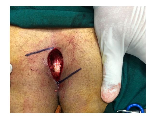
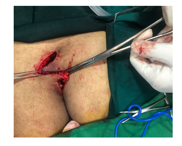
Despite the various surgical and nonsurgical methods for the treatment of pilonidal sinuses, no standard procedure has yet been proposed. The most appropriate treatment has long been a subject of debate. Although nonsurgical methods have gained popularity in recent years and, in some cases, have yielded acceptable results, the employment of different surgical techniques remains the mainstay of treatment in most surgical departments. (17, 18) Most of these surgical techniques utilize a common approach consisting of complete excision of all sinus tracts and surrounding tissues until the sacro-coccygeal bone is reached. Until this stage of treatment, almost all surgical procedures use the same technique. After this stage, however, the differences in treatment methods become evident. (2)
Traditionally, pilonidal sinus disease has been treated by excision of the sinus tract and leaving the wound open to heal by secondary intention. Due to the secondary wound healing method’s higher monetary costs, longer duration of wound healing, pain, discomfort, and patient dissatisfaction, surgeons have considered using primary surgical wound closure techniques (1, 19, 20). A wide variety of surgical techniques have been reported in the treatment of pilonidal sinuses. These techniques range from the simplest (initial closure with simple stitches) to complex methods of transferring surrounding tissue as flaps. These methods consist of off-midline techniques, such as an asymmetric or oblique incision as performed in the Karydakis-flap (4, 5, 21), Limberg flap (22, 23), Dufourmentel, rhomboid flaps (24, 25), and other asymmetric procedures (26–28). Full-thickness skin flap techniques, such as VY-plasty (29) or Z-plasty methods (14, 30), utilize full-thickness skin and subcutaneous tissue to cover the midline defect (1, 20). For pilonidal sinus treatment, the current research compares the results of two methods: secondary wound healing and the Z-plasty surgical technique.
4.1- Operation time
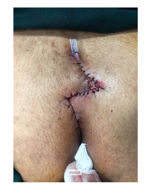
In the present study, the duration of surgery for the secondary wound healing group was significantly shorter than that of the Z-plasty group. The main reasons for the Z-plasty technique’s lengthier operation are 1- two more incisions are needed, 2- homeostasis must be established to prevent hematoma under the flaps and 3- pressure on the suture line is not reduced by closure of the flaps in one layer, so the flaps must be repaired in several layers with absorbable PDS sutures (poly-dioxanone suture) to prevent suture line pressure. In the Z-plasty method, extreme care must be taken to minimize the chance of necrosis of the flaps, a disastrous outcome. In the current work, no cases of flap necrosis occurred. The methods employed to ensure a well-vascularized flap are as follows: 1- flaps made with the fewest number of cuts and as low as possible, 2- electrocautery used as sparingly as possible, 3- no deep and bulky stitches in the flap part of the wound, and 4- flaps repaired in several layers without tension. The length of operation for the Z-plasty patients was significantly longer than that of the secondary wound healing group (traditional method). Even though the Z-plasty procedure is not very complex, it is technically more demanding. Shavinder Dogra et al. reported an average operating time of 75 minutes for Z-plasty, which is longer than the open method but similar to other primary closure techniques. (6) A review of the literature reported an average time of 34.59 minutes for the traditional method. (20) In Sughra Praveen et al.’s study, the operation length for Z-plasty ranged between 30–45 minutes, which is closer to the findings of the current study (52.97 ± 7.89 mins). (31) Yong-Ping Yang et al. reported that the operation length for Z-plasty was significantly longer than that for the simple excision technique since Z-plasty requires tissue release to create the flap and more suturing. (32)
4.2- Wound healing duration
The duration of complete wound healing in the Z-plasty group was shorter than that in the secondary wound healing group. In Z-plasty patients, the present study considered healing as the time when wound stitches were removed. However, for secondary wound healing patients, healing requires that the wound be filled with granulation tissue and then covered by epithelium that migrates from surrounding normal skin to completely cover and close the wound. From the patient’s perspective, the most challenging part of pilonidal sinus disease is the recovery time. In the traditional secondary wound healing method, it takes weeks to months to attain complete wound healing. The present study showed that the wound healing process was significantly shorter for Z-plasty patients, who were able to resume normal life activities sooner. In 2014, S. Priyadarshi et al. studied a total of 50 pilonidal sinus cases divided into two analogous groups. The mean hospital stay and total recovery time were reported to be significantly longer with the open method. (15) Yamini Sorate et al. also reported that the total recovery time and hospital stay were longer for the open technique than for the Limberg flap or Z-plasty methods. (33) In Yong-Ping Yang et al.’s study, the hospital stay was significantly shorter for patients in the Z-plasty group. Although the complete recovery time was not measured, it was concluded that Z-plasty patients needed less time for total recovery than patients who underwent a simple excision.(32) B.N. Anandaravi et al. compared two techniques in pilonidal sinus treatment: primary closure and laying open. For primary closure, the Limberg flap, Karydakis technique, and Z-plasty surgeries were studied. A shorter duration for wound healing and an earlier return to work were statistically significant in the primary closure group. (34) Fazeli et al. reported the same results. (3)
4.3- Pain
Compared to the Z-plasty group, the severity of pain for patients in the secondary wound healing group was clearly greater and harder to bear (P < 0.0001). One of the hypotheses explaining the lower amount of pain in Z-plasty patients is the lower amount of tension on the repaired tissues, which lessens tissue stimulation and thus pain. In the study of Priyadarshi et al., the VAS was significantly higher (more pain) in the open technique group than in the Z-plasty group. (15) Arvind et al. showed a significant improvement in pain severity after Z-plasty repair. The mean VAS score for coccygeal pain decreased from 7.33 ± 0.5 to 2.11 ± 1.2 (P < 0.05).(35) Elshazly reported significantly lower postoperative VAS scores in the Limberg group (2.1 ± 1.2 versus 5.2 ± 1.4), presumably because of the lower wound tension in the Limberg flap procedure.(36)
Fazeli et al., on the other hand, found no significant difference in the severity of pain experienced by patients. (3) Some studies do not support the findings that Z-plasty patients experience less severe pain. For example, Yong-Ping Yang et al. reported that postsurgical pain by the VAS score was significantly higher in patients treated by Z-plasty than in those treated by simple excision. However, at the end of the third postsurgery week, there was no significant difference in pain between the two methods. (32)
4.4- Dressing
The number of dressings applied in the Z-plasty group was significantly fewer than that in the secondary wound healing group. In Z-plasty, wound care after hospital discharge was performed by the patient at home. In contrast, patients in the secondary wound healing group required a health care professional for wound care after discharge. Therefore, Z-plasty appears to be more cost effective than traditional secondary wound healing. Fazeli et al. observed similar results and concluded that Z-plasty required significantly fewer dressing changes and posthospitalization health care, which suggested lower postoperative care costs despite lengthier hospitalization and longer operations.(3) In K.K. Hameed's research showed that fewer dressing changes in the primary closure technique significantly lowered the total cost of treatment in comparison to the secondary wound healing group. (37) M. Rao et al. reported that closed wounds required significantly fewer dressings than open wounds and remarkably lower costs. (30)
4.5- Recurrence and complications
As reported in the literature, the wound infection rate after off-midline closure techniques is approximately 6.3%. (2) The current study observed a 7.1% wound infection rate among Z-plasty patients and a 4.8% rate among patients in the secondary wound healing group. The recurrence rate was 7.1% in the Z-plasty group and 14.3% in the secondary wound healing group. These results indicate no significant difference between the two techniques in regard to wound infection and recurrence rates, which are the two most critical factors when considering an ideal course of treatment.
Similar observations have been made in the literature. In their seven-year study, Abdul Hakeem Jamali et al. utilized Z-plasty primary repair to manage 55 patients suffering from pilonidal sinuses. The majority of patients (74.5%) recovered with no complications. (10) In 2015, Jagdeep Rao et al. treated 40 pilonidal sinus patients by performing the Z-plasty technique. No tissue necrosis or recurrence in a six-month follow-up period was observed. (30) Yong-Ping Yang et al. reported a 5.88% and a 6.06% infection rate in their Z-plasty and simple excision groups, respectively; this was not statistically significant. In its six-month follow-up period, this same study saw only one case of recurrence, which belonged to the simple excision group.(32) A meta-analysis indicated that, compared to primary midline closure, secondary healing of a pilonidal sinus is associated with a 58% lower risk of recurrence, which is statistically significant. Similar to the present work, the meta-analysis study reported no significant difference in the recurrence rate of open healing and off-midline primary closure techniques, such as Z-plasty. (2) The 30-case study by B. N. Anandaravi reported two cases of infection in its off-midline primary closure group and one case in the open healing group, results that are close to the present work’s findings. There was also no recurrence. (34) In Sughra Parveen et al.’s research of 40 patients treated with a Z-plasty flap, six (15%) had a superficial wound infection that was treated conservatively, and 5% experienced a recurrence reported four months after surgery. (31) In Siddhartha Priyadarshi et al.’s work, recurrence was found in 5.88% of the open technique group and none at all in the Z-plasty group. For wound infection, the difference between the two study groups in early postoperative complications was not statistically significant. (15)
Fazeli et al. reported an infection rate of 13.9% in open wound patients and 9.7% in the Z-plasty group along with a recurrence rate of 4.2% in both groups(3). In Jagdeep Rao et al.’s study of 40 patients treated with the Z-plasty method, there were no recurrences in the 6- to 12-month follow-up. Regarding postoperative complications, 5% of patients experienced numbness over the flap, 7.5% suffered from wound infection, and 12.5% developed wound seroma. Necrosis of the flaps did not occur. (30) Of the 20 patients treated by Z-plasty flaps in Shavinder Dogra et al.’s study, only two were diagnosed postoperatively with wound infection. No recurrence was reported in the 12- to 24-month follow-up period. (6)
The limitations of the present study are as follows: 1- the number of patients was limited, 2- all patients were treated in one center, while it was better to have used several centers, and 3- the duration of the follow-up period (6 months) was not sufficient and should have been at least two years.