Free-living amoebae (FLA) are unicellular eukaryotes distributed worldwide, in various environments, and are known to phagocyte microorganisms or to transport microorganisms that have acquired the ability to survive or even to multiply within FLA 1. Thus, FLA have multiple roles in the ecology of microorganisms: replicative niche, widespread reservoir, protective armour, mode of dissemination, gene exchange, selection of virulence traits and adaptation to macrophage1. FLA are considered as a place where horizontal gene transfer occurred frequently between amoeba-resisting microorganisms present inside FLA and between FLA themselves 2,3.
Cell connectivity has been shown to be essential to maintain homeostasis in metazoans. Intercellular communication can occur both direcly (contact-based communication) and indirectly (mediated communication)4,5. From the early 2000’s, a new way of cell-to-cell communication was identified and called tunneling nanotubes (TNTs) 6,7. Described as cytoplasmic bridges, TNTs may allow both uni- and bilateral transfer of information mostly between eukaryotic mammalian cells 8,9. Various types of exchange through TNTs have been reported including transmission of organelles, pathogens, ions, genetic material or misfolded proteins 10–12. TNTs formation is stimulated by inflammatory and stress conditions such as acidity, exposure to reactive oxygen species, ischemic condition, inflammatory signals or pathogen infections suggesting that TNTs are strongly implicated in survival mechanisms of cells 4,9,10,11. TNTs are also involved in misfolded proteins transport in neurodegenerative diseases such as Alzheimer, Parkinson or Huntington 8. Interestingly, some proteins from viral origin could enhance TNTs formation in eukaryotic mammalian cells. Human immunodeficiency virus type (HIV-1) nef protein first promotes actin remodelling, initiates consequently TNTs formation and then uses TNTs to transport itself to another cells 8. Protected from external environment in human hosts including humoral response, viruses can therefore use TNTs to safety promote their intercellular spreading 8. Several other virus including herpes, influenza, and porcine reproductive and respiratory syndrome viruses have been described as TNT cargoes 8. SARS-CoV-2 induces the formation of TNTs and exploits this route to invade non permissive cells and potentiate infection in permissive cells 15. Besides viruses, TNTs also mediated propagation of bacteria and even parasites 16 indicating that TNTs can be hijacked by microorganisms for dissemination among eukaryotic cells. In the early 2010’s, intercellular nanotubes between prokaryotes have also been reported 17 but TNTs were not described so far in any unicellular eukaryotics until now.
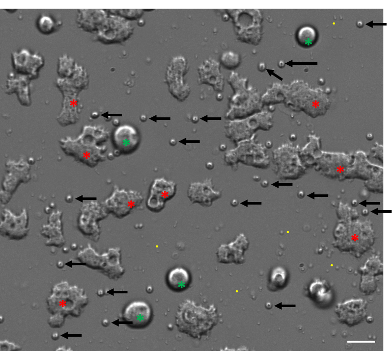
During work in progress, we studied potential interactions between Acanthamoeba castellanii and Cryptosporidium parvum oocysts in a pre-formed (Pseudomonas aeruginosa) biofilm conditions. In such conditions, FLA, C. parvum oocysts and P. aeruginosa could be observed through phase gradient contrast light microscopy (supplementary Figure 1). Interestingly, pairs of isolated FLA trophozoites were connected through long single membrane protrusion with features corresponding with type 1 of TNT 18. Single-long protrusions (from 16 to 82 µm) exhibited trumpet-shaped origins with a thinner elongated central portion (Figure 1 A1-A3) and a mean length of 37 µm. Time-lapse imaging revealed dynamic aspects of TNT1 structures including morphological variations possibly considered as “intermediate states” (Figure 2C and supplementary movie 1). Closed FLA firstly separated to each other leading to the formation of TNT1 structure. Then, successive “coming and going” of connected FLA cell bodies resulted in variable length and thickness of TNT1 structure (Figure 2C and supplementary video 1). Less frequently, short multiple connections with similar features with type 2 of TNT (< 5 µm) 18, were detected between very closed isolated trophozoites or within a cluster of FLA (Figure 1B1-B3, supplementary movie 2). Such TNT1/2 connections were restricted to trophozoites and were not detected in FLA cysts or bacteria or oocysts. The TNT structures were also observed when FLA were cultured alone (in control condition). Three criteria are currently proposed for the identification of protrusions as TNTs i.e. i) connecting at least two cells, ii) containing F-actin and iii) floating over the substratum in 2D cultures. Fluorescent labeling of cytoskeleton components combined to Z-stack acquisitions (x, y, z) through confocal microscopy and image analysis allowed a 2D/3D representation of intercellular communication. Indeed, actin labeling with Alexa Fluor 488-coupled phalloidin revealed in fixed amoebae the presence of F-actin in both categories of TNTs namely TNT1 (Figure 2A) and TNT2 (Figure 2B). In contrast to actin, tubulin revealed by an immunocytochemical procedure was only present in TNT1 observed in methanol-fixed trophozoites (Figure 2A and 2B). However, actin and tubulin contents within TNT1 were variable among amoebae pairs suggesting “intermediate states” of TNT1 and/or TNT1 subtypes (Figure 2A). Projections of tubulin labeling revealed that protrusion is really hovering freely above substratum, parallel to the surface of culture dish (Figure 2C). In addition, phase gradient contrast light microscopy time-lapse also demonstrated that FLA can pass under TNT1 (supplementary movie 3).
In this study, two types of TNTs namely TNT1 and TNT2 connected long and short distance motile FLA trophozoites. All TNTs detected so far with wide-field and confocal imaging approaches were only homo-TNTs i.e. between amoebae and any other connection could not be detected in Cryptosporidium oocysts and Pseudomonas aeruginosa. Cell connectivity through TNTs appeared as a feature maintained during evolution highlighting its crucial role in intercellular communication. As example, inter-bacteria nanotubes were observed in Bacillus subtilis model with length ranged up to 1 µm with width ranged from 30 to 130 nm and could represent a major form of bacterial communication in nature, especially in biofilms where cells proximity could favour such interactions17. Another interesting characteristic is the mechanism of TNT1 formation in A. castellanii which resulted from cell dislodgement while it is generally described as a directed filopodia-like protrusions in mammalian cell lines18. Possible involvement of specific amoeboid cell surface structures including pseudopodia, acanthopodia, lobopodian and filipodia in the formation of TNTs deserves further investigations. Taken together, these data suggest for the first time that FLA are forming spontaneously homo-TNTs representating therefore a unique model to determine the mechanisms of formation and transfer in unicellular eukaryotics.
The characterization of TNTs between FLA suggest enhanced communication between FLA and microorganisms that they carry and even more potentiality of genetic exchanges. Interestingly, FLA resistant-microorganisms are able to resist to macrophages and conversely1. Interestingly, TNTs were previously described between macrophages 19 . Thanks to the simplicity of culture, FLA may represent therefore a relevant organism to study i) TNT formation, ii) microbial exchanges iii) genetic transfer and iv) large-scale screening of molecules on TNT formation. It could facilitate knowledge about macrophage resistance. The discovery of connectivity between unicellular eukaryotic FLA through TNT offers many perspectives to study and understand microbial pathogenicity mechanisms and resistance.