Materials, bacterial strain, plasmids, and culture conditions
Media components of LB and 2xYT (yeast extract, tryptone) were obtained from Ibresco (Life Science, Iran) and n-dodecyl β-d-maltoside (DDM) was purchased from Molekula GmbH (Germany). UDP-GlcUA, UDP-GlcNAc (U6751 and U4375, Sigma-Aldrich), and other reagents were supplied by Bio Basic Inc. (Canada).
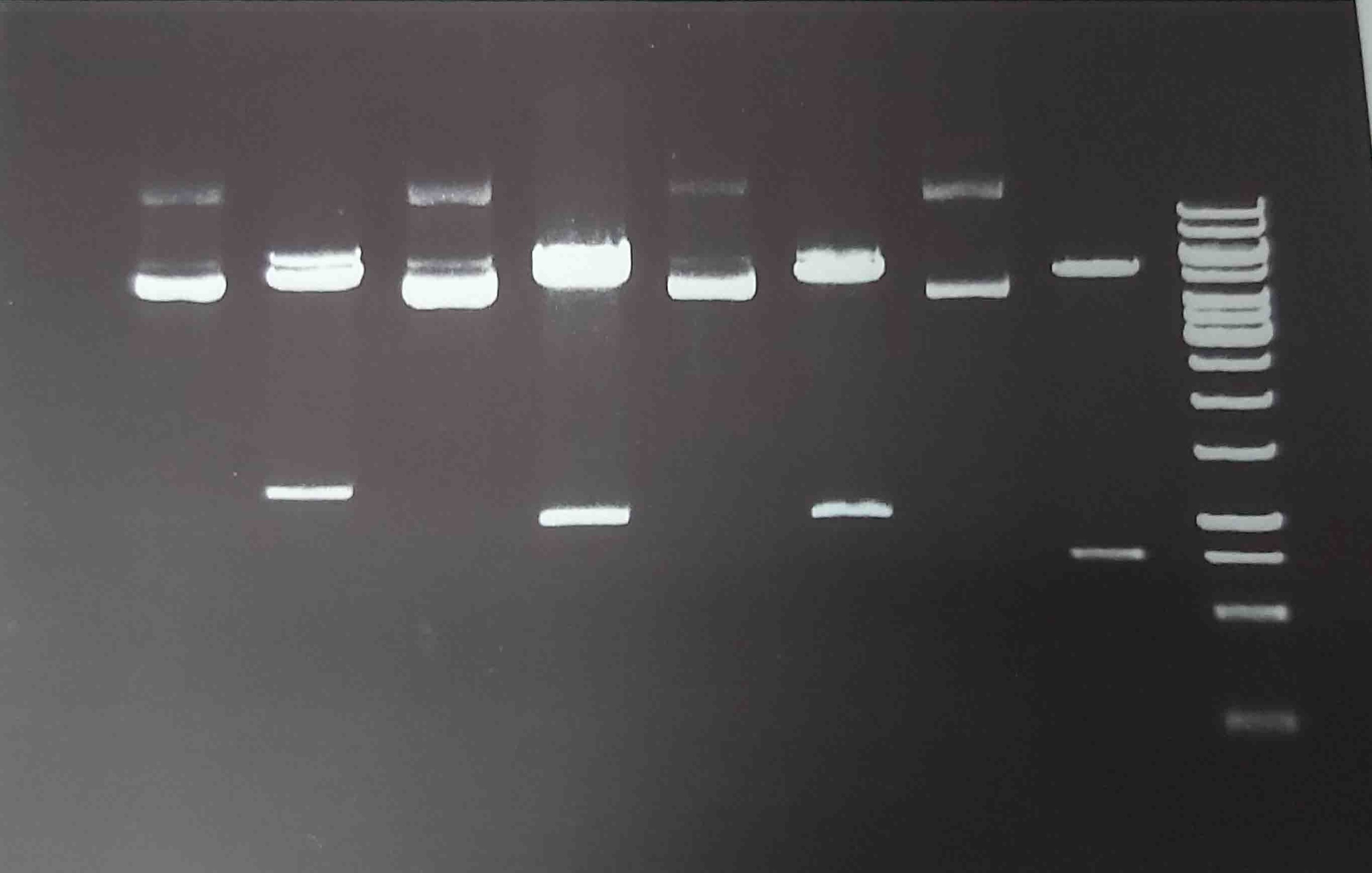
Cloning of SeHAS and variants
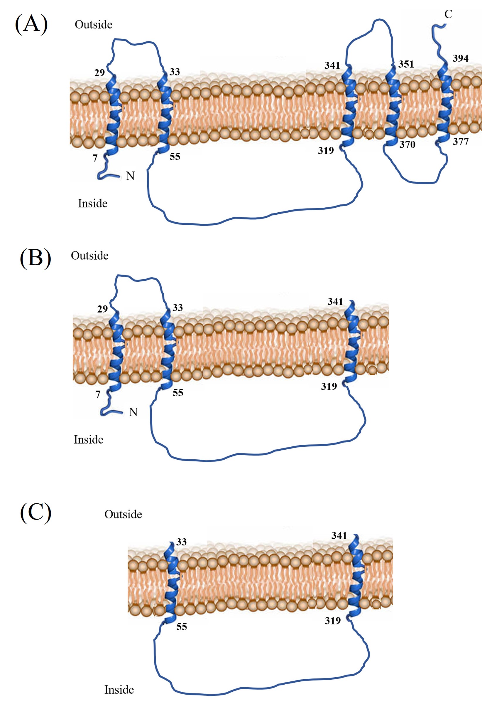
S. equisimilis isolate S88 group G (GGS-S88) was obtained from the previous study (25). This strain was used for DNA genomic extraction (23, 24). The genes of SeHAS and variants were amplified using specific primers (Table S1, Additional file 1). The genes were then cloned into pET-28a (+) (Novagen, Germany) and transformed into E. coli BL21(DE3) strain (Novagen, Germany) (Fig. S1, Additional file 2). To facilitate the purification of SeHAS variants, a C-terminal fusion of 6-His residues was added to each construct. The recombinant hosts were grown on LB broth or LB agar containing 50 µg/mL kanamycin. The summarized properties of variants used in the present study were shown in Table 1. The schematic membrane topology of variants is depicted in Fig. S2 (Additional file 3).
Table 1: The characteristics of SeHAS and variants.
|
Enzyme
|
Transmembrane membrane domain
|
Start
|
End
|
Length (aa)
|
MW (kDa)
|
|
SeHAS
|
1, 2, 3, 4, 5
|
1
|
417
|
417
|
42.0
|
|
HAS123
|
1, 2, 3
|
1
|
341
|
341
|
39.1
|
|
HAS23
|
2, 3
|
33
|
341
|
308
|
35.9
|
|
HASIntra
|
None
|
55
|
319
|
264
|
30.0
|
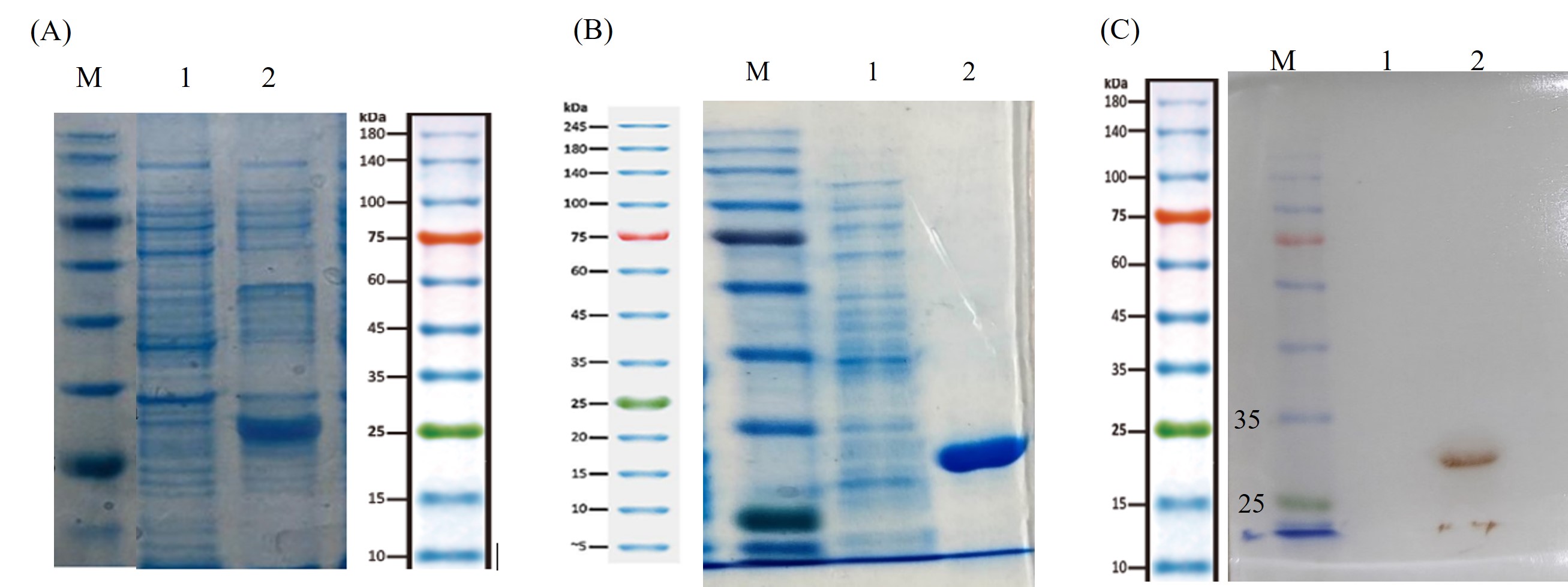
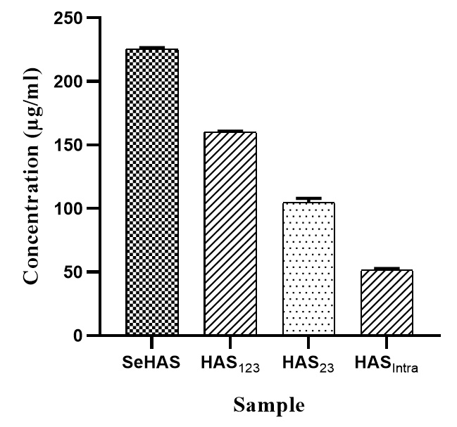
Expression and purification of variants
The cultures were induced with 1 mM IPTG and incubated for 18 h at 37 ° C. The cells were centrifuged and the pellets were washed twice with phosphate-buffered saline (PBS). For purification of HASIntra, a hybrid method was used. First, the pellet was resuspended in 12 mL lysis buffer (8 M Urea, 0.5 M NaCl, 72 mM K₂HPO₄, 17 mM KH₂PO₄, pH 7.5) and incubated on ice for 40 min. The sonicated cell (three times, 30s at 20 watt) was centrifuged at 12 000 g for 20 min. The supernatant was transferred to Ni-NTA resin (Sigma Inc., USA) equilibrated with lysis buffer. The mixture was incubated for 90 min at 4 °C with constant mixing. After incubation, the mixture was then washed two times with wash buffer I (lysis buffer containing 3 M Urea, pH 7.5) and wash buffer II (lysis buffer with 100 M NaCl, pH 7.5), respectively. The protein was eluted with elution buffer (72 mM K₂HPO₄, 17 mM KH₂PO₄, 300 M NaCl, 250 mM Imidazole, pH 7.5). Total protein concentration for HASIntra was determined by Bradford assay using bovine serum albumin (BSA) as standard. The expression of HASIntra was confirmed by 12% SDS-PAGE and western blot (anti-His antibody, Sigma, USA) according to standard procedures (26). In our previous study, we used an ionic detergent, sodium dodecyl sulfate (SDS), for the solubilization of membrane proteins (25). However, SDS has a deleterious effect on the protein conformation that leads to the denaturation of protein. Therefore, for membrane proteins (SeHAS, HAS123, and HAS23), we examined the effects of non-ionic detergents such as DDM, Triton X-100, and Tween 20. The concentration of purified variants was carried out using Bicinchoninic acid (BCA) protein assay kit (Pierce) using bovine serum albumin as a standard (27). Unlike Bradford method, BCA assay is compatible with a broad range of detergents at high concentrations (28).
Activity measurements
Enzyme activity of SeHAS and the variants was performed in a reaction (100 µL) containing 25 mM sodium and potassium phosphate, 50 mM NaCl, 20 mM MgCl2, 1.0 mM DTT, 1.0 mM EDTA, 2M glycerol, 1mM DDM, 1 mM UDP-GlcUA, and 1.0 mM UDP-GlcNAc, adjusted to pH 7.0. To initiate the reaction, 0.1 µmol of each variant was added and the mixture was gently mixed at 30 °C. After 60 min incubation, the reactions were terminated by the addition of SDS at a final concentration of 2% w/v (29).
HA purification
At first, 0.15 M NaCl solution was added to the reaction mixtures. Then, the mixtures were cooled to 4 °C for 60 min to form the HA salt. The HA salt was precipitated by adding three volumes of ethanol followed by incubation at 4 °C for 24 h. The resulting HA salt was collected by centrifugation (12 000 g, 20 min at 4 °C) and resuspended in distilled water (30, 31). The protein impurities were determined using a UV/Vis spectrophotometer (Thermo Fisher, USA). To investigate the effect of the purification process on HA Mw, the purification step was also performed on a control HA with a MW of 760 kDa (Bloomage Corporation).
HA quantification
HA titer was determined by carbazole assay with some modifications (32, 33). Briefly, a serial dilution of standard (0-500 µg/mL, D-glucuronic acid, Sigma) and sample solutions (50 µl) was prepared in a 96-well microplate. Then, 200 µL sodium tetraborate solution (0.025 M in saturated sulfuric acid) was added to the wells and mixed gently. The microplates were heated for 20 min at 80 °C. After cooling at room temperature, 50 µL carbazole (0.125% in absolute ethanol) was added to each well and mixed well. The microplate was read in a microplate reader (Metertech, Taiwan) at a wavelength of 550 nm after heating at 80 °C for 20 min in an oven and cooling at room temperature for 15 min.
FIIR spectroscopy
The identification of purified polymers was performed by Fourier transform infrared spectroscopy (FTIR spectroscopy). For this purpose, the samples and control were analyzed by a FTIR apparatus (Termo, USA,) in a wavenumber range of 4000-400 cm-1 under the same operational conditions.
Polydispersity and MW determination
The polydispersity of HA samples and the controls (HA10kDa and HA760kDa) (Bloomage Corporation) was evaluated by dynamic light scattering (DLS) technique (Malvern, Nano-Zs) (34). The HA MWs were determined by polyacrylamide gel electrophoresis (PAGE) with a combined Alcian blue and silver staining reported by Min and Cowman with some modifications (35). The mini-slab gels of 8 × 9 × 0.1 cm containing 15% acrylamide, 0.5% N, Nˊ -methylene bis-acrylamide in 0.1 M Tris-borate-1 mM Na2EDTA, pH 8.3 (Tris/borate/EDTA) were used. The purified samples were dissolved in water and mixed with a one-fifth volume of 2 M sucrose in Tris/borate/EDTA buffer and loaded on the gel. Bromophenol blue (0.005% in Tris/borate/EDTA buffer containing 0.3 M sucrose) was used as a tracking dye and LMW-HAs (Echelon Biosciences Inc., HYA-LOLAD) marker was used as a ladder. The gels were run at 170 V for 30 min, then at 250 V for 10 min, and finally at 200 V for ~40 min until the tracking dye reached within 1 cm of the gel bottom. The electrophoretic process was carried out at 4°C (36). Immediately after electrophoresis, the samples were fixed in the gel by soaking the gel in 0.5% w/v Alcian blue in 2% w/v acetic acid, for 30 min far from light. After destaining in water for 30 min, the gel was subjected to silver staining, beginning from the oxidation step, using the Bio-Rad silver stain kit according to the manufacturer’s protocol (35). In brief, the gel was soaked for 10 min in a 200 mL solution of 0.0034 M potassium dichromate and 0.0032 N nitric acid. It was washed two times for 20 min in 200 mL of deionized water and placed in 100 ml of 0.012 M silver nitrate for 20 min. This step was followed by rapid rinsing with 100 mL of the image developer solution, which contains 0.28 M sodium carbonate and 0.5 ml of commercial formalin per liter. The gels were gently shaken in this solution until the HA bands appeared. The development was stopped by discarding the developer and addition of 100 ml 5% w/v acetic acid. The gels were washed twice with 200 ml of water before storage and sealed in plastic bags (37). Estimation of the Mw was performed by Rf value using an online graphing program (https://www.bio-rad.com/webroot/web/pdf/lsr/literature/Bulletin_6210.pdf).
Proliferation assay
MTT assay was used to investigate the effect HA products on cell proliferation. Adherent human umbilical vein endothelial cells (HUVEC, NCBI code: C554) were seeded at a density of 1.0 × 10⁴ cells/well in the specific medium containing 10% fetal bovine serum (FBS, Gibco U.S.A). After the cell growth, the medium was replaced with HA products, o-HA (HA10kDa), and n-HA (HA760kDa) at different concentrations (180 μL preparations in medium supplemented with 10% FBS). The plates were then incubated for 48 and 72 h. After that, 20 μL methylthiazolyldiphenyl-tetrazolium bromide (MTT, T0793 Bio Basic Canada) reagent was added to each well, and the plates were incubated for 4.0 h at 37°C. The supernatants were discarded and isopropanol was added to each well. The microplates were shaken in an oscillator at room temperature for 15 min and the absorbance was measured using an Epoch microplate spectrophotometer (BioTek, USA) at 570 nm. The experiment was repeated three times and the results were expressed as the percentage of viability when compared to untreated cells (Negative control is considered as 100% of viability).
Wound healing assay
HUVEC were plated at a density of 1.0 × 10⁵ cells/well in a specific medium containing 10% FBS in 12 wells plates. After reaching a cell density of 1.0 × 10⁹ per well, the medium was removed and the cells were rinsed with phosphate-buffered saline (PBS, pH 7.0). Then, a sterile pipette tip was held vertically to scratch a line in each well. The detached cells were removed by washing with PBS. After that, the HA products at final concentrations of 10 and 200 µg/mL were added to the wells. The controls were 180 µL culture medium without HA, n-HA (HA760kDa), and o-HA (HA10kDa). The scratch closure was monitored and imaged at 5-time points for 24 h using a BEL INV-2 microscope (BEL Engineering, Italy) (38). The results were expressed as the percentage of wound closure with respect to starting time point, quantified using an image-analysis program (ImageJ, version v1.54d) (39).
Statistical analysis
Statistical analysis was performed using Student’s t-test to compare two groups or one-way ANOVA in the case of three or more groups with GraphPad Prism. All data are presented as mean ± SD from three independent experiments. A p-value less than 0.05 was considered a statistically significant difference between the samples.