5.1 Cell culture
This study was approved by the Ethics Committee at the Tongji Medical College of Huazhong University of Science and Technology (No.
2018-S288). ADSCs were isolated from the subcutaneous fat from patients without cancers. The adipose tissue was cut into small pieces and the connected fascial tissue was removed. Then the adipose tissue was digested for 1 h with 0.2% collagenase type I (Sigma, St Louis MO, USA) and centrifuged for 4 min at 1000 rpm. The cell pellets obtained were passed through a 70-µm filter (Corning, Rochester NY, USA), and then cultivated in Dulbecco’s modified Eagle’s medium (DMEM; Gibco, Gaithersburg MD, USA) with 10% fetal bovine serum (FBS, Serapro, Naila, Germany). The human colon cancer cell line HCT116 and mouse colon cancer CT26 were preserved in our laboratory (Hubei Province Key Laboratory of Molecular Imaging) and propagated in an RPMI-1640 medium (Gibco) supplemented with 10% FBS.
5.2 ADSCs-EV isolation
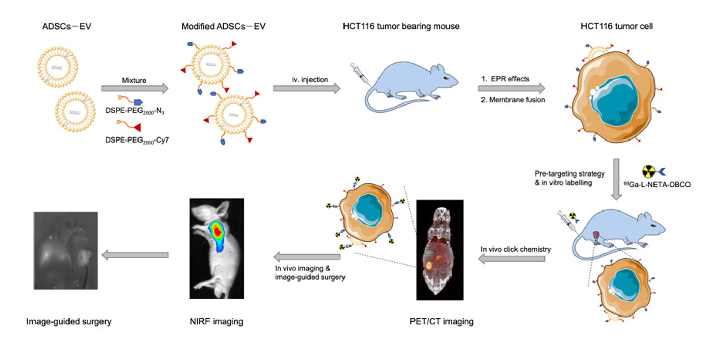
Extracellular vesicles from serum-free ADSCs culture supernatant were obtained by differential centrifugation. Dead cells and cell fragments were removed by centrifugation at 3000 g for 30 min. Then the supernatants were centrifuged at 13,000 g for 70 min. The supernatants were concentrated by an Amicon® Ultra-15 Centrifugal Filter Device (100 kDa molecular weight, Millipore, USA). Finally, the supernatants were centrifuged at 120,000 g for 70 min to obtain ADSCs-EV, which were subsequently suspended in PBS, passed through a 0.22 µm filter, and quantified by surface proteins with a BCA Protein Assay Kit (Beyotime, Shanghai, China) and stored at − 80°C.
5.3 ADSCs-EV characterization
Extracellular vesicles were examined by transmission electron microscopy (TEM, Hitachi, Japan), dynamic light scattering (DLS, Malvern Instruments Ltd., Worcestershire, UK), and western blot analysis. Changes in hydrodynamic diameters were monitored for 8 d by DLS to test the stability of the ADSCs-EV in vitro. A Cell Counting Kit-8 (CCK8) kit (SAB, College Park, MD, USA) was used to identify the cytotoxicity of different concentrations of ADSCs-EV in HCT116 human colon cancer cells.
5.4 In vitro cell binding
The modification of extracellular vesicles was adapted from a literature reported method[31]. Briefly, we introduced 1mg DSPE-PEG per 1mg extracellular vesicles. ADSCs-EV and DSPE-PEG2000-Cy5 (Ruixi, Xi’an, China) were incubated at room temperature for 30 min, and Cy5-labeled ADSCs-EV (EV-Cy5) were obtained. EV-Cy5 (100 µg /mL) were added onto HCT116 cells grown in a confocal dish and incubated at 37℃for different time periods (6 h, 12 h, 24 h, 48 h). The cell nuclei were counterstained with 4′,6-Diamidino-2-phenylindole (DAPI) (Boster, Wuhan, China). Cells were fixed with paraformaldehyde and observed under a Fluorescence microscope. Confocal microscopy was used to observe the tumor-binding ability of ADSCs-EV. ADSCs-EV and Cy5-labeled DSPE-PEG2000 (DSPE-PEG2000-Cy5; Ruixi, Xi’an, China) were incubated for 30 min, and then the ADSCs-EV labeled with Cy5 (EV-Cy5) were extracted. EV-Cy5 (100 µg/mL) were added into the HCT116 cells cultured in a confocal dish and incubated for 12 h. Additionally, the skeleton of tumor cells was stained with FITC-phalloidin, and nuclei were stained with 4′,6-Diamidino-2-phenylindole (DAPI; Boster, Wuhan, China). Finally, the confocal dish was fixed with paraformaldehyde and observed by confocal microscopy (LSM 880, ZEISS).
5.5 Tumor-bearing nude mouse models
The protocol of mouse experiments was reviewed and approved by the Animal Care Committee of Tongji Medical College, Huazhong University of Science and Technology.
HCT116 cells (5 × 10
6) suspended in 100 𝜇L PBS were subcutaneously injected into the right upper limb of BALB/C nude mice (male, 4 weeks old, Beijing HFK Bioscience co., Ltd, China). CT26 cells (5 × 10
6) suspended in 100 𝜇L PBS were subcutaneously injected into the right upper limb of BALB/C mice (male, 4 weeks old, Beijing HFK Bioscience co., Ltd, China). After the tumor size reached approximately 0.8 cm, the mice were prepared for study. Orthotopic colon cancer models were also prepared with the following protocol. The 5-week BALB/C nude mice were laparotomized to expose the cecum, then HCT116 cells (5 × 10
6) suspended in 50 µL PBS were injected into the serosal layer of the colon. Four weeks after cell injections, the mice were prepared for study.
5.6 The modification of ADSCs-EV
Using hydrophobic insertion approach [32], DSPE-PEG2000-Cy7 (Ruixi, Xi’an, China) and DSPE-PEG2000-N3 (Ruixi, Xi’an, China) were incubated with ADSCs-EV for 30 min at 37°C to form N3(Cy7)-PEG2000-DSPE-ADSCs-EV (Cy7-EV-N3). The samples were passed through centrifugal filter devices (100 kDa molecular weight, Amicon®Ultra-15) before further use.
5.7 Synthesis and identification of 68Ga-L-NETA-DBCO
68GaCl3 was obtained from the 68Ge/68Ga generator with HCl (0.05 M) as eluent. Sodium acetate (1.25 M, pH = 8.6) was added to 500 µL of 68GaCl3 (187MBq) to adjust the pH to 3.7. L-NETA-DBCO (5 nmol) was used to chelate the radionuclide 68Ga, and the reaction was maintained 10 min at 100°C. After the mixture was cooled, a C18 column was used to purify 68Ga-L-NETA-DBCO. 68Ga-L-NETA-DBCO was conjugated with N3-modifed ADSCs-EV by in vivo click reaction, which enables PET imaging. The radiochemical purity and in vitro stability of the probe (2 h in fetal bovine serum) were measured by high-performance liquid chromatography (HPLC).
5.8 Cellular uptake
To assess cell uptake, we incubated 1 × 10⁶ of HCT116 cells with RPMI-1640 medium supplemented with 10% fetal bovine serum containing Cy7-EV-N3 (100 µg/mL) at 37°C for 0, 6, 12, 18, 24, 36 and 48 h. Then the supernatants were removed. 68Ga-L-NETA-DBCO (37 kBq/well) were added to the HCT116 cells. After incubation for 0.5, 1, 2, and 3 h, the supernatants were removed and washed with PBS. The remaining cells were lysed in NaOH. Cell lysates and supernatants were collected. Radioactivity was measured using a γ-counter (PerkinElmer, USA).
5.9 In vivo PET Imaging
In order to identify the best time points, Cy7-EV-N3 (200µg) was intravenously injected into the HCT116 tumor-bearing nude mice at different pre-targeting time points (10, 20, and 30 h). 68Ga-L-NETA-DBCO (3.7MBq) were injected into the mice (n = 3 per group) via the tail vein. Mice were anesthetized with 2% isoflurane, and micro-PET static imaging was performed at 1 and 2 h after the injection of 68Ga -L-NETA-DBCO. Static PET images were collected for 10 min using a small-animal PET scanner (TransPET BioCaliburn 700, Raycan Technology Co., Ltd, Suzhou, China). PET images were reconstructed with the ordered-subset expectation maximization three-dimensional/maximum a posteriori probability algorithm, and then image analysis was performed using Amide (http://amide.sourceforge.net) and Carimas 2.10 (Turku PET Centre, Finland) software.
5.10 NIRF imaging
Cy7-EV-N3 was injected into the mice bearing HCT116 tumor grafts (n = 3 per group) via the tail vein for NIRF imaging. Mice were anesthetized with 2% isoflurane, and NIRF imaging was performed at different times (1, 5, 10, 20, 30, and 50 h). The static NIRF images were acquired with 750-nm excitation and 790-nm emission filters using an IVIS spectrum imaging system (In-Vivo FX PRO, Bruker, Germany). NIRF images were analyzed by Bruker MI (Germany).
5.11 Multimodal PET/CT and NIRF imaging of orthotopic colon transplantation tumor
Twenty hours after Cy7-EV-N3 injection, 68Ga-L-NETA-DBCO were injected into the orthotopic colon transplantation tumor model mice via the tail vein for click chemistry in vivo. 2 h after 68Ga-L-NETA-DBCO injection, the mice’s bladders were emptied by compression and the mice were anesthetized using 2% isoflurane. Static PET/CT images were collected for 10 min using a small-animal PET scanner (TransPET Discoverist 180, Raycan Technology Co., Ltd, Suzhou, China). PET/CT images were reconstructed with the ordered-subset expectation maximization three-dimensional/maximum a posteriori probability algorithm, and then the analysis of images was done using Amide (http://amide.sourceforge.net) and Carimas 2.10 software. After PET/CT imaging was completed, NIRF imaging was subsequently performed and analyzed as described above. The mice were then sacrificed and the tumor tissue was collected for HE staining of pathological sections.
5.12 Immunohistochemistry analysis
The CD31 immunohistochemistry analysis was performed to evaluate the vascularization of the tumors. The HCT116 tumor tissues were collected, fixed in 4% paraformaldehyde and embedded in paraffin. The tumor sections (5 µm) were dewaxed, rinsed with EDTA buffer (pH 9.0), and blocked with 3% hydrogen peroxide. The tumor sections were incubated with anti-CD31 antibody (Abcam, Cambridge MA, USA) at 4°C overnight. Then the tumor slices were incubated with secondary antibody (HRP-labeled goat anti-rabbit IgG, Abbkine, Redlands CA, USA) at room temperature for 30 min. The sections were stained with 3, 3’-diaminobenzidine (DAB, Beyotime, Hangzhou, China) for 8 min, subsequently by counterstaining with hematoxylin (Beyotime) for 2 minute and were observed under microscopy.
5.13 Real-time NIRF imaging for intraoperative guidance
Twenty hours after the injection of Cy7-EV-N3, resection of the subcutaneous tumors in mice was performed using a real-time IVIS spectrum imaging system (Premium Imaging FB800, Premium imaging, California, USA).
5.14In vivo toxicity studies
BALB/c mice (n = 4 per group) received an i.v. injection of N3-EV-Cy7 (200µg) +68Ga-L-NETA-DBCO (7.4MBq) or PBS. Mice were euthanized on 1st and 7th day after the injection. Their blood samples and major organs were collected. The function of liver and kidney, such as alanine amino transferase (ALT), aspartate aminotransferase (AST), and alkaline phosphatase (ALP), blood urea nitrogen (BUN), and creatinine (CRE) were measured by the blood biochemical autoanalyzer (Chemray 240, Rayto Life and Analytical Sciences Co., Ltd, China). Hematoxylin and eosin (H&E) of major organs (hearts, livers, spleens, lungs and kidneys) were examined using an optical microscope (IX73, Olympus, Japan).
5.15 Statistical Analysis
Data are shown as the mean ± standard deviation (SD). Comparisons between groups were evaluated with the unpaired Student’s t-test. 𝑝 < 0.05 was considered to be statistically significant. Statistical analysis was conducted using GraphPad Prism v8.0 software.