2.1 Animals and SAH model
A total of 105 adult Sprague-Dawley (SD) rats (male, 260–300 g, 7–8 weeks old) were used in this study. All experimental procedures were reviewed and approved by the Institutional Animal Care and Use Committee of Shandong First Medical University and conformed to the protocols set forth by the National Institutes of Health in the United States.
The SAH model was established using the intravascular puncture technique, as previously described[18, 19]. To summarize, rats were anesthetized with isoflurane (5% for induction and 2.5% for maintenance). The right common carotid artery (CCA) was surgically exposed and the internal carotid artery (ICA) and external carotid artery (ECA) were then isolated from their bifurcations. A nylon wire was inserted into the ICA via the ECA and advanced approximately 21–22 mm from the arterial bifurcation until a distinct sensation of penetration persisted for 15 seconds. In the sham-operated group, the same procedure was followed, excluding the puncturing of the vessel.
2.2 Neurological function assessment
Neurologic function was evaluated blindly at 1, 3, and 5 days post-SAH employing a modified Garcia score encompassing six dimensions including spontaneous activity, spontaneous movement of all limbs, forelimb movement, climbing wall of wire cage, reaction to touch on both side of trunk, and response to vibrissae touch. Each dimension was scored on a scale of 0–3 or 1–3, with a minimum total score of 3 and a maximum of 18[18].
At 5 days after the procedure, the severity of SAH bleeding was evaluated [18]. Briefly, the basal cistern of the rat cranial base was divided into six segments, and each segment was rated on a grade of 0–3 based on the amount of bleeding. The sum of the six regions was the SAH score (0–18), and a score of 0–7 was excluded from the study.
Additionally, the Hang wire test was employed to evaluate somatosensory motor function[20]. Briefly, rats were placed on a strip of wire with supports at both ends, and their behavior on the wire within 30 s was recorded for scoring: 0, falling; 1, two front paws hanging from the wire strip; 2, front paws hanging from the wire and attempting to climb up; 3, two front paws and one or two hind paws hanging from the wire strip; 4, four paws hanging from the wire strip with the tail wrapped around the wire strip; and 5, escaping to the supports. The experiment was repeated three times and the average score was taken for calculation.
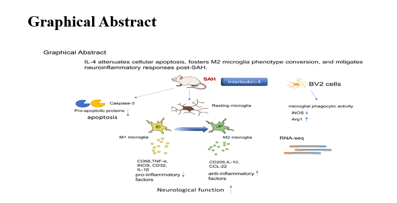
2.3 Experimental design in vivo
The experimental design employed is depicted in Fig. 1a. In the in vivo experiments, aimed at investigating the alterations in IL-4 expression levels subsequent to SAH, rats were randomly assigned to two groups: the sham group(n = 6) and the SAH group(n = 36). Peripheral blood and CSF samples were taken for enzyme-linked immunosorbent assay (ELISA) at 12 hours, 24 hours, 3 days, and 5 days post-SAH.
To examine the impact of IL-4 on neuroinflammation after SAH, rats were randomly divided into three groups: the sham group (administered with phosphate-buffered saline (PBS), n = 15), the SAH group (subjected to SAH and administered with PBS, n = 24), and the SAH + IL-4 group (subjected to SAH and administered with IL-4, n = 24). All rats were euthanized 5 days post-SAH, and subsequent analyses included immunofluorescence staining and quantitative real-time polymerase chain reaction (qRT-PCR).
2.4 Enzyme-linked immunosorbent assay
Sham and SAH rats were anesthetized with isoflurane and placed on a stereotaxic apparatus, and CSF samples were collected at the site of the occipital pool[21]. Blood samples were taken by cardiac puncture and then centrifuged for serum (3000 × g for 10 min at 4°C). IL-4 levels in the CSF and serum samples were assessed using a rat IL-4 ELISA kit, following the manufacturer's instructions (Mlbio, China). The absorbance density (OD) values were measured at a specific wavelength of 450nm using a spectrophotometer, and the concentrations of each sample were calculated accordingly.
2.5 Administration of IL-4
IL-4 administration was performed as shown previously[22]. After isoflurane anesthesia, animals were placed on a brain stereotaxic instrument and immobilized. According to the in vivo experimental design, an implantable slow-release pump (RWD Life Science, China) containing recombinant rat IL-4 (Peprotech, 60ng/d) or PBS was positioned into the lateral ventricle of the lesion (coordinates: AP 1.0 mm, ML 1.5 mm, DV 4.5mm) immediately after the onset of SAH. The pump was then started and the infusion was continued at a constant rate of 0.5 µ l/h for 5 days until the rats were euthanized[22].
2.6 Immunofluorescence
Immunostaining was conducted following established protocols[23]. Briefly, brain tissues were collected after cardiac perfusion with cold 4% paraformaldehyde (PFA), placed in 4% PFA overnight, and then dehydrated using 30% sucrose/PBS for 4 days. Tissues at distances of -2.5 to -5 mm from bregma were selected for serial sections of 25-µm thickness. Sections were incubated overnight with primary antibody, including anti-NeuN (1:200, MAB377, Abcam), anti-cleaved caspase-3 (1:400, #9661, CST), anti-Iba-1 (1:1000, 019-19741, Wako, Japan), anti-Iba-1 (1:500, ab5076, Abcam), anti-CD68 (1:1000, ab125212, Abcam), and anti-CD206 (1:500, AF2535, R&D, USA), and then incubated for 2 hours at room temperature with the appropriate fluorescent secondary antibody (Jackson ImmunoResearch Laboratories). DAPI (Southern Biotech) was utilized for nuclear staining and mounting. Micrographs were taken using a confocal microscope (Olympus, Japan), and immunopositive cells in the basal cortex were quantified using Image J software.
2.7 Cell culture and treatment
In the in vitro experiments, BV2 cells were divided into three groups for cytophagocytosis assay, qRT-PCR and RNA-seq analysis: (1) control group, (2) oxyhemoglobin group, and (3) oxyhemoglobin + IL-4 group. The microglial BV2 cells (purchased from BULEFBIO, China) were cultured in high-glucose DMEM (GIBCO, USA) media supplemented with 10% fetal bovine serum (FBS) and 1% penicillin-streptomycin, and incubated in a humidified environment at 37°C with 5% CO2. Oxyhemoglobin (10µM, Shanghai yuanye Bio-Technology Co., China) was added to the medium for 24 h to induce in vitro SAH [24]ཡ For the oxyhemoglobin + IL-4 group, 20 ng/ml IL-4 was administered for 24 h of treatment with reference to previous descriptions before[25] proceeding to the next step of the study. DMSO was used as a control for the treatment conditions.
2.8 Phagocytosis assay
As described above, BV2 cells were inoculated into 24-well plates and incubated for 24h, and then the in vitro SAH model was induced, followed by 24 h of incubation with or without IL-4. Fluorescent microbeads with a diameter of 1µm (Invitrogen, diluted 1:15000) were introduced into the culture medium and incubated with the cells for 4 hours. The BV2 cells were then fixed on cover slips using 4% paraformaldehyde. Immunofluorescent staining with phalloidin was performed to label the cell cytoskeleton, while DAPI was used to stain the cell nuclei for 5 minutes. Laser confocal microscopy was used for imaging, and Image J software was used to count the number of microglia phagocytosed microspheres in different groups by randomly selecting 3 fields of view within 4 wells in each group[26, 27] .
2.9 qRT-PCR
Total RNA was extracted from rat brain basal cortex tissue and BV2 cells using the Total RNA Kit (Qiagen, Santa Clara, CA). The isolated RNA was then reverse transcribed into complementary DNA (cDNA) through the reverse transcription process, employing the First Strand cDNA Synthesis Kit (Yeasen, Shanghai, China) according to the manufacturer's instructions. Amplification was subsequently performed as per the manufacturer's protocol. The endogenous reference gene primers for GAPDH in rats and mice, as well as other qRT-PCR primers, were obtained from Sangon Technology (Shanghai, China) and are listed in Table 1. All experiments were repeated three times. The relative expression levels were calculated using the 2 − ΔΔCt method, with GAPDH mRNA serving as an internal control for normalization.
Table 1
Primers used in the qRT-PCR reaction.
| |
Gene
|
Forward primer (5′-3′)
|
Reverse primer (5′-3′)
|
|
For Rat experiments
|
Tnf α
|
CCCAGACCCTCACACTCAGATCAT
|
CAGCCTTGTCCCTTGAAGAGAA
|
|
iNOS
|
CAAGCACCTTGGAAGAGGAG
|
AAGGCCAAACACAGCATACC
|
|
CD32
|
AATCCTGCCGTTCCTACTGATC
|
GTGTCACCGTGTCTTCCTTGAG
|
|
IL-1β
|
TCTCACAGCAGCATCTCGACAAG
|
CCACGGGCAAGACATAGGTAGC
|
|
IL-10
|
AAGGCAGTGGAGCAGGTGAAG
|
CACGTAGGCTTCTATGCAGTTGATG
|
|
CCL-22
|
CTGATGCAGGTCCCTATGGT
|
GCAGGATTTTGAGGTCCAGA
|
|
For BV2 cells experiments
|
iNOS
|
CAAGCACCTTGGAAGAGGAG
|
AAGGCCAAACACAGCATACC
|
|
Arg1
|
TCACCTGAGCTTTGATGTCG
|
CTGAAAGGAGCCCTGTCTTG
|
|
Aqp1
|
TTGACTACACTGGCTGCGGTATC
|
GTTTGAGAAGTTGCGGGTGAGC
|
|
Mgl2
|
CTAACAGTTCCTTCCCAGTCCTTCC
|
CACGGAGATGACCACCAGTAGC
|
|
Mmp13
|
CTTCCTGATGATGACGTTCAAG
|
GTCACACTTCTCTGGTGTTTTG
|
|
Cybb
|
GACAGGAACCTCACTTTCCATA
|
TGAAGAGATGTGCAATTGTGTG
|
|
Mmp12
|
TGTACAGCATCTTAGAGCAGTG
|
TATGTAGTCTACATCCTCACGC
|
|
Cx3cr1
|
TCGGTCTGGTGGGAAATCTGTTG
|
CAGGTTCAGGAGGTAGATGTCAGTG
|
|
Ccl9
|
CTGCCCTCTCCTTCCTCATTCTTAC
|
TGCTGTGCCTTCAGACTGCTC
|
2.10 RNA sequencing (RNA-seq) analysis
Total RNA was isolated from BV2 cells of both the oxyhemoglobin group and oxyhemoglobin + IL-4 group, with subsequent sequencing analysis primarily carried out by BGI Corporation (China) [28]. Differential expression analysis was carried out using DESeq2 with a log-fold change (FC) threshold of > 0.5 and q value (p-adjusted) < 0.05 to identify differentially expressed genes (DEGs). Gene ontology (GO) analysis, KEGG pathway analysis and protein-protein interaction (PPI) network analysis were all performed on the Dr. Tom network platform of BGI (online analysis and visualization website: http://report.bgi.com), and q value < 0.05 was considered pathway enrichment. The protein-protein interaction (PPI) was mapped by String (https://string-db.org/).
2.11 Statistical analysis of data
All data in this study were presented as mean ± standard deviation (SD). Unpaired Student t-test was used to compare the two groups, and one-way analysis of variance (ANOVA) was used between multiple groups followed by Tukey’s post hoc tests. Two-way ANOVA with Bonferroni post hoc test was performed for modified Garcia score and hang wire test. P < 0.05 was considered statistically significant.