2.1. Tissue samples
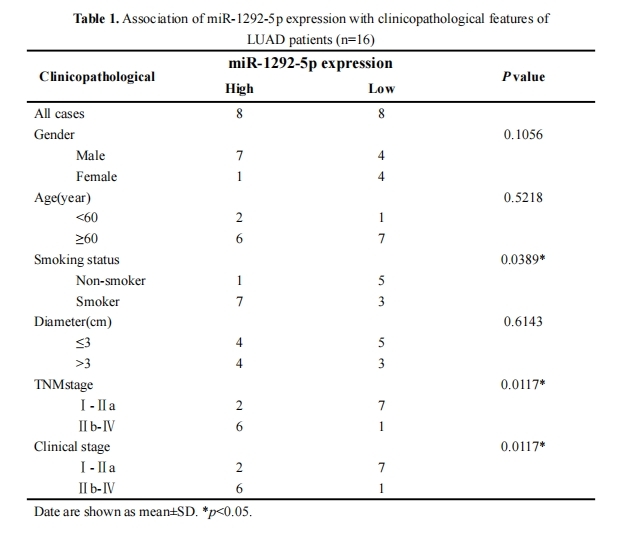
We acquired 16 pairs of LUAD and matched tumor-adjacent tissues from patients who underwent surgery without preoperative chemotherapy or radiotherapy at Affiliated Hospital of Guangdong Medical University between 2010 and 2021. All patients signed informed consent forms. All tissues were stored in liquid nitrogen. This research was ap-proved by the Ethics Committee of Affiliated Hospital of Guangdong Medical University.
2.2. Cell culture, transfection and stable cell lines
LUAD cell lines (A549, H1299, H838, PC9), human normal lung epithelial cells (BEAS-2B) and 293A cells were purchased from the Cell Bank of the Chinese Academy of Science (Shanghai, China). All cell lines were cultured with DMEM medium (Gibco, CA, USA) containing 10% fetal bovine serum at 37°C in 5% CO2. In some experiments, the BMP pathway inhibitor LDN193189 (Beyotime, Shanghai, China) was added at a concentration of 30 nM and incubated for 48 h. miR-1292-5p mimics, inhibitor, SMURF1 overexpression plasmid and corresponding negative controls were synthesized by GenePharma (Shanghai, China). RNAs were transfected into cells with Lipofectamine RNAiMAX (Invitrogen, CA, USA), while plasmids were transfected with Lipofectamine 3000 (Invitrogen). To establish stable cell lines, miR-1292-5p or control mimics were transfected into A549 cells, and cells were selected with puromycin (Sigma-Aldrich, USA) for 3 weeks.
2.3. RNA extraction and quantitative real-time polymerase chain reaction (RT-qPCR)
Total RNA was extracted from tissues and cells using Trizol reagent (Invitrogen) following the manufacturer’s instructions. miR-1292-5p and U6 were polyadenylated using a poly-A polymerase-based first-strand synthesis kit (AG, Hunan, China) following the manufacturer’s protocol. For SMURF1 mRNA and β-actin, cDNAs were synthesized by Evo M-MLV RT Premix (AG). RT-qPCR was performed on an ABI7500 (Applied Biosystems, Foster City, CA, USA) or LightCycler 480 (Roche Applied Biosystems, CA, USA). U6 and β-actin mRNA were used as internal controls for miRNA and mRNA, respectively. Primers were synthesized by Sango Biotech (Shanghai, China) and sequences are as follows: β-actin forward CACAGAGCCTCGCCTTTGCC and reverse ACCCATGCCCAC-CATCACG; U6 forward CGCTTCGGCAGCACATATACT and reverse CGCTTCAC-GAATTTGCGTGTC; miR-1292-5p UGGGAACGGGUUCCGGCAGACGCUG; SMURF1 forward TTTGGAACTGGTGGCTATG and reverse GCTGCTGGGATGTGAAA.
2.4. Transwell assays
Transfected cells were inoculated into the upper chamber of a Transwell chamber in 200 µl serum-free medium (BD Biosciences, Franklin Lakes, NJ, USA), with or without Matrigel (BD Biosciences) for invasion and migration assays, respectively. The lower chambers were filled with 750 µL medium containing 20% fetal bovine serum. After cultivation for 24 h, cells were fixed with 4% paraformaldehyde and stained with crystal violet (Beyotime). Migrated and invaded cells were imaged using a Leica fluorescent micro-scope (Leica Microsystems GmbH).
2.5. Western blot
Proteins were extracted from transfected cells using radio immunoprecipitation assay buffer containing Phenylmethanesulfonyl fluoride (Beyotime), electrophoresed on 10% SDS-PAGE gels and transferred onto polyvinylidene fluoride membranes (Millipore, Billerica, MA, USA). After blocking the membranes with 5% defatted milk for 2 h, the membranes were incubated with the following primary antibodies at 4°C overnight: anti-E‐cadherin (20874-1-AP, Proteintech, Chicago, IL, USA), anti‐N‐cadherin (22018-1-AP, Proteintech), anti‐BMPR2 (AF6327, Beyotime), anti‐P-Smad5 (AF1375, Beyotime), anti‐SMURF1 (PB0937, Boster, Wuhan, China) and anti‐GAPDH (AF1186, Beyotime). Mem-branes were then incubated with HRP-labeled secondary antibodies (PR30011, Proteintech) for 1 h. Protein signals were detected using BeyoECL star (Beyotime).
2.6. Dual-luciferase assay
Cells were inoculated into 24-well plates and cultured to 60% confluence. The 3′ UTR of SMURF1 mRNA (wild-type (Wt) or mutant-type (Mut)) was cloned into a luciferase plasmid to generate Wt-SMURF1 or Mut-SMURF1, respectively, and the plasmids were co-transfected with miRNA mimics or miRNA negative control into cells. Firefly and Renilla luciferase levels were detected using the Dual-Luciferase Assay System (Promega).
2.7. Immunofluorescence staining
Transfected cells in a confocal dish were fixed with 4% paraformaldehyde, washed with PBS and permeabilized with 0.5% Triton X-100 for 30 min. Cells were then incubated with Phalloidin (Yeasen, Shanghai, China) for 60 min at 37°C. Antifade mounting medium with DAPI (2-(4-Amidinophenyl)-6-indolecarbamidine dihydrochloride, Beyotime) was added to cells, and signals were examined and imaged by Olympus Laser confocal microscopy (Olympus Corporation, Tokyo, Japan).
2.8. Animal experiments
Four-week-old male nude mice (BALB/c) were purchased from the Medical Experimental Animal Center of Guangdong province (Guangdong, China). Mice were randomized into two controls (n = 5/group) and stably transfected A549 cells expressing miR-1292-5p or control were injected into the tail veins of the mice (1 × 10⁸ cells/ each mouse). After 4 weeks, the lungs and livers of mice were removed and weighed. Animal experiments were approved by the animal ethics committee of Guangdong Medical University.
2.9. Statistical analysis
GraphPad Prism 8.0 (GraphPad Software, USA) and SPSS ver. 26.0 (IBM, USA) were used for statistical analysis. Differences between groups were analyzed with Student’s t test or one-way analysis of variance (ANOVA). Data from at least three independent experiments were determined as mean ± standard deviation (SD). The P less than 0.05 was considered to indicate statistical significance.