3.2.1. SEM
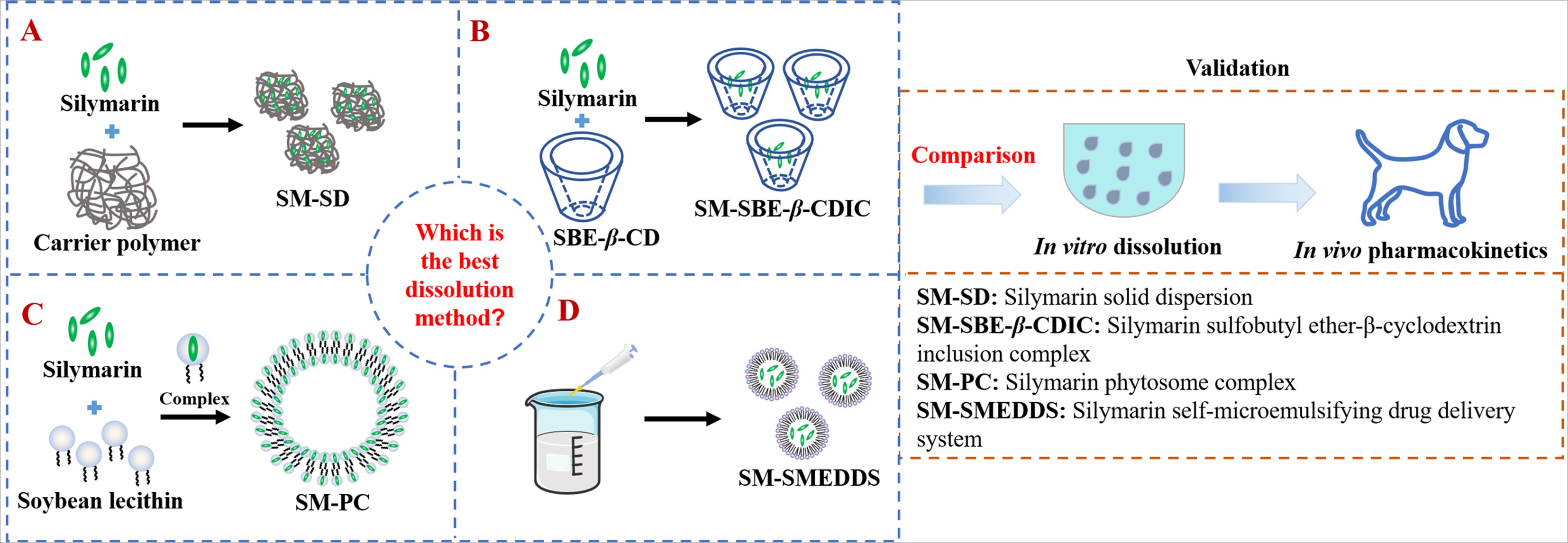
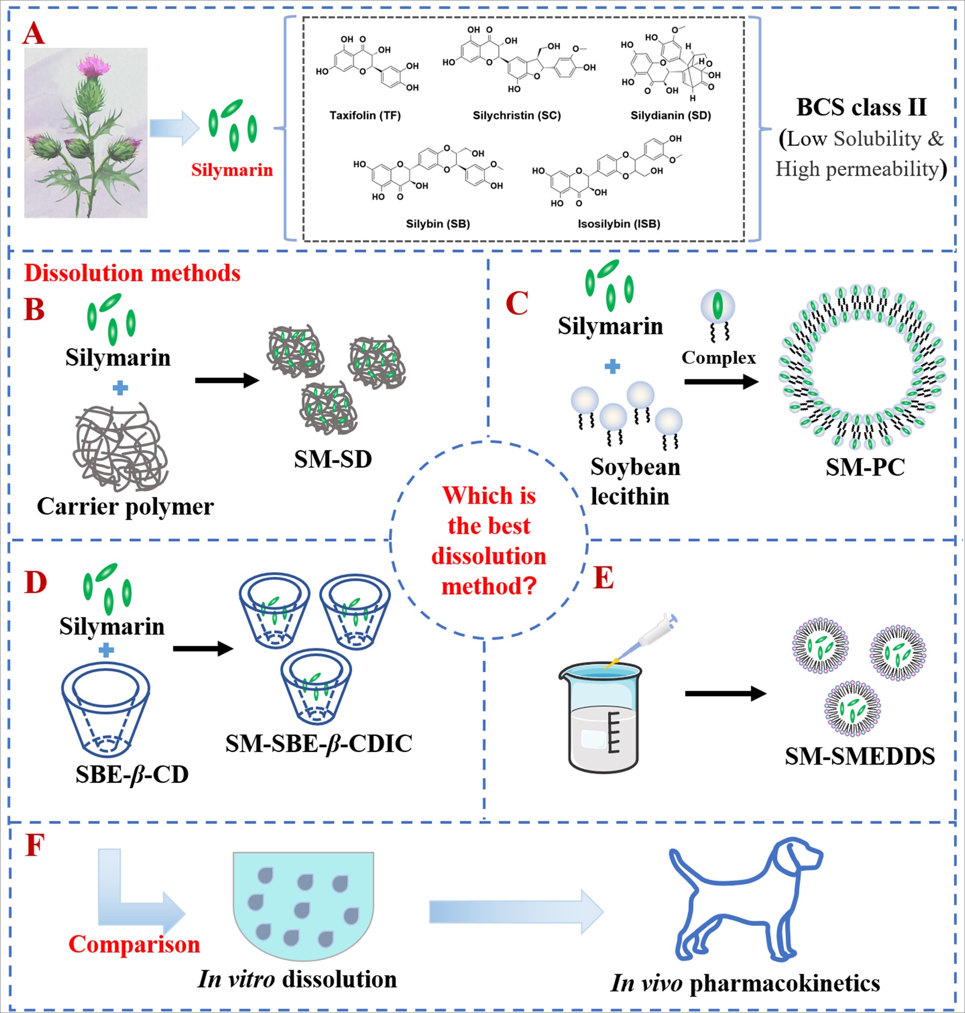
Several techniques, such as spray-drying, solvent evaporation, and hot-melt extrusion, can be employed to prepare SD[34]. Among these methods, the solvent-evaporation is particularly effective in enhancing the aqueous solubility and dissolution rate of BCS class II drugs by uniformly dispersing or encapsulating drug molecules into polymer matrices[35, 36]. Hydrophilic polymer matrices, including PVP, PEG, and F68, are frequently employed in the formulation of SD for insoluble drugs. PVP, known for its anti-crystallization properties, effectively inhibits the recrystallization of SD during storage, thereby preserving the amorphous state of the drug. Consequently, PVP is commonly utilized as a polymer of choice in the preparation of amorphous SD[37, 38]. F68, a nonionic surfactant commonly utilized in the preparation of SD, is extensively employed for enhancing the dissolution and solubilization of insoluble drugs and ensure optimal drug dispersion[16, 39]. PEG 4000, 5000, and 6000 are frequently employed in the preparation of SD due to their advantageous properties such as low toxicity, non-absorption in the gastrointestinal tract, absence of interference with drug content analysis, and ability to significantly enhance the dissolution rate of insoluble drugs[40, 41]. Nevertheless, the high viscosity of PEG poses challenges in the desiccation of the solution, thereby restricting its potential applications. Therefore, the solvent evaporation method was employed in conjunction with PVP and F68 as joint carriers to prepare SD in this study.
The morphology of SM, PVP K30, F68, physical mixture of PVP K30, F68 as well as SM, and SM-SD was assessed through SEM (Fig. 1). SM exhibited various block shapes with wide particle size distribution (Fig. 1A). PVPK30 showed amorphous character with round shapes[42] (Fig. 1B). F68 presented in irregular shapes (Fig. 1C). In the images of the physical mixture (Fig. 1D), the drug demonstrated superficial interaction, whereas in the images obtained from SM-SD (Fig. 1E), a complete alteration in morphology was observed, indicating the presence of SM in the carriers in either a molecular or amorphous form.
PC have a similar structure to liposomes, but differ in terms of pharmaceutical agent localization. In liposomes, water soluble agents are entrapped within the inner aqueous core, while in PC, the active constituents are complexed with the phospholipid head through polar and hydrogen-bonding interactions[43, 44]. In the presence of ethanol, the hydrophilic polar head of soybean phospholipids engages in hydrogen bonding interactions with the active hydrogen atom of SM (such as -COOH and -OH)[18]. This interaction promotes the integration of the SM molecule into the polar head of the phospholipid, while the hydrophobic tail portion of the phospholipid ensures the proper orientation of this complex, leading to the formation of an amphiphilic complex that enhances transportation from an aqueous environment to a lipid-soluble environment[45]. Following the formation of the PC, the constituents' membrane permeability and oil-water partition coefficient would experience a substantial increase. Moreover, drug's solubility and dissolution would be significantly enhanced[46].
The representative SEM images of SM, soybean lecithin, physical mixture of SM and soybean lecithin, and SM-PC were shown in Fig. 2. In the physical mixture (Fig. 2C), the presence of SM (Fig. 2A) and soybean lecithin (Fig. 2B) were easily distinguishable from each other. However, SM-PC (Fig. 2D) presented a significant alteration in both shape and surface morphology when compared to SM and soybean lecithin, which could be attributed to the complete miscibility of SM and soybean lecithin[47], suggesting that SM may exist in the PC in an amorphous state.
CDs exhibit high biocompatibility and are considered “generally recognized as safe” (GRAS) by the Food and Drug Administration (FDA). Among CDs, β-CD is the most commonly used. Its derivatives, such as 2-hydroxypropyl-β-cyclodextrin (HP-β-CD), 2,6-dimethyl-β-cyclodextrin (DM-β-CD), and sulfobutyl ether-β-cyclodextrin (SBE-β-CD), which substitute hydroxyl groups in the molecule's rings, have demonstrated improved solubility than β-CD. Phase solubility studies, which are widely used to determine the effect of CDs and auxiliary substances on drug solubility[48], were employed to investigate the impact of complexation on SM solubility in the presence of β-CD, HP-β-C, DM-β-CD or SBE-β-CD (Supplementary Method). The solubility concentrations of TF, SC, and SD were relatively similar among the aforementioned CDs, whereas the solubility of SB and ISB varied significantly depending on the type of CD used, with SBE-β-CD > DM-β-CD > HP-β-CD > β-CD (Fig.S2). Consequently, SBE-β-CD was selected as the preferred CD for the preparation of the inclusion complex in this study. The solubility of each component of SM exhibited a non-linear increase with increasing concentration of SBE-β-CD, within the range of 0 to 1 mM, indicating a 1 : n (n ≈ 6) complex formation (Fig.S3). The inclusion rate of TF, SC, SD, SB, and ISB was 28.18%、26.56%、28.55%、72.95%, and 27.79%, respectively. And the yield of the inclusion complex was 85.34%.
The morphology of the prepared SM-SBE-β-CDIC was observed by SEM (Fig. 3). The physical mixture of SM and SBE-β-CD (Fig. 3C) exhibited distinct characteristics of SM (Fig. 3A) and SBE-β-CD (Fig. 3B), suggesting the absence of any interactions between these two compounds following physical mixture. Conversely, a significant alteration in morphology was observed in SM-SBE-β-CDIC (Fig. 3D), manifesting as compact and uniform plate-like crystal particles, wherein the original morphology of both components was no longer discernible. This observation serves as evidence that SM and SBE-β-CD have indeed formed an inclusion complex. The alteration in morphology was also observed in the inclusion complexes of another poorly water-soluble drug with SBE-β-CD[49].
3.2.2. FTIR
FTIR, a potent technique employed for structural analysis, was utilized to analysis the interactions between drugs and polymers, as it provides information on various functional groups that exhibit unique characteristics in terms of band number, position, shape, and intensity. As depicted in Fig. 4A, the FTIR spectra of SM, PVPK30, and F68 exhibited a resemblance to the previously reported patterns[50, 51]. The SM exhibited prominent peaks at 3453 cm− 1 (-OH stretching vibration), 2936 cm− 1 (O-H stretching ), 1640 cm− 1 (C = O stretching), 1511 − 1467 cm− 1 (skeleton vibration of aromatic C = C ring stretching), 1365 cm− 1 (-OH in plane bending), 1271 cm− 1 (C-O-C stretching), 1165 − 1033 cm− 1 (in plane = C-H bending), and 996 cm− 1 (O-H out plane bending, benzopyran ring)[50, 52]. The FTIR spectrum of PVPK30 exhibited distinct absorption bands at 1670 cm− 1 (C = O) and 2954 cm− 1 (C-H stretch), respectively[53]. The spectrum of F68 shows distinct function groups at 2887 cm− 1 (C-H stretching), 1112 cm− 1 (C-O-C stretching vibrations). In both the physical mixture and SM-SD spectra, the drug and excipients displayed the characteristic absorption peaks without any emergence of new functional groups, indicating a lack of significant interaction between SM, PVP30, and F68. However, small peak shifts (1 to 3 cm− 1) were observed, suggesting the presence of weak interactions in these compounds, possibly due to hydrogen bonding or hydrophobic interactions[54].
In the FTIR spectrum of SM-PC group (Fig. 4B), soybean lecithin exhibited its characteristic absorption peaks at 2925 cm− 1 and 2854 cm− 1 (C-H, fatty acid chain), 1236 cm− 1 (P = O), 1730 cm− 1 (C = O, fatty acid ester). The spectrum of the physical mixture of SM and soybean lecithin was primarily a superimposition of the major peaks from both components. No new peaks were observed in the SM-PC sample compared to the physical mixture. However, there was a slight shift in the stretching vibration peaks at P = O and C = O, which shifted to 1276 cm− 1 and 1743 cm− 1, respectively. What’s more, the stretching vibration of -O-H (3453 cm− 1) in SM was broadened and shifted in the FTIR spectrum of SM-PC, indicating the formation of a hydrogen bond between SM and phospholipids[55]. The results suggest that the SM-PC complex does not form new chemical bonds, but rather exhibits weak intermolecular interactions, specifically hydrogen bonding, between the hydroxyl groups of SM and the carbonyl group and phosphoryl group of phospholipids[56].
As for SM-SBE-β-CDIC group (Fig. 4C), the characteristic bands of SBE-β-CD in the infrared spectra were 3423 cm− 1 (O-H stretching vibration), 2800–3000 cm− 1 (-CH and CH2 stretching vibration), 1647 cm− 1 (H-O-H bending), 1161 cm− 1 (C-O-C stretching vibration), 1043 cm− 1 (sulfoxide stretch)[57]. The spectral analysis of the physical mixture demonstrated similarity to the straightforward combination of SM and SBE-β-CD. In contrast, the SM-SBE-β-CDIC exhibited alterations in the frequency, broadening, and disappearance of the infrared bands that are distinctive to the pure drug. These changes suggest modifications in the strength and length of the drug's bonds due to the activation of certain "host-guest" interactions, indicating the formation of inclusion complexes[58].
3.2.3. PXRD
PXRD was used to determine the degree of crystallinity and amorphous nature of the components in the formulation. The PXRD patterns of SM, PVPK30, F68, physical mixture (SM, PVPK30, and F68), and SM-SD were depicted in Fig. 5A. SM exhibited sharp diffraction peaks at 18–32°(2θ), indicating that its high degree of crystallinity. PVP K30 did not generate any peak, suggesting its amorphous nature. F68 exhibited some crystallinity, as indicated by the two peaks of high intensity at 19.3° and 23.5°and other peaks of lower intensity[59]. The physical mixture exhibited peaks similar to those of F68 and SM, albeit with lower intensity. The low-intensity peaks of SM in the physical mixture may be attributed to the high percentage of F68, resulting in a dilution effect. Furthermore, the absence of any new peaks in the physical mixture indicates a lack of interaction, which aligns with the findings from the FTIR analysis. However, the peaks of SM were absence in SM-SD, suggesting that SM presented in the amorphous state in SD, which was beneficial to increase its solubility[60]. The diffraction peaks of F68 were still clearly visible, indicating that F68 remained in its crystalline form in SD, which is consistent with previous research[61].
Similarly, the SM crystal diffraction peaks were absence in both SM-PC (Fig. 5B) and SM-SBE-β-CDIC (Fig. 5C), indicating SM exists in an amorphous state within PC and SBE-β-CD.