1. Lf coupled versus uncoupled SLNs for targeting lung cancer
1.1 Colloidal properties of targeted SLNs and Morphology
The size measurements indicated gradual increase in diameter from 75.28 to 120 nm with an increase in concentration of Lf from 0 to 4 %w/v, Formula F1-F9, Figure 1A, together with a corresponding increase in PDI. This change in size might be allocated to Lf attachment on the surface of NPs. Our results were consistent with those of Shilpi et al. [15] who reported an increase in SLN size by 35 nm when Lf was coupled to rifampicin-loaded SLNs, to achieve drug targeted delivery to the lungs for tuberculosis treatment. Similarly, coumarin-6 SLNs were 66.3±0.91 and 81.16± 0.97 nm in diameter with PDI of 0.216 and 0.318, for Cou-SLNs and Lf-Cou-SLNs, respectively.
The zeta potential of MYR-CPX-SLNs was (-26.5±4.38 mV), while, for Lf-coupled SLNs the ZP values are shifted from negative to positive by increasing Lf conc. ranging from -17.7±2.73 (F1) to 0.6±0.66 mV (F10) containing 6% w/v Lf. This change could be due to the presence of positively charged groups of amines groups of Lf which is covering SLNs’ surface (Figure 1B) [27]. The reverted weak ZP reflects the doubling in PS that can be attributed to SLN agglomeration.
The surface morphology for both targeted (Lf-MYR-CPX-SLNs) and non-targeted SLNs (MYR-CPS-SLNs) appeared spherical with smooth surface as the TEM photomicrographs show (Figure 1C) while MYR-PH-CPX showed irregular vesicular structure as previously reported [21]. Interestingly, ligand-coupled SLNs (F6) were darker in accordance with photomicrographs were obtained by Shilpi et al.[15].
1.2 Confirmation and quantification of Lf coupling to SLNs
SLNs were loaded with MYR-PH complex and prepared by the hot homogenization technique as reported by Gaber et al.[21]. Lf was attached to SLNs via electrostatic interaction between anionic SLNs and cationic Lf.
Lactoferrin coupling efficiency to SLNs was confirmed by FT-IR. The FT-IR analysis of free Lf showed the distinctive protein band peaks; at 3288 cm-1 represents –N–H stretching of the amide I at 1635.815 cm-1 due to C=O stretching vibration of the peptide group and amide II at 1508.298 cm-1 due to N–H bending with contribution of C–N stretching vibrations. The amide peak has disappeared in the spectra of Lf-MYR-CPX-SLNs which was considered as further confirmation of Lf attachment and there was no shift in peak positions compared with free Lf (Figure 2). Similar observations were noticed by Yao et al. [26] who developed, bovine Lf-loaded Liposomes and SLNs.
Further, from Bradford test, in case of Lf-MYR-CPX-SLNs (F6) the concentration of unbound Lf was 0.47 mg/ml reflecting Lf coupling efficiency of 95.17 %.
1.3 Drug entrapment efficiency and in vitro release study
MYR entrapment efficiency was 97.9±0.14% to 95.3±0.5% for uncoupled and Lf-coupled SLNs (F6), respectively. This slight decrease in entrapment efficiency (%EE) may be attributed to slight leaching of the drug during the incubation process at the time of Lf attached with SLNs. Similarly, the encapsulation of Cou-6 in SLNs was found to be promising (96 and 98.4%) for both Lf-coupled and non-non coupled SLNs, respectively. These results highlighted the ability of these NPs (Cou-SLNs/ Lf-Cou-SLNs) for diagnostic and cellular trafficking purposes.
The in vitro drug release from Lf coupled and uncoupled MYR-CPX-SLNs was studied using the dialysis technique. The percent MYR release was recorded to be 71.5±1.33 % and 67.7±2.01% from MYR-CPX-SLNs and Lf-MYR-CPX-SLNs, respectively (Figure 1D). Lf might provide considerable shielding on the NP surface. Similar release behavior was reported for rifampicin and paclitaxel release from uncoupled and Lf-coupled SLNs [15,27] as well as methotrexate release from lactoferrin–dendrimer conjugates (Lf-PPI) and plain dendrimer (PPI) [28].
2. In vitro assessment on cell culture models
2.1 Anti-tumor activity
Previous data revealed 33% reduction in IC50 for MYR-phospholipid complex instead of free MYR [20]. Herein, the antitumor activity of Lf-MYR-CPX-SLNs (F6) was explored relative to MYR-phospholipid complex and MYR-CPX-SLNs.
Lf functionalized SLNs were found to exhibit significant superior growth inhibition of A549 cells (Two-way, ANOVA, p =0.000002) in comparison to non-functionalized SLNs and MYR-phospholipid complex. The viability profiles showed high % viability ranging from 60 to 89 % at low MYR concentration (20 µM), Figure 3. By comparing our results to results obtained by Rajendran et al. [29], who performed cytotoxicity study for MYR on A549 cells in concentration range from 20 to 315 µM. it was found that at low MYR content (20 µM), 92% cell viability was obtained which decreased by increasing the drug concentration and the obtained IC50 was 229 µM [29]. Further, gradual increase in concentration resulted in a corresponding reduction in viability to 10 % in case of Lf-MYR-CPX-SLNs compared to 43 % for MYR-phospholipid complex at 200 µM MYR concentration. Indeed, 2-3 fold lower IC50 value was recorded for Lf-functionalized SLNs (35.01 µM) compared to 67.29 and 113.8 µM for MYR-CPX-SLNs and MYR-phospholipid complex, respectively.
The potential toxicity of targeted SLNs might be attributed to improved cellular uptake via ligand–receptor interaction. These findings were in good agreement with research published by Pandey et al.[27] who reported that, Lf-coupled SLNs encapsulating paclitaxel (PTX) exhibited considerable higher cytotoxicity as compared to PTX-loaded SLNs. Further, PTX solution was found to be less cytotoxic and IC50 values were 7.5±0.4, 4.6±0.1 and 1.1±0.03 µg/ml for Free PTX solution, PTX-loaded plain SLNs and PTX-loaded Lf-coupled SLNs, respectively.
2.2 In vitro cellular uptake and colocalization studies
2.2.1 Effect of incubation time
The uptake of Cou-SLNs and Lf-Cou-SLNs as well as free dye solution in DMSO was investigated on A549 cells for 4 and 24 h by CLSM.
After 4 h incubation period, very weak green fluorescence comparable to control cells could be recognized in case of Cou-SLNs and free dye (Figure 4A). In comparison, Lf-Cou-SLNs exhibited distinct green spots even within this short incubation period.
Longer incubation with A549 cells (24 h) enabled better uptake of all samples including free dye, Cou-SLNs and Lf-Cou-SLNs (Figure 4A). Interestingly, stronger fluorescence signals denoting higher level of internalization could be depicted in case of Lf-Cou-SLNs. Nafee et al.[20] previously reported that the total fluorescence intensity of Cou-SLNs internalized was >2.5 times higher than that of free dye. Herein, Lf-Cou-SLNs showed ~ 4 folds higher fluorescence intensity in the cells than Cou-SLNs (Figure 4B) confirming the direct effect of ligand targeting on cellular uptake.
2.2.2 Localization of labeled SLNs in A549 cells
In order to distinguish between surface binding of SLNs and concrete internalization, 3D time laps imaging was carried out allowing imaging of 40 stacks inside the cells in the z-direction along 14 µm (Figure 4C). The Z-stacks confirm particle localization inside the cells namely within the cytoplasm and seldom dispersed in the nuclei. Lf-Cou-SLNs could be clearly detected with intense green fluorescence intensity and sharper spots within the cell vicinity indicating their possible entrapment in intracellular vesicles. This reveals the vital role of active tumor targeting via ligand-receptor binding in improved cancer nanotherapeutics. Similarly, Liu et al.[30] demonstrated that, (RRWQW) a cell penetrating peptide obtained from bovine lactoferrin, was non-covalently complexed with plasmid DNA, is able to efficiently deliver the plasmid DNA into A549 cells.
3. Nano-embedded microparticles for inhalation (Physical characterization)
For better drug deposition deeply in the lung, the nanoparticles should be converted to microparticles to overcome clearance by exhalation. Thus, in this study a two-step process successfully prepared inhalable SD-MPs with aerodynamic diameter between 1 and 5 μm.
We previously formulated SD-MPs [20] by spray drying MYR-CPX-SLNs with carrier mixture consisting of mannitol: maltodextrin and L-leucin (1.5:0.5:0.75 %w/v) (SD-MP3). A prefect yield of 89% w/w of excellent flow powder was achieved with aerodynamic diameter 2.39 µM and drug recovery of 95% [20]. The aforementioned optimized formulation was selected for the current study.
Matrix formers namely sugars (mannitol) or polysaccharides (maltodextrin), were selected because of their ability to act as drying protectants for drugs during water removal process and to shape the MP modulating particle/particle interactions [31,32]. The amino acid L-leucine was added to act as an aerosolization enhancer forming a coat (shell) on the dry particle surface preventing any particle fusion and therefore preserving the individual MP as collected from the dryer [31]. Moreover, it was employed due to its powder dispersibility enhancing effect as previously reported in many studies [32,33].
3.1 Percentage of yield recovered and drug content
It was found that, the % yield of spray-dried powder varied between 28.75 - 89.05% w/w during spray drying processing as illustrated in the supplementary material, Nevertheless, by combining maltodextrin with mannitol in either SD-MP3 or SD-MP4, an increase in the yield was slightly significant when compared to SD-MP2, (One-way ANOVA, p < 0.05) (Table S.1- supplementary material). This observation was in accordance with previous literature demonstrating that the presence of dextran as an example of oligosaccharides with mannitol. It was found that dextran was able to suppress the shrinkage and particles collapse due to the change in glass transition temperature Tg (collapse temperature). This is attributed to suffering of mannitol from low Tg besides its rubbery state might result in sticking to the spray dryer [32]. Similarly, a study reported by Kumar et al. [34] where, a combination of a low molecular weight sugar with a high molecular weight one was used to achieve higher spray drying yields. Further, MYR content in the different formulations (SD-MP1 to SD-MP4) ranged from 90.5 to 95.15 % (Table S.1-supplementary material). Thus, the active ingredient seemed to be uniformly distributed in the different powder formulations.
3.2 Particle size measurement
The size of microparticles (SD-MP1 to SD-MP4), (Dv50) was remarkably small ranging from 4.5 to 6.90 μm indicating their suitability for lung deposition. The span values (1.24 – 1.84) reflect the narrow particle size distribution as depicted in (Table S.2-supplementary material).
3.3 Morphological characterization
SEM photomicrographs of raw MYR, and SD-MPs powders (SD-MP1 to SD-MP4) were demonstrated in Figure 5A-E. The scanning electron micrograph of raw MYR (Figure 5A) revealed the presence of large crystals aggregated together with a particle size that is too large for inhalation. Spray drying MYR-CPX-SLNs with different water-soluble matrix formers resulted in change in the particle appearance. It was depicted that, presence of mannitol in (SD-MP1) showed separated spherical particles with moderately corrugated surface (Figure 5B), whereas SEM images of leucine-containing particles (SD-MP2) revealed the enhancement in both surface roughness and corrugation (Figure 5C). By virtue of leucine hydrophobicity it probably acted as a water repellent and thus reduced moisture uptake by sugars, reducing their particle cohesiveness [32,35]. By combining the high molecular weight maltodextrin with the low molecular mannitol in either SD-MP3 or SD-MP4, spherical particles with less corrugated surface were obtained (Figures D-E). The use of different ratios between mannitol and maltodextrin showed the same effect on the surface morphology of SD-MPs. This mixture succeeded in suppressing particles shrinkage and collapse owing to the change in their Tg as discussed above. Furthermore, a hard layer on the nano embedded MP surface is formed and was capable to prevent their collapse during the spray drying procedure [20].
3.4 Flow properties
Flow property is considered as one of the promoting parameters for efficient aerosolization of the spray-dried powders. The results for angle of repose, Hausner ratio and Carr’s index are shown in detail in (Table S.3-supplementary material). Further, the use of carrier formers enhanced significantly the flow properties compared to spray dried MYR-CPX-SLNs without any carrier (supplementary material). In addition, the combination of maltodextrin with mannitol in SD-MP3/SD-MP4 showed the best flow characteristic as evidenced by low θ, CI and HR values.
3.5 Drug-Excipient Compatibility Study
3.5.1 Differential Scanning Calorimetry (DSC)
DSC analysis was employed to investigate any physical change in the crystalline state and thermal behavior of MYR during the solidification process. The DSC thermograms of raw MYR powder, maltodextrin, mannitol, leucine, selected microparticles formulations (SD-MP2 and SD-MP3) were depicted in (Figure S.1A-F Supplementary material). It was shown that, no characteristic peak of drug was observed as drug is dissolved in SLNs as previously discussed in our previous work [20] while, peaks of mannitol and leucine were detected after spray drying.
3.5.2 Fourier transform infrared (FTIR) spectroscopy
For further detection of any possible change and chemical interaction between MYR and the SD-MPs in the solid state, IR spectra of MYR, mannitol, maltodextrin, leucine, SD- MPs (SD-MP1-SD-MP3) and spray dried MYR-CPX-SLNs are illustrated in (Figure S.2-Supplementary material). The FT-IR spectrum of SD-MPs showed the disappearance of characteristic MYR peaks. This could be due to overlapping of the drug by the presence of excess carriers. The same observation was exhibited by Ishak and Osman [32], where the characteristic peaks of atorvastatin disappeared in the IR spectrum of spray-dried self-micro emulsifying powders.
3.6 In vitro deposition
For efficient inhalation therapy of lung cancer, the microparticles should be delivered to tumor site with minimum/no exposure to systemic circulation. Accordingly, the aerosol performance of all MP formulae was monitored, and the results are shown in the supplementary material (Figure S.3A )SD-MP3 achieved the most promising aerosolization pattern in line with previously reported formula characterized by MMAD of 2.77 μm, an FPF of 81.23 and an EF of 93% [20]. For the in vivo deposition experiments, fluorescently labeled microparticles were prepared (SD-Cou MP) analogous to formula SD-MP3 but replacing MYR with cou 6. MPs showed an MMAD ~ 2.5 µM, FPF > 80%, and EF > 90%.
4. In vivo Pharmacodynamics (tissue deposition and organ distribution) of fluorescent labeled SLNs & SD-MPs after intravenous and pulmonary administration, respectively
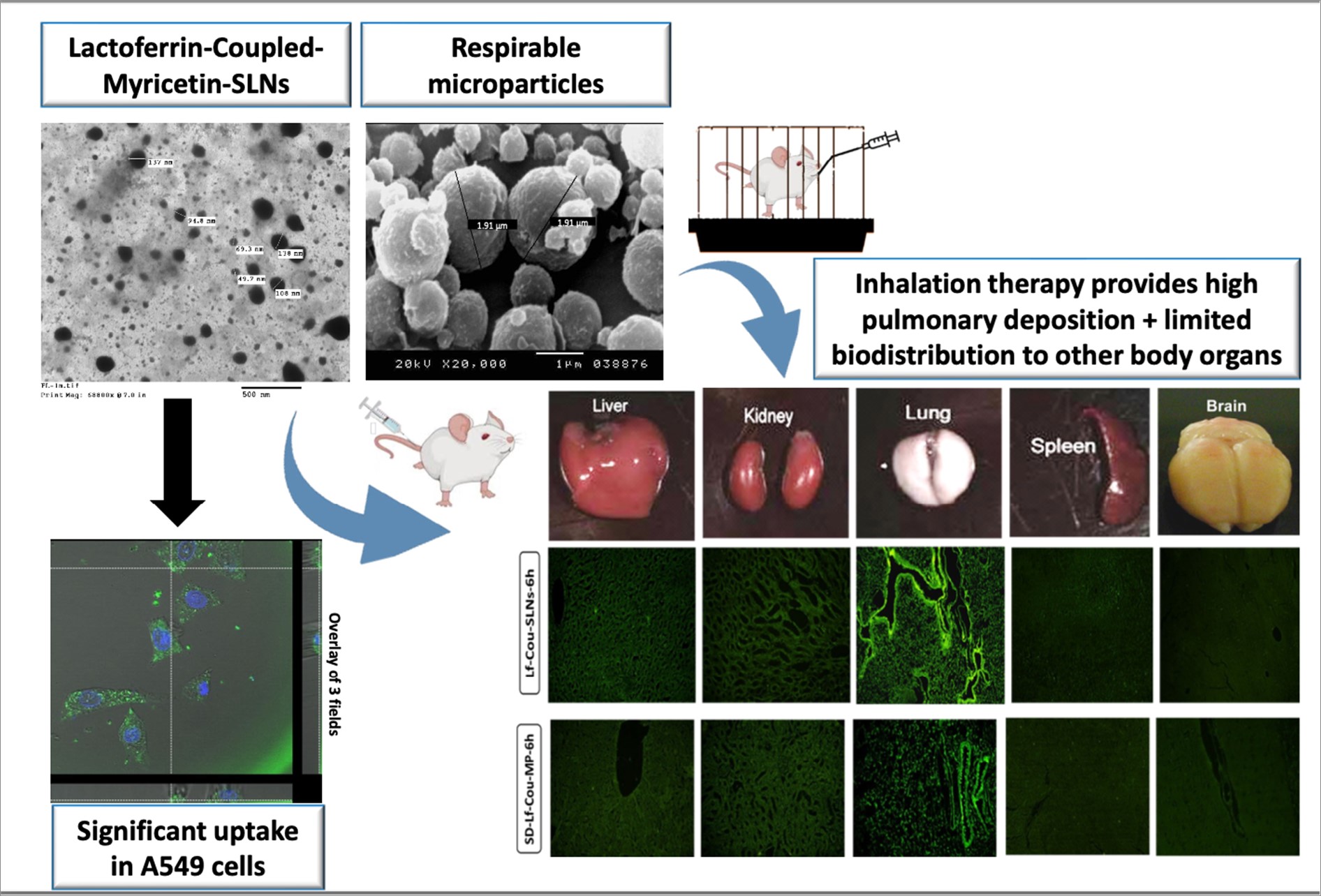
A point of interest is to explore the extent of pulmonary distribution as well as other organ biodistribution following IV and pulmonary oropharyngeal administration of both Lf-coupled and uncoupled labeled SLNs and SD-MPs in addition to free dye as control. Thus, in this section, two factors will be explored, first; the impact of the nanosystem as well as lactoferrin on pulmonary deposition. Second, comparing the two routes of administration (Local Vs. Systemic) in terms of the efficiency of in vivo lung deposition. Figures 6 and 7 represent the fluorescent photomicrographs of sections from different organs (Liver, kidney, lung, spleen and brain) 1- and 6h- following IV and pulmonary administration of the aforementioned samples.
Noteworthy, after 1 h, it was found that both IV administered SLNs (Cou-SLNs, Lf-Cou-SLNs) and inhaled nano-embedded microparticles (SD-Cou MP and SD-Lf-Cou MP) showed distinct localization in the bronchial and alveolar tissues (Figure 6A and 7A). This could be accredited to the presence of soy lecithin phospholipids in the SLN composition analogous to pulmonary surfactants phospholipids that might serve as storage bank for the inhaled lipid-based nanosystem [36]. In comparison, control MP and control SLNs loaded with the free dye also demonstrated notable accumulation in the same regions (Figure 6A &7A). this might be due to lipophilic nature of Coumarin-6, (log P 55.43), which is meant to be subtle across the trachea, airways and alveolar tissue among minutes once insufflation of the dry powder [37].
However, photomicrographs taken after 6 h revealed faster migration of the free dye (Figure 6B & 7B) followed by non-targeted fluorescently-labeled particles (Cou SLNs & SD-Cou MP). This was revealed by the remarkable increase of fluorescent particles in liver, kidney and spleen, while diminished in lungs. Noticeable, functionalizing surface of SLNs with lactoferrin (specific targeting ligand to lungs) (Lf-Cou-SLNs & SD-Lf-Cou MP) showed superior retention in the alveolar cells and protected them from clearance to other organs, which highlights the role of Lf in both targeting and retention [30].
Our results were in agreement with Pandey et al. [27] who developed, Lf-functionalized SLNs encapsulating paclitaxel (PTX) for the treatment of lung cancer. The in vivo biodistribution studies showed that concentrations of PTX accumulated in lungs was higher via Lf-SLNs > plain SLNs > free PTX after intravenous administration. These studies suggested that Lf-coupled SLNs could be used as potential targeting carrier for delivering potent drugs directly to the lungs.
In comparison, the local delivery of the free dye via inhalation was expected to place the Cou 6 in the vicinity of lung cells with less systemic exposure resulting in less toxic side effects. But unfortunately, free dye was rapidly cleared from the lung and reached other organs in high concentrations after 6h (Figure 6B). Further, loading Cou 6 into SLNs with/without Lf significantly reduced the dye deposition in other organs relative to free dye, Figure 6B. The enhanced tolerability of Cou-loaded SLNs inhalable MP over the inhalable free Cou MP might be a reason for making the pulmonary delivery of anticancer drugs loaded in nanosystems a viable option and significantly increased the therapeutic effect of the treatment. On the other hand, a higher accumulation of dye was recognized in the liver, kidney and spleen 1 and 6 h after IV administration than local application which might be a reason for organs toxicity associated with systemic application.
As a proof of concept, for more quantitative comparison between pulmonary and IV administration, the mean fluorescence intensity in the photomicrographs was analyzed using Image J software and illustrated in Figures 8A-B. After 1 h, the fluorescence intensity was found to be 259 and 345 gray for SD-Cou MP and SD-Lf-Cou MP, respectively compared to 187.9 and 221.15 gray for Cou-SLNs and Lf-Cou-SLNs. This gives an idea that % of drug deposition in lung after pulmonary administration was ~ 1.5 folds higher than IV injection. However, fading in intensity was noticed after 6 h in case of SD-Cou MP (Figure 8B). This might be related to the fate of SLNs that might be taken by alveolar macrophages which, are responsible for clearance of materials deposited in the alveolar region, in which mucociliary clearance is absent [38,39]. Another explanation could be related to transcytosis of SLNs into the epithelial cells and/or across the epithelia of the respiratory tract into the interstitium and then to blood and lymph might be involved till they reach extra-pulmonary organs [40,41].
This confirmed our findings that by functionalizing surface of SLNs with specific targeting ligand to the lungs aided in maintaining the particles in the lung and protected them from clearance to other organs. Similarly, the fading in fluorescence intensity in lungs occurred in case of Cou-SLNs while, that of Lf-Cou-SLNs remained almost unchanged (Figure 7A). Therefore, form the previous results it could be concluded that the role of Lf wasn’t only exerted on specifying the targeting of nanosystem to lungs in shorter time but also, maintaining it for longer duration and evading their clearance from lungs by macrophages.
Thus, the present study showed that SD-MP loaded with SLNs and coupled with Lf could be effective in treating lung cancer and without targeting ligand, it might have the potential to be used in cancer treatments of organs other than the lung with less invasive procedure than intravenous administration.

