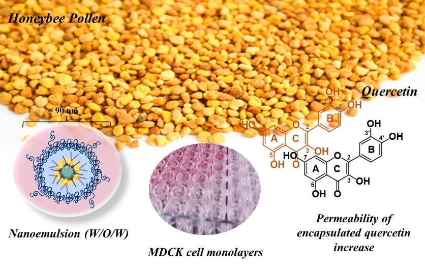
HBP samples were collected in Chile's V Region during the dry seasons of 2021 and handed to our research team still fresh in vacuum packs. Then, the samples were frozen at -20°C and the palynological method described in the Chilean Normative (NCh3255, 2011) was followed to elucidate the botanical origin of the samples (Bridi et al., 2019). The HBP extract was prepared using ethanol and ultrasonication according Bridi et al. (2019; 2022) and is described in (Supporting Information, S1). W/O/W nanoemulsions were developed in two emulsification steps and optimized using an iterative mathematical model (Cortés-Ríos et al., 2020). The detailed procedures of the assay are provided (Supporting Information, S2). The formulations with HBP were referred to as HBP-MNE, whereas those HBP-free (only ethanol as the inner aqueous phase) were referred to as MNE. HBP and HBP-MNE samples were analyzed using HPLC-DAD as previously described by Bridi et al. (2019; 2022). The compounds were quantified based on a calibration curve and a quercetin standard. Details of the method are provided (Supporting Information, S3).
MDCK-wt cells were cultured at 90% relative humidity, 37°C, and 5% CO2 atmosphere. The culture medium consisted of DMEM (Dulbecco´s Modified Eagle´s Media) supplemented with 10% FBS (Fetal Bovine Serum), 1% antibiotic/antimitotic solution, 1% NEAA, and sodium pyruvate (1mM). Spent media was replaced every two days and cells were subcultured every four days or when 90% confluence was reached. Cells were seeded onto 96-well plates (200 µL) at 35,000 cells/well and incubated for 24 h at 37°C, 5% CO2, and 90% humidity. The spent medium was removed, and monolayers were washed three times with PBS (100 µL) followed by the addition of 200 µL of either HBP (20 mg/mL) or HBP-MNE (30 mg/mL). The transport buffer was used as a positive control and the culture medium alone (no cells) served as a negative control. Monolayers were incubated either for 30 min or 2 hours. Then, the treatment solution was aspirated, cells were washed three times with 100 µL of PBS, fresh culture media (200 µL) was added, and the plates were incubated for an additional 24 hours. Next, 20 µL of Alamar Blue® commercial kit were added to each well. After 3 hours of incubation, the absorbance was read at 620 nm in a microplate reader (800TS, Biotek).
Wt-MDCK cells were seeded onto Transwell® inserts (1.12 cm2, 60,000/insert) and placed in 12-well plates. The tight-junction competence was monitored by comparing atenolol permeability (Papp) values obtained from parallel transport and uptake studies. Cell monolayers were considered suitable for use after 7–10 days post-seeding. For the assay, the culture media was aspirated, and cells were washed three times with Hank´s Balanced Saline Solution (HBSS) pH 6.8, 10 mM HEPES (transport buffer) in both compartments. Then, 500 µL of a donor solution (n = 9) was pipetted into the apical compartment of the monolayers, and 1.5 mL of transport buffer was added in the basolateral compartment. Plates were incubated at 37°C, 50 rpm, for 2 hours in a shaker TOU-120, MRC. Inserts were transferred into an empty plate to stop the transport. A sample of each compartment solution was collected, transferred to an HPLC vial, and diluted either in 100X (donor solution) or 2x and 10x (receiver solution) at 50:50 (mobile phase A: mobile phase B). Quercetin was quantified by UPLC-MS/MS is included (Supporting Information, S4).
Statistical Analysis
The apparent permeability of each analyte was calculated based on Fick's first law for steady-state systems in sink conditions as per the following Eq. (1):
$${P}_{app}=\frac{{C}_{R}}{{C}_{D}}\bullet \left(\frac{{V}_{R}}{dt\bullet A}\right)$$
1
where:
CR is the receiver concentration after incubation (µM), CD is the donor concentration at time zero (µM), VR is the volume of the receiving compartment (1.5 mL), dt is the duration of the experiment (7200 s), and A is the area of the monolayer (1.12 cm2).
Mean Papp values, standard deviations (SD), and the standard error of the mean (SEM) were determined (n = 9). The mean Papp of HBP-MNE was compared to that of the control (HPB) by a two-tailed paired Student’s t-test. A p-value of < 0.05 was considered statistically significant.